Author: Logan Young

  • New CMS Quality Measure for CT Radiation Dose Draws Scrutiny

    New CMS Quality Measure for CT Radiation Dose Draws Scrutiny

    An intersocietal panel of experts in CT convened by the American Association of Physicists in Medicine (AAPM)—with representation from clinical practice, academia, and industry input from Siemens Healthineers and Canon—examined a new performance measure in the quality-based payment programs of the Centers for Medicare & Medicaid Services (CMS). Publishing their findings in the American Journal of Roentgenology [1], the panel identified 20 important issues and ambiguities with the new measure, which became effective this year.

    Collectively, these issues reflect unclear definitions, opaque methodologies, technical and legal barriers, and potential misalignment with clinical realities—posing significant obstacles to consistent, equitable, and scientifically valid implementation across diverse care settings.

    Ambiguity surrounds where reporting is required versus optional and exactly which adult study types qualify, compounded by difficulties in consistent inpatient versus outpatient categorization. Terminology inconsistencies and unclear mapping of studies to dose and image quality categories add to the confusion. Meanwhile, patient size assessment methodology and calculation of size-adjusted dose diverge from established standards, while noise measurement lacks a recognized protocol. Criteria for excluding studies and handling combination studies remain undefined.

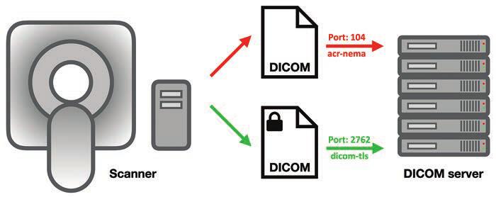
    Then, there are the tech queries: is HL7 EHR connectivity mandatory, are alternative data transfer mechanisms even feasible, what potential IT burdens and/or security liabilities will radiology practices have to shoulder? Also, performance expectations for compliance thresholds are unspecified, as are methods for comparing diverse protocols under a single set of thresholds. Identical thresholds across different categories raise additional questions.

    “Transparency and stakeholder engagement are essential for effective quality initiatives in medicine,” said Mahadevappa Mahesh, MS, PhD, president of AAPM.

    Dr. Mahesh | President, AAPM

    “We wrote this paper to call attention to issues and ambiguities with the CMS measure, and we look forward to working with CMS to address these issues and continue the culture of quality and safety that has developed in CT imaging over the past two decades.”

    Balancing Image Quality and Patient Safety

    One of the benefits to patients that will come from “The New CMS Measure of Excessive Radiation Dose or Inadequate Image Quality in CT: Issues and Ambiguities—Perspectives from an AAPM-Commissioned Panel” in AJR is that the expertise of the entire imaging community will be used to develop quality improvement initiatives that will keep radiation doses as low as possible while maintaining the quality of medically essential CT imaging. From physicians and physicists to technologists, regulators, and business leaders, “we’re confident that we can get this right by working together,” said Dr. Mahesh.  

    Technology Has Already Lowered Doses

    A lifesaving technology used to diagnose disease and guide treatment, CT is the first-line imaging technique in many cases, especially in emergency departments and cancer centers. Concerns have been raised about the increased utilization of CT in medicine because the modality uses ionizing radiation, which at very high doses is known to increase a patient’s risk for developing cancer. However, at the low doses of radiation utilized in medical imaging, including in CT, the risk is extremely small—perhaps negligible.

    Over the past two decades, imaging and allied health professionals have collectively worked to reduce CT doses. New scanner technologies have played a starring role in decreasing doses, including features that automatically measure the size of the patient and adjust the radiation dose to the right value. This is especially important for children, who require lower doses than adults due to their smaller size.    

    Dr. McCollough | Prior President, AAPM

    “Some authors multiply the very small potential risk of a CT scan by the millions of patients who receive one and predict that we will see an increase in cancer,” said Cynthia McCollough, PhD, past president of AAPM.

    “This can lead to alarmist stories and patients who really need a CT refusing to get one. Further, at the low doses we are talking about, it is debated whether the risk is even real. CT has been around for over 50 years and the predicted increases in cancer just aren’t being seen.”

    Editorials Stress Ticking Clock, Call for Clarity

    In her accompanying AJR editorial, Stephanie Leon, PhD, of the University of Florida in Gainesville, noted that “quality-based payment programs will be impacted starting in January 2027,” which means that imaging has two years and counting to figure all of this out [2].

    CMS Quality Reporting ProgramCMS Payment SystemReporting RequirementTimeline
    Hospital IQR ProgramHIPPSOptional. Hospitals are required to report three eCQMs self-selected from a list and three eCQMs mandated by CMS. The measure will be available on the self-selection list and thus its reporting is optional.Reporting will begin in CY 2025; CY 2025 results will impact FY 2027 payments.
    Hospital OQR ProgramHOPPSRequired. Once the measure is fully implemented, hospitals will be required to report the measure.Reporting will be voluntary in CY 2025 and mandatory in CY 2027; CY 2027 performance will impact CY 2029 payments.
    MIPSᵃMPFSOptional. Participants are required to report six MIPS quality measures, including at least one outcome measure, that are self-selected from a list (possibly a specialty-defined measure set depending on the reporting mechanism). If more than six measures are available, then reporting the measure is optional.Reporting will begin in CY 2025; CY 2025 results will impact FY 2027 payments.

    IQR: Inpatient Quality Reporting; OQR: Outpatient Quality Reporting; MIPS: Merit-based Incentive Payment System; HIPPS: Hospital Inpatient Prospective Payment System; HOPPS: Hospital Outpatient Prospective Payment System; MPFS: Medicare Physician Fee Schedule; eCQM: electronic clinical quality measure; CY: calendar year; FY: fiscal year

    aApplies to clinicians and clinician groups

    Another AJR editorial written by Kishore Rajendran, PhD, of the Mayo Clinic in Rochester, MN, and chair of the working group on the physics of quantitative imaging at AAPM, called for improved transparency, too. “A nonproprietary, community-based approach is imperative to ensure full transparency, achieve consensus among CT stakeholders, and provide reliable clinical diagnoses at the lowest radiation dose possible,” wrote Dr. Rajendran [3].  

    Watch as AJR senior author Ehsan Samei, PhD, and first author Jered R. Wells, PhD, call for a fundamental shift toward open-source, open-access, consensus-based, and community-owned strategies and resources to ensure quality and safety of CT: YouTube.com/@AJR_Radiology


    References:

    1. Wells JR, Christianson O, Gress D, et al. The new CMS measure of excessive radiation dose or inadequate image quality in CT: issues and ambiguities—perspectives from an AAPM-commissioned panel. AJR 2025 May. doi: 10.2214/AJR.24.32458
    2. Leon, SM. CMS measure on CT dose and image quality: good intentions, but not quite ready for prime time. AJR 2025 May. doi: 10.2214/AJR.25.32908. 
    3. Rajendran K. Transparency and stakeholder engagement as cornerstones for effective quality initiatives in medical imaging. AJR May. doi: 10.2214/AJR.25.32859

  • The Fishbowl Test: Grading Cervical Canal Stenosis

    The Fishbowl Test: Grading Cervical Canal Stenosis

    When it comes to the cervical spine, cord integrity matters most. Even mild changes can spell trouble if the cord is compromised.

    The Big Picture

    Cervical canal stenosis isn’t just about the degree of narrowing; it’s about whether the spinal cord, itself, is at risk, too. Even without measurable stenosis, cord flattening can cause myelopathy. Understanding Dr. Lea Alhilali’s fishbowl analogy helps clarify how to distinguish mild, moderate, and severe cases.

    Key Takeaways

    • Cord first: Regardless of canal narrowing, deformity or signal changes in the cord point to a higher risk of myelopathy.
    • Not just static: Static imaging may underestimate the impact; dynamic forces, repetitive microtrauma, or microischemia may drive symptoms.
    • Dr. Alhilali’s fishbowl analogy . . .
      • Mild stenosis:
        • Either ventral or dorsal CSF is effaced, but the cord still has room to “swim.”
      • Moderate stenosis:
        • Both ventral and dorsal CSF are lost, restricting cord movement.
      • Severe stenosis:
        • No CSF remains—cord is compressed, “fish” crushed.

    Challenges Ahead

    • Why cord flattening causes myelopathy without stenosis remains unclear, and mechanisms are still debated.
    • Dynamic assessment may offer better insight than static MRI but isn’t standardized.
    • Management depends on correlating imaging with clinical findings, which are often nuanced.

    Bottom Line

    Think of the cervical cord like a fish in a bowl: it needs space to move. Once the CSF “water” is gone, the cord, as well as the patient, suffers. Classifying stenosis by available space—not merely narrowing—sharpens diagnostic accuracy and clinical relevance.

    This lecture comes from The ABC’s of Degenerative Spine Imaging by Lea Alhilali, MD, part of ARRS’ Neuroradiology Longitudinal Course: Essential Topics and Cases for Everyday Practice.


    Next in the series: THURSDAY, OCTOBER 30


    Seizure, dementia, and metabolic disorders—an intersection of neurology, psychiatry, and radiology.

    Lecture Title  Speaker NameLecture Start Time 
    Welcome Charlotte Taylor12:00 PM
    Altered Mental Status, Seizure and Beyond Abdel Mahammedi12:05 PM
    Epilepsy Detection Erik H. Middlebrooks12:30 PM
    Non-AD Dementia Disorders Petrice Cogswell12:55 PM
    Alzheimer’s Disease and New Therapeutics Suzie Bash1:20 PM
    Q and A 1:45 PM

  • Delegate Decisions: Three Key Takeaways From the AMA Meeting

    Delegate Decisions: Three Key Takeaways From the AMA Meeting

    Reiterating, the house of radiology’s influence in shaping our nation’s health care policy writ large, the American Medical Association (AMA) House of Delegates (HOD) advanced several measures with significant implications for American Roentgen Ray Society (ARRS) members during its own annual meeting in Chicago this June.

    In short: expect DICOM mandates to simplify imaging transfers, elevated oversight of AI, and more rigorous validation for CT-based calcium scoring [1].

    Finally, Federally Interoperable DICOM

    The HOD passed a pivotal resolution calling on AMA advocacy for federal health IT interoperability standards to include the DICOM format, a critical “missing link” that delegates have long championed. Despite over two decades of EHR development and federal mandates, DICOM has been excluded from formal interoperability frameworks. As a result, radiological images frequently cannot travel seamlessly through EHR systems, frustrating patients and providers alike. For one example, patients arriving for mammograms at new facilities are often dumbfounded that previous studies cannot be accessed digitally from elsewhere. The absence of interoperable imaging standards contributes to delayed care, redundant exams, unnecessary radiation exposure, and burdens for patients. And the security risks are legion (Fig. 1).

    Fig. 1—Schematic shows DICOM server, computers that can exchange and store DICOM objects. Server offers DICOM service, which is software that can send and receive DICOM messages, running via specific computer ports (i.e., communications channels). Secured DICOM service is known as dicom-tls (port 2762), which uses transport layer security for negotiations, authentication, and encryption. A service that cannot be queried by hackers because it uses strong authentication mechanisms, this service sends and receives encrypted DICOM messages that cannot be read by hackers either. However, this is only true for manufacturers that have chosen to implement its strong authentication and encryption features. Arrows show direction of data transmission.

    Spearheaded by neurology and orthopedic associations, this resolution urges inclusion of DICOM in the U.S. Core Data for Interoperability (USCDI) and seeks regulatory action requiring EHR and imaging archive vendors to support secure, efficient exchange of DICOM data. Testimony also highlighted policy fissures stemming from the Health Information Technology for Economic and Clinical Health (HITECH) Act of 2009, which exempted radiologists—alas, not viewed as patient-facing—from certain data-sharing requirements, thereby keeping imaging outside USCDI [2]. A significant win for medical imagers across the country, delegates did have to amend the resolution to get it passed, but this is meaningful progress toward closing the interoperability gap that hampers timely, coordinated, and secure care.

    More Oversight and Transparency for AI

    With AI digging even deeper into the specialty—at last count, over 75% of the more than 1,000 algorithms cleared by the Food and Drug Administration target radiology [3]—the HOD continued sounding the alarm on the “black box” nature of so many machine-learning, deep-learning, and radiomic systems. Resolution 519, though not adopted thanks to too much overlap with AMA’s existing AI policies, successfully highlighted acute issues of explainability, advocating for evidence-based, transparent AI within a deliberately structured framework [4]. Aligning with AMA’s stance that the physician’s expertise remains central to clinical decision-making, everyone in Chicago agreed that today’s radiologists must be able to comprehend and articulate how generative AI, agentic AI, or some future proprietary amalgam of the two arrives at any given verdict. All too often, seemingly slight updates to vendor hardware, scanning protocol, or patient demographics end up altering algorithmic performance, further underscoring the need for responsible vetting and robust monitoring of AI.

    No LDCT for Coronary Calcium, Yet

    AMA also tabled a resolution regarding expanded promotion and usage of low-dose CT (LDCT) to screen both lung cancer and coronary artery disease via coronary calcium scoring. Emphasizing the modality’s value as a public health tool for high-risk individuals, particularly those with pack-year history of smoking, LDCT delivers far less radiation than standard CT and can detect small lung nodules early. Indeed, crucial research from the National Lung Screening Trial shows it can reduce lung cancer mortality by up to 20% [5].

    And yet, uptake is still cripplingly low; fewer than 6% of eligible patients receive LDCT screening. To buttress the resolution’s goals, the American College of Radiology is launching complementary efforts, including expanding its early lung cancer registry to capture incidental findings from routine CTs, not just formal screening exams [6]. Such distinction will deepen insights into nodule detection and follow-up.

    Fig. 2—73-year-old patient who underwent lung cancer screening by LDCT of the chest. Axial CT image shows coronary artery calcification (CAC). CAC was assessed as severe by consensus visual analysis.

    More broadly, many hospitals have begun offering low- or no-cost LDCT screenings as an entry point for preventive care. Apropos, this resolution specifically solicits a coordinated national effort of public awareness campaigns and provider education to ensure affordable, widespread access to this potentially life-saving tool.

    For further details about the 2025 Annual Meeting of the HOD, click here.


    References:

    1. ACR highlights key AMA meeting measures. Aunt Minnie website. www.auntminnie.com/practice-management/associations/news/15749013/acr-highlights-key-ama-meeting-measures. Published June 20, 2025. Accessed August 13, 2025
    2. Fornell D. Radiologists call on AMA to push for new federal IT interoperability standards. Radiology Business website. radiologybusiness.com/topics/medical-imaging/radiologists-call-ama-push-new-federal-it-interoperability-standards. Published June 24, 2025. Accessed August 13, 2025
    3. Carey L. Radiology drives July FDA AI-enabled medical device update. Aunt Minnie website. www.auntminnie.com/imaging-informatics/artificial-intelligence/article/15750598/radiology-drives-july-fda-aienabled-medical-device-update. Published July 14, 2025. Accessed August 13, 2025
    4. Specialty and Service Society (SSS) 2025 Annual Meeting of the House of Delegates. AMA website. www.ama-assn.org/system/files/a25-sss-agenda-resolution-grid.pdf. Accessed August 13, 2025
    5. National Lung Screening Trial. NIH National Cancer Institute website. www.cancer.gov/types/lung/research/nlst. Accessed August 13, 2025
    6. Fornell D. AMA resolution backs expanded low-dose CT screenings for lung cancer, heart risk. Radiology Business website. radiologybusiness.com/topics/medical-imaging/computed-tomography-ct/ama-resolution-backs-expanded-low-dose-ct-screenings-lung-cancer-heart-risk. Published June 20, 2025. Accessed August 13, 2025
  • Honoring the Life and Work of Leonard Berlin, MD (1935–2025)

    Honoring the Life and Work of Leonard Berlin, MD (1935–2025)

    Leonard Berlin, MD (1935–2025), professor emeritus at Rush Medical College and the University of Illinois College of Medicine, was a truly towering figure among radiologists, uniquely celebrated for his clinical acumen and his groundbreaking medicolegal editorial.

    His peaceful passing on Wednesday, September 3, aged 90, was mourned widely across the imaging community.

    Hired by the 10th Editor in Chief of the American Journal of Roentgenology (AJR), the late Lee Rogers, to usher in a new era for the publication, Dr. Berlin’s more than 225 AJR articles repeatedly addressed the delicate intersection of radiologic ethics and errors—fundamentally shaping our specialty’s approach to risk and responsibility.

    Among the 2002 ARRS Gold Medalist’s most cited contributions is “Radiologic Errors and Malpractice: A Blurry Distinction,” published in the September 2007 issue of AJR. In this reflective review, Dr. Berlin deftly unpacks the nuanced differences between unintentional diagnostic lapses and actionable negligence, prompting readers to consider both systems-based improvements and individual accountability.

    Equally notable is another AJR manuscript from that year advocating for greater transparency in “Communicating Results of All Radiologic Examinations Directly to Patients: Has the Time Come?.” Here, Dr. Berlin asks a simple, albeit pointed question: whether direct communication with our patients is long overdue. In the signature style he cultivated within the pages of “the yellow journal,” he answers dually that clarity and timeliness when conveying findings enhances care and defensibility alike.

    Of course, Dr. Berlin’s commitment to ethical reflection continued well into recent years. His 2020 AJR manuscript, “Medicolegal–Malpractice and Ethical Issues in Radiology,” offers an updated meditation on evolving legal and moral challenges facing radiologists in the age of digital record-keeping and shifting standard-of-care expectations. His myriad thought pieces on topics as diverse as outcome bias and the perils of defensive medicine reminded ARRS members, as well as the whole House of Radiology, that imaging isn’t solely about what’s on the film or the screen in front of us; indeed, it is also about understanding responsibility and the very human impact of our mistakes.

    Dr. Berlin’s name endures, too, through the Leonard Berlin Scholarship in Medical Professionalism, which was administered by ARRS’ own The Roentgen Fund®. From more than a decade, the “Lenny Scholarship” helped to cultivate so many leaders in the field, including AJR’s present Editor in Chief, Dr. Andrew Rosenkrantz.

    “Dr. Berlin’s made an immeasurable impact on radiology through his decades of commentaries, editorials, talks, and other contributions, to share his deep knowledge and experience on medicolegal issues, communications, and ethics,” said Dr. Rosenkrantz. “All of us in radiology learned and benefitted from his unique insights and perspectives in these areas. He will be greatly missed.”

    An “extraordinary leader, educator, mentor, and friend to so many,” 2019 Berlin Scholar Dr. Richard Duszak wrote in remembrance.

    Leonard Berlin’s funeral will be held this Sunday, September 7, at 1:30 PM Central at Beth Hillel Bnai Emunah in Wilmette, IL. In lieu of flowers, the family has requested that contributions be made to the Jewish United Fund.