Chief Radiology Resident, Western University Chair, ARRS Resident Advisory Subcommittee
Published July 15, 2020
Like many facets of life, radiology residency has not been immune to a new reality wrought by the coronavirus disease (COVID-19) pandemic. As one of the first cities in Canada to have a confirmed case of COVID-19, the novel coronavirus was certainly top of mind among our residents, fellows, faculty, and staff at Western University in London, Ontario, Canada. As residents, we worked closely with our program director to advocate for necessary changes to our program to ensure our safety while continuing our education during this pandemic.
As our government mandated physical distancing and temporary business closures, our department also acted in concert and canceled most outpatient imaging. As a result, we noticed a sudden drop-off in cases for residents to report during the day. While after-hours resident call duties remained, we modified our daily work routine for two months by implementing a schedule of alternating a week of working in hospital and a week of self-studying at home. Importantly, we continued to receive dedicated radiology teaching as our “hot seat” case conferences and academic half-day lectures proceeded as scheduled via video conferencing.
There was also the looming anticipation of our redeployment to other clinical services. This has not yet transpired, largely due to public health policies that have slowed community transmission for the time being. Fortunately, we have had access to appropriate personal protective equipment when caring for patients. Stress management has indeed been especially crucial to our wellness during this trying time. I believe that constant, albeit distanced, social interaction with colleagues and friends has helped emphasize our shared experiences and that “we are all in this together.”
The Greek philosopher Heraclitus once said, “change is the only constant in life.” During this harrowing time, this axiom still rings true with our residency training as we adapt to this pandemic.
Professor of Radiology NYU School of Medicine Center for Advanced Imaging Innovation and Research Laura and Isaac Perlmutter Cancer Center
Published June 22, 2020
Breast cancer is the most common cancer in women worldwide with approximately 2 million cases diagnosed each year. In the United States, breast cancer is the second leading cause of cancer-related mortality among women. Multiple studies showed that regular screening mammography reduced breast cancer mortality by 40% or more. However, this year, we learned that men at high risk of developing breast cancer may benefit from mammography.
Screening of Men at High Risk for Breast Cancer
Researchers at NYU School of Medicine conducted the largest review in the United States of the medical records of men who have had a screening mammogram. The study involved 1,869 men, ages 18 to 96, who had a mammogram between 2005 and 2017. Some men sought testing because they felt a mass in their breast, while others had no symptoms and wanted to be screened because a family member had recently received a breast cancer diagnosis. In total, 41 men were found to have breast cancer, as confirmed by breast tissue biopsy. Among the 271 men who had screening exams, 5 had the disease. All those with breast cancer had surgery (mastectomy) to remove their tumor. A key finding was that mammography was more effective at detecting cancer in men with high risk than is the norm for women with average risk of breast cancer. For every 1,000 exams in these men, 18 had breast cancer. By contrast, the detection rate for women is roughly 5 for every 1,000 exams.
Among the study’s other main findings was that men who had already had breast cancer were 84 times more likely to get it again than men who had no personal history of the disease. Men with an immediate relative who had breast cancer, such as a sister or mother, were three times more likely to develop the disease. Other men with elevated risk of breast cancer included those of Ashkenazi descent, an ethnic group widely known for high rates of some cancers (who were 13 times more likely to get breast cancer than non-Ashkenazi men) and those who had genetic mutations, such as BRCA1 or BRCA2 (up to 7 times more likely than men with no genetic risk). Current National Comprehensive Cancer Network Guidelines only recommend checking for breast cancer as part of annual physical exams, not using more sensitive imaging tests like a mammogram, for men age 35 and older with BRCA mutations. The take-home point is that men need to be more aware of their risk factors for breast cancer and that they, too, can develop the disease.
Contrast Enhanced Spectral Mammography
Another exciting development is that the armamentarium that radiologists have to detect breast cancer continues to expand. The latest imaging tools focus on functional imaging tests that reveal physiological activities within the breasts. Functional imaging includes measuring changes in metabolism (e.g., PET/CT or PET/ MRI), changes in the tumor cellularity (diffusion weighted imaging on MRI), regional chemical composition (MR spectroscopy, sodium MRI) and changes in blood flow. The increase in the number of blood vessels (neoangiogenesis) and the increased permeability of blood vessels that feed a tumor are the two main factors that account for uptake of gadolinium that leads to the enhancement of breast cancers on MRI. But MRI is expensive, and we know that iodine based contrast may also be used to detect breast cancer. As a result, contrast enhanced spectral mammography (CESM) is being used in both the screening and diagnostic setting. Similar to breast MRI, CESM identifies the increased blood flow associated with breast cancer and is largely independent of breast density.
Compared with MRI, clinical implementation of CESM is much easier and at much lower costs. CESM requires an intravenous injection of iodinated contrast (dose 1.5 ml/kg) and a dual energy mammography system. A “low-energy” acquisition image resembles a normal mammogram, whereas a “high-energy” image, using a keV above the k-edge of iodine, will enhance with the contrast agent signal. Images are performed in the standard craniocaudal and mediolateral mammographic views. The postprocessing, recombined iodine-only mammograms will identify enhancing lesions. Studies show that CESM has good diagnostic accuracy when used to evaluate the extent of disease in women newly diagnosed with breast cancer. Like breast MRI, CESM outperforms combined mammography and ultrasound in the detection of additional disease and in assessment of tumor size, compared with pathology. The literature reports that CESM has a small reduction of sensitivity, while providing a higher specificity compared with breast MRI in the evaluation of tumor extent.
In addition, CESM works well as a screening exam with an additional cancer detection rate 6.6 – 13.1/1,000 over conventional mammography. Therefore, it may be feasible to screen a larger population of women (e.g., women at moderately increased risk for breast cancer), and it may be advantageous to use CESM over breast MRI in this subset of women. Overall, CESM is a safe technique with a modest increase in the radiation dose compared to conventional mammography. Also, serious adverse contrast reactions are infrequent. Currently, the lack of a CESM-compatible biopsy device for lesions exclusively seen on CESM is a limitation of this new technique.
Abbreviated Breast MRI and Ultrafast MRI
Recently, many authors have evaluated the potential of an abbreviated breast MRI to increase the accessibility of breast MRI, especially for the screening of women at above-average risk for breast cancer. The conventional breast MRI exam is a 30-minute examination that is expensive and not well tolerated by some patients. These factors, along with the limited availability of MRI scanners, preclude population-wide screening with breast MRI. Abbreviated MRI, with shorter image acquisition and interpretation times, may increase the availability of breast MRI and reduce the costs. The basic abbreviated breast MRI protocol includes a pre-contrast and one post-contrast T1-weighted imaging, along with subtraction images and maximum intensity projection images. Multiple variations on this basic protocol have been evaluated. These protocols exploit the high sensitivity of MRI, while reducing acquisition and interpretation times. A recent review of 21 studies on abbreviated breast MRI, performed in eight different countries and in over 4,500 women, confirmed the diagnostic accuracy was similar to the full breast MRI protocol.
With stronger magnets and improvements in breast coils and MRI software, ultrafast sequences have been developed to measure the rapid arterial perfusion and the rapid venous drainage of breast cancers. The temporal resolution of ultrafast protocol is typically less than 10 seconds/frame and may be incorporated into abbreviated or full breast MRI protocols. The hope is that imaging faster may allow radiologists to better distinguish between benign enhancing lesions and background parenchymal enhancement from breast cancers. Ideally, ultrafast MRI sequences may allow for increasing the specificity of abbreviated breast MRI, without increasing the scan time.
Artificial Intelligence
Without a doubt, artificial intelligence (AI) is the most talked about new diagnostic development in the field of radiology. Breast imaging is at the forefront of this research because we have decades of experience using computer-aided detection (CAD). Further, similar to chest radiography, large numbers of screening mammograms are available to be converted into datasets to train these AI algorithms. These new deep learning–based CAD models are proliferating due to recent breakthroughs in computer technology, data science, and algorithm development. Computer processing speed and memory have increased exponentially, owing to faster graphics processing units and parallel processing. Simultaneously, there have been mathematical advances that enabled the use of complex and multilayered neural networks, which led to a markedly improved performance of machine interpretation of highly standardized imaging tasks (e.g., predictions of cancer or no cancer).
New CAD platforms will differ from traditional CAD in several important ways. Some of these deep learning models no longer require manual feature design and minimize training with humans (i.e., radiologists). These AI algorithms learn discerning features that are best predictive of outcomes independently and may identify novel imaging features that are imperceptible to the human eye. The capacity for continuous feedback and learning will allow deep learning–based CAD to improve over time. In theory, deep learning algorithms can be trained for pattern recognition of image data (pixel-related information), correlate that data to tumor registry data (the truth), and assess risk when it recognizes a similar pattern (predict likelihood of cancer). Further feedback into the AI algorithm of whether that prediction is correct and truth-based will improve its performance in the future. New CAD systems may eventually be able to identify novel features associated with more relevant cancers by incorporating patient- and tumor-level variables—a task that is now performed in small groups of patients, usually in the research setting. This design has the potential to maximize the mortality benefit of breast cancer screening and to address the issues of overdiagnosis and overtreatment. Therefore, there is hope that these deep learning algorithms may hold real potential to improve clinical care.
Clinical Case-Based Review of Breast Imaging
Focus on honing your breast imaging diagnostic skills. This special course combines didactic lectures and rapid fire case review sessions to provide deeper insights into breast imaging.
The literature on these AI algorithms for mammography shows that their performance for lesion detection and classification are approaching that of radiologists. A preliminary study showed a similar diagnostic accuracy for an AI algorithm that evaluated screening digital breast tomosynthesis exams. It is anticipated that these AI algorithms will play a major role in screening in the near future, both to improve the quality of the screening programs and to assist with the increasing workload of interpreting screening mammograms. Furthermore, early studies suggest that when AI models predict a very low likelihood of malignancy, these mammograms may be triaged and interpreted by the algorithm alone, saving time and resources.
Other potential applications for screening mammography deep learning models beyond lesion detection and classification include assessment of mammographic breast density. This quantitative analysis of breast density is important because supplemental screening is recommended in women with mammographically dense breasts. A more recent development is utilizing these AI algorithms to predict a woman’s risk for developing breast cancer in the future by incorporating the normal mammographic parenchymal pattern (density, texture, etc.). Risk assessment may be further personalized when information from the electronic health record is included in deep learning risk models. The addition of radiogenomics, which combines radiologic phenotypes with underlying genetic signatures, has the potential to add relevant tumor and patient predictive and prognostic information using information extracted from images.
Although there is a lot of excitement about AI, many experts urge caution because these AI tools haven’t been evaluated in a wide variety of clinical settings. Most studies on AI and breast imaging are retrospective enriched reader studies. To increase the generalizability of the results, prospective studies in different patient populations should be performed.
It is clear that the “one size fits all” approach may no longer be relevant. Instead, the standard mammograms, ultrasounds, and breast MRI exams are being tailored for specific clinical indications, often augmented with AI tools.
On Thursday, March 19, the governor of California issued a mandate for state citizens to stay at home and not congregate, except for essential and emergency needs. I manage the radiology department at the University of California-San Diego, and needless to say, this mandate was therefore of particular and great interest to me. At the time, I saw this as a brave and decisive move on the governor’s part. Given the lack of other governors showing the wisdom of consensus in such an action, I also felt this instance of decisiveness demonstrated a high degree of confidence in his advisors and his own perspective. Many of us were paying attention to a number of news stories that suggested our lives were about to be significantly disrupted.
Ever since December 2019, as with many of my acquaintances and family members, I had been following at a distance and with one eye the epidemic in Wuhan. My attention and interest increased exponentially as December proceeded and Wuhan locked down tighter and tighter, then as December turned to January with mounting casualties, and more so as various reports regarding the impact of the epidemic and control measures on citizens came to light. Perhaps many of us even preceding this pandemic have a personal story regarding the invisible erstwhile masters of the universe, or something related. I certainly did. My personal story dealt with the swine flu vaccination of 1976. I was very close to my maternal grandparents. My maternal grandfather was on a ventilator for two weeks, and at one point, he could only blink due to Guillain-Barré syndrome. We held our own breaths as he very slowly improved. It took him two long years to fully recover. As a consequence, I was fascinated by virology in medical school as a result of my grandfather’s misadventure, which regrettably was about 34 years in the past. Perhaps I paid just a little more attention to epidemics and cures than the average citizen.
Somehow, seeing two hospitals erected in one week—the first with 1,000 beds and the second with 1,300 beds—was a firm nudge in the ribs for me. Firstly, I felt that was an impossibility for any of the environments that I have worked, meaning if we ever had to add a thousand hospital beds in a week, we couldn’t possibly do it. Second, it underscored the seriousness of the situation and that the epidemic may well be headed to our shores.
So, I read up on the popular culture sounding boards dealing with the 1918 influenza epidemic, SARS in 2002, H1N1 in 2009, and MERS in 2012. Naturally, I also went back to the bubonic plague. Reading did not build up my confidence.
As the shelter edict was issued in California, immediate and rapid changes in our delivery of health care took place. These changes influenced and affected our faculty, our residents and trainees, our patients, our clinical operations, our research, and our staff. Research came to a near grinding halt, and clinical volume dropped by 75% overnight, levelling off at 45% down. We wrestled with a span of consequences, from “how do we teach remotely?” to “what do we do with idle staff?”
Dr. Norbash explains how The Roentgen Fund® supports innovation in the radiology field.
Our elective cases nearly instantaneously and precipitously dropped in number as patients sheltered at home, and we realized how much of our volume was truly nonurgent and nonemergent. Our residents and technologists and faculty bravely and selflessly provided the same exceptional level of service they had always provided, only now with the understanding and belief that there is increased risk of harm to self. We watched the calendar and counted the days, preparing to the best of our abilities for the inevitable tsunami to hit. Our leadership assembled emergency plans with exceptional sophistication and creativity, doubling our ICU beds and creating standard operating procedures overnight in droves. Nearly three weeks later, the tsunami hasn’t hit us. We have, however, watched closely and day by day as our worst nightmares unfolded across the country, in New York City, where unparalleled compassion, ingenuity, and creativity have been demonstrated by our remarkable colleagues. The rest of us are petrified that what New York City is experiencing could be our future, and we convince ourselves that our lesser population density and lack of success with mass transit somehow protects us.
We really don’t know when and how this is going to end. Maybe, by the time you read this, the whole thing is over and solved. Maybe we are seeing multiple tsunamis scattered across the country battering us on a daily basis. As I write this, we certainly don’t know if San Diego is relegated to a gentle slope for an infinity, necessitating masks and social distancing forever, rather than a bump or a tsunami with an implied and potential resolution of sorts. In the meantime, we are relieved to have been spared massive carnage up until now, although there still are lost lives with the accompanying scarring, sorrow, and regret one would expect. In the meantime, we are trying to understand how to provide our faculty and staff some semblance of a paycheck when revenue is down by 40%, giving them a heightened sense of purpose with a sprint that is turning into a marathon, which will be progressively more difficult if and as this crisis stretches into the summer, fall, and possibly beyond. Not knowing is the most difficult part. We need to salvage the critical, scientific, and teaching missions that distinguish us, in preventing irreparable damage to what keeps us distinct and gives us unique value: teaching and discovery.
Many predict that the lockdown will be extended to the end of May, and some predict the end of June. Just today, a newsfeed quoted an extremely influential billionaire stating we won’t be over COVID-19 until Fall 2021. There’s much discussion regarding how much unemployment and financial instability we collectively will tolerate. We, as a people, are not in all instances risk averse. After all, we have states where motorcycle riders can ride without helmets, and legions of humans still smoke cigarettes. We also have too many people who suspect the value of immunizations. There is also much discussion regarding how we will deal with multiple recurrent waves of COVID-19 rising proportionately with our inevitable societal lapses in vigilance and awareness, if COVID-19 becomes an annual affair.
In the moment, I am inspired by my colleagues throughout the health system. My fellow radiologists are optimistic and creative, perpetually showing their innovative and flexible spirit. Every day there are new solutions and new approaches percolating among them, as they do their best to ensure optimal deployment of our tripartite mission. All this in graceful partnership with technologists, nurses, front desk staff, and trainees. Our brilliant departmental resident AI scientists have even deployed an intelligent tool in our PACS that will catch pneumonias, which may be too subtle for the naked eye to see. In the middle of this crisis, there are blinking flashes of creativity going off like lightbulbs. Everywhere under this roof, radiologists as tinkerers and creative spirits who are solving problems, as we elevate and illuminate each other’s vision.
I can’t imagine a better group of compatriots to have in my lifeboat.
Come July, the future of the 113-year-old American Journal of Roentgenology (AJR) will rest in the hands of “one of the most widely published researchers in academic radiology” (Radiology Business Journal).
As prolific as his city is sleepless, Andrew B. Rosenkrantz of New York University has edited the textbook MRI of the Prostate: A Practical Approach and authored or co-authored more than 350 peer-reviewed publications, all while training some 40 clinical fellows and mentoring over 80 residents resulting in publication. As NYU Grossman’s Professor of Radiology and Urology, Director of Prostate Imaging, Director of Health Policy, and Section Chief of Abdominal Imaging, he thrives in every last one of those professional titles, too.
For this professional society, in particular, Rosenkrantz remains so much more.
An ARRS member since 2004, he has received both the 2014 Melvin M. Figley Fellowship in Radiology Journalism and the 2017 Leonard Berlin Scholarship in Medical Professionalism. In addition to starring roles with ARRS’ Publications and Practice Improvement Committees, Rosenkrantz serves on the Scientific Program Subcommittees for Genitourinary Imaging, Efficacy, Administration, and Informatics.
Speaking on his appointment to AJR’s chief chair, Deborah Baumgarten, ARRS Publications Committee chair, said, “It became clear during the selection process that Andy Rosenkrantz is visionary, dedicated, proactive, and really quite brilliant.”
InPractice spoke with AJR’s soon-to-be editor in chief—a creative and affable man who, despite being aged much closer to the left side of 40, was named AuntMinnie’s Most Influential Radiology Researcher of 2018 and can already measure his CV in plain-text kilobytes.
InPractice: You will be just the 13th chief editorial officer of the world’s oldest continuously-operating radiological journal. For context, when did you first encounter “the yellow journal?” And what does taking the reins from someone like Thomas Berquist mean to you now?
Andrew B. Rosenkrantz: I began reading AJR early in residency, around the time that Robert Stanley began as Editor in Chief. At the time, given their educational value, I was drawn to the journal’s clinically-oriented research and image-rich review articles. Indeed, it was quickly clear that radiologists could rely on each issue to provide a wealth of practical content and that staying abreast of the journal’s latest articles would help in learning to be a clinical radiologist. I’ve remained an avid reader since that time, including throughout Berquist’s tenure. During his many years at the helm, Berquist has worked tirelessly on behalf of the quality and integrity of the journal’s content and launched a staggering array of pilots and new initiatives to the benefit of the journal’s authors, reviewers, and readers. It is an enormous privilege, though also humbling, to now have this opportunity to follow Berquist in this role.
IP: From 2012–2015, you were AJR’s CME Consulting Editor for Genitourinary Imaging; currently, you’re one of the journal’s five Genitourinary Imaging Assistant Editors, a position you’ve held with distinction since 2014. To what do you attribute your success at AJR?
ABR: I’ve benefitted greatly from the AJR as a practicing radiologist, and I have felt that it’s been important to give back and serve the journal as opportunities to do so have arisen. Over the years, I’ve been fortunate to have been provided chances to support the journal in these various editorial board capacities, and I have sought to make the most of these roles. I’ve also come to recognize the importance of the entire editorial team in enabling the journal to thrive, and I look forward to empowering a new generation of editorial board members to continue to shape the journal.
IP: You published your first article in AJR in 2010, and since that February issue, you’ve authored and co-authored some 60 articles, letter-to-editor replies, and guest editorials for the journal. Given your wide-ranging interests, as well as that “Most Influential Radiology Researcher of 2018” laurel from AuntMinnie, what is it about AJR, specifically, that’s drawn and kept your attention?
ABR: Even as my own research interest have evolved, the AJR has remained a primary journal in which to try and publish. AJR publishes articles on a wide range of topics, covering all areas of radiology practice. Despite this breadth of the journal’s content, it has maintained a compelling track record of publishing articles that are clinically impactful and will make a difference in radiologists’ practice. The journal’s editorial board has done an impressive job of staying in touch with its readership and knowing what articles its readers will find interesting and relevant to their day-to-day work.
IP: Meanwhile, you’ve been “Rocking the Review” for AJR for more than a decade, receiving the Top, Outstanding, and two Distinguished Reviewer Awards. How does the implied dichotomy here (author vs reviewer) influence your overall approaching to medical publishing?
ABR: Authors and reviewers need to work together to produce the highest-quality final accepted manuscripts. Reviewers must recognize their role as not just advising whether to accept or reject a submitted paper, but to provide the critical feedback that will fundamentally improve the paper. Authors must take the reviewer feedback seriously and be as responsive as possible in revising their work. The AJR will focus on strategies for best engaging and serving both of these important groups.
IP: Similarly, as the recipient of ARRS’ Figley Fellowship and Berlin Scholarship, how have these two Roentgen Fund® accolades— the first for journalism, a second for professionalism—informed your subsequent research and practice?
ABR: The Figley Fellowship provided a unique opportunity to learn the inner workings of the journal and its operations. I was invited to spend time at ARRS headquarters in Leesburg, Virginia and work closely with the journal staff—observing all the steps in the review and production pathway, from manuscript submission to publication. That experience laid a key foundation for an even deeper level of involvement with the journal in the following years. I dedicated the Berlin Scholarship to exploring issues relating to diversity among radiologists pursuing research and publication, encompassing projects seeking to not only understand challenges and barriers, but also strategies and opportunities for change. Likewise, this work will be important in guiding the journal in the coming years.
IP: As the incoming Editor in Chief, do you foresee a more equitable union of, say, the types of informatics research you’ve been pursuing at the Neiman Health Policy Institute with the more diagnostic content for which AJR has long been heralded?
ABR: No question, AJR has been a home for outstanding research and reviews in health policy, along with the journal’s more traditional diagnostic content. A large part of the journal’s appeal has been the inclusion in each issue of articles addressing policy, quality, informatics, and other aspects of modern radiology practice management. More recently, the journal has introduced “Best Practices” articles that provide an evidence-based assessment to guide radiologists in addressing focused clinical questions. These articles have quickly become very popular with the journal’s readership and will become an even more frequent component of the journal in the coming years.
IP: These days (and especially with AJR), a scientific journal’s impact factor is a lot more than just a number. Can you explain your philosophy concerning impact factor at large?
ABR: The impact factor reflects the number of citations in a given year to the journal’s contents in the prior two years, divided by the total number of citable items in the journal in those two prior years. As citations by subsequent investigators indicate that an article is influencing future researchers, the AJR will seek to publish high-quality, innovative articles that will contribute to a growth in its impact factor. At the same time, this metric is only one component of a journal’s overall reach, not necessarily reflecting interest by broader audiences. Thus, the journal will need to complement impact factor with other measures, including those relating to social media and online communication platforms, in tracking its influence.: These days (and especially with AJR), a scientific journal’s impact factor is a lot more than just a number. Can you explain your philosophy concerning impact factor at large?
IP: An abdominal imaging specialist yourself, what would be on the not-too-distant horizon for AJR regarding your primary research focus: prostate MRI? Relatedly, how close are researchers to something like an optimal MRI for targeted prostate biopsy and risk assessment?
ABR: As it turns out, a good number of the landmark papers in prostate MRI were published in AJR over the past decade, a testament to authors’ recognition of the journal’s role as a leader in clinically-oriented radiological research. While I’ve largely pulled back on my own research efforts in prostate MRI, I continue to be amazed by the tremendous work being pursued in this area by numerous research teams across the globe. In the next few years, I anticipate that we’ll see research in this field seeking to validate shorter and more streamlined prostate MRI protocols, establish paradigms that leverage prostate MRI results to reduce the overall number of biopsies performed, and support wider adoption of MRI-guided minimally invasive therapies for prostate cancer.
IP: Given all that has happened in medical imaging since AJR was established—and particularly what’s happening in the field right now—what would you mark as the biggest challenges to and opportunities for radiology here in the 21st century?
ABR: Radiology is inherently a technology-driven specialty, and radiologists have always been leaders in embracing new technologies and quickly translating these to clinical practice. A critical challenge now facing radiology is to continually ensure the value of such technologies—beyond, say, incremental improvements in image quality. As a specialty, we must be prepared to address deeper questions, such as how our latest technological advances alter care pathways and improve outcomes that are meaningful to patients. Patients, payers, and policy makers are expecting us to provide a strong evidence basis to support the clinical adoption of the new imaging methodologies that we develop. This creates an exciting opportunity for researchers in the field to take the lead and pursue the kind of novel, high-quality work that will provide this important evidence to support our clinical practices.
Associate Professor of Radiology and Biomedical Informatics,
Division of Cardiothoracic Imaging, Nuclear Medicine and Molecular Imaging
Department of Radiology and Imaging Sciences, Emory University
Dr. De Cecco is a consultant for/receives institutional research support from Siemens.
Published April 22, 2020
On January 30, 2020, the 2019 novel coronavirus disease (COVID-19) was declared to be a global health emergency by the World Health Organization. Four months later, the virus is still spreading all over the globe—more than 3.3 million confirmed cases and 235,000 deaths worldwide—with the United States the most affected nation, numbering more than 1.1 million cases and over 65,000 deaths. Dramatic containment measures have been put into place to halt the diffusion of the virus, yet worldwide health care systems are still struggling with the massive influx of COVID-19 patients.
Currently, reverse transcription–polymerase chain reaction (RT-PCR) serves as the gold standard for the diagnosis of COVID-19. However, chest radiography and CT play an important role in the management of patients affected by COVID-19 from diagnosis to treatment response assessment, depending on the clinical situation and particularly in the early days of the outbreak and in specific geographic areas where RT-PCR tests are not readily available. In these situations, chest radiography as first-line imaging and chest CT in complex cases can provide assistance to clinicians by identifying suspicious findings for COVID-19.Xu Z, Shi L, Wang Y, et al. Case report pathological findings of COVID-19 associated with acute respiratory distress syndrome. Lancet Respir 2020; 8:420–422
Wong HYF, Lam HYS, Fong AH-T, et al. Frequency and distribution of chest radiographic findings in COVID-19 positive patients. Radiology 2019; 27:201160
Zhong B-L, Luo W, Li H-MH, et al. Sensitivity of chest CT for COVID-19: comparison to RT-PCR. Lancet 2020; 395:A1–A2
Lee YP, Jin Y, Fangfang Y, et al. Imaging profile of the COVID-19 infection: radiologic findings and literature review. Radiology 2020 Feb 13 [Epub ahead of print] Besides diagnosis, these images can be used to analyze or predict disease progression and severity. In the long term, chest CT imaging will likely play a role in the follow-up of patients with COVID-19, with possible development of long-term sequela, such as pulmonary fibrosis.
Artificial intelligence (AI) algorithms applied to patients with confirmed COVID-19 or subjects under investigation offer the potential to develop a more accurate automated approach for early detection and prognostication using the combination of clinical and imaging data. At the moment, several AI solutions are being developed for application in different stages of the COVID-19 diagnostic workflow, from diagnosis to prognosis.
AI for Classification of COVID-19 Pneumonia
In the early COVID-19 outbreak, radiographic and CT evaluations have been extensively utilized for diagnostic purposes due to their fast acquisition times. AI can be applied to develop algorithms that quickly learn COVID-19 pulmonary patterns from large datasets, as well as using similar manifestations from other types of pneumonia.
Radiography-Based AI Classification
Chest radiography is often used as an initial imaging test. Although generally considered less sensitive than chest CT, chest radiography can provide important information about the pulmonary status of COVID-19 patients, especially in more severe cases. A study by Wong et al. reported that abnormal chest radiographic examinations were found in 69% of patients at admission and 80% of patients at a later time during hospitalizationWong HYF, Lam HYS, Fong AH-T, et al. Frequency and distribution of chest radiographic findings in COVID-19 positive patients. Radiology 2019; 27:201160. COVID-19 presents itself mainly as airspace opacities, ground-glass opacity (GGO), and consolidation at a later stage. Bilateral, peripheral, and lower-zone involvement is observed in 90% of cases, while pleural effusion is rarely described. There are a few AI studies using radiographic images to detect and diagnose COVID-19-related pneumonia from other types of pneumonia and healthy subjects. Wang et al. proposed a deep convolutional network to classify COVID-19-related pneumonia using the largest COVID-19-related database so far, including radiographic examinations in 1,203 healthy patients, 660 patients with viral pneumonia, and 45 patients with COVID-19 Wang L, Wong A. COVID-Net: a tailored deep convolutional neural network design for detection of COVID-19 cases from chest x-ray images. arXiv website. arxiv.org/abs/2003.09871. Published Mar 22, 2020. Updated Apr 15, 2020. Accessed May 7, 2020 . They achieved an overall accuracy of 83.5%. Ghoshal et al. reported the use of a Bayesian convolutional neural COVID-19 classification using 70 chest radiographic images of patients with COVID-19, obtained from an online COVID-19 dataset, and images of patients without COVID-19 obtained from Kaggle’s Pneumonia Chest X-Ray Challenge Ghoshal B, Tucker A. Estimating uncertainty and interpretability in deep learning for coronavirus (COVID-19) detection. arXiv website. arxiv.org/abs/2003.10769. Published Mar 22, 2020. Updated Mar 27, 2020. Accessed May 7, 2020. This study showed heat maps to visualize the locations used by the network to classify COVID-19-related pneumonia, increasing the transparency of the AI process, and they obtained a 92.9% accuracy for severe acute respiratory syndrome coronavirus 2 (SARS-CoV-2) detection.
From a recent review paper, the overall accuracy of AI-based radiographic algorithms for the classification of COVID-19-related pneumonia was pretty good, ranging between 83.5% and 98% Shi F, Wang J, Shi J, et al. Review of artificial intelligence techniques in imaging data acquisition, segmentation and diagnosis for COVID-19. IEEE Rev Biomed Eng 2020 Apr 16 [Epub ahead of print].
CT-Based AI Classification
Chest CT images are considered more sensitive for the visualization of COVID-19-related pulmonary manifestations. Several studies have described radiological chest CT patterns, characterizing different stages of the disease. Early signs of the disease are ground-glass nodules, especially subpleural in the lower lobes, which can be found both unilaterally and bilaterally. In the following stages, diffuse ground-glass nodules, “crazy-paving” pattern, and even consolidation can be found, often bilaterally in distribution encompassing multiple lobes Lee YP, Jin Y, Fangfang Y, et al. Imaging profile of the COVID-19 infection: radiologic findings and literature review. Radiology 2020 Feb 13 [Epub ahead of print]
Pan F, Ye T, Sun P, et al. Time course of lung changes on chest CT during recovery from 2019 novel coronavirus (COVID-19) pneumonia. Radiology 2020 Feb 13 [Epub ahead of print]
Bernheim A, Mei X, Huang M, et al. Chest CT findings in coronavirus disease-19 (COVID-19): relationship to duration of infection. Radiology 2020 Feb 20 [Epub ahead of print]. At the most severe stage, dense consolidations become more prevalent. At the recovery stage, consolidation patterns are gradually resolved, while GGOs are still present for a longer time.
Studies on the AI-based classification of COVID-19-related pulmonary manifestations on chest CT are more prevalent than the ones on radiographic images. One of the largest studies performed by Shi et al. Shi F, Xia L, Shan F, et al. Large-scale screening of COVID-19 from community acquired pneumonia using infection size-aware classification. arXiv website. arxiv.org/abs/2003.09860. Published Mar 22, 2020. Accessed May 7, 2020 used chest CT images of 2,685 patients, of which 1,658 patients tested positive for COVID-19, while 1,027 images represented patients with non-COVID-19-related pneumonia. A Size Aware Random Forest method (iSARF) was used to train the algorithm to not only classify the different pneumonia causes, but also segment the image to calculate the involved lung volume. With an accuracy of 87.9%, additionally, their results showed that small volumes have a lower sensitivity for detection. Another large study performed by Li et al. Li L, Qin L, Xu Z, et al. Artificial intelligence distinguishes COVID-19 from community acquired pneumonia on Chest CT. Radiology 2020 Mar 19 [Epub ahead of print] of 4,356 chest CT images (1,296 COVID-19, 1,735 community-acquired pneumonia, and 1,325 non-pneumonia) using a pre-trained deep convolutional network (ResNet50) showed an excellent accuracy rate of 96% for the classification of COVID-19-related pneumonia.
AI Prediction of Disease Severity and Progression
With increasing laboratory test availability for COVID-19 diagnosis, the focus of medical imaging is shifting to the assessment of disease severity and disease progression, which can be used for treatment planning optimization and treatment efficiency evaluation Pan F, Ye T, Sun P, et al. Time course of lung changes on chest CT during recovery from 2019 novel coronavirus (COVID-19) pneumonia. Radiology 2020 Feb 13 [Epub ahead of print]
Bernheim A, Mei X, Huang M, et al. Chest CT findings in coronavirus disease-19 (COVID-19): relationship to duration of infection. Radiology 2020 Feb 20 [Epub ahead of print]
Zhao W, Zhong Z, Xie X, Yu Q, Liu J. Relation between chest CT findings and clinical conditions of coronavirus disease (COVID-19) pneumonia: a multicenter study. AJR 2020 Feb 19 [Epub ahead of print]
Li K, Wu J, Wu F, et al. The clinical and chest CT features associated with severe and critical COVID-19 pneumonia. Invest Radiol 2020; 55:1
Li M, Lei P, Zeng B, et al. Coronavirus disease (COVID-19): spectrum of CT findings and temporal progression of the disease. Acad Radiol 2020; 27:603-608. Specific manifestations and affected lung volumes can be used as an indication of disease severity. Tang et al. Tang Z, Zhao W, Xie X, et al. (2020) Severity assessment of coronavirus disease 2019 (COVID-19) using quantitative features from chest CT images. arXiv website. arxiv.org/abs/2003.11988. Published Mar 26, 2020. Accessed May 7, 2020 proposed a random forest model to quantify disease severity using chest CT images of 176 patients with confirmed COVID-19. They reported an accuracy of 87.5% with 0.91 AUC. More interestingly, they showed that specific quantitative features, such as the volume of GGO and its ratio with respect to the whole lung volume, are good indicators of the severity of COVID-19.
A study by Huang et al. Huang L, Han R, Ai T, et al. Serial quantitative chest CT assessment of COVID-19: deep-learning approach. Radiol Cardiothorac Imaging 2020 Mar 30 [Epub ahead of print] used a deep learning algorithm to automatically quantify CT lung opacification percentage, evaluating longitudinal changes of these quantitative parameters in sequential examinations and taking into account the clinical parameters and disease severity. A total of 126 patients were included, representing mild (6), moderate (94), severe (20), and critical (6) cases. They showed that the opacification progression was mainly present between baseline and first follow up, but not in later stages, and they observed that the opacification percentage increased with worsening disease severity.
Emory AI Project: The PREDICTION Study
At Emory University, in collaboration with the Georgia Institute of Technology, we have started an AI project on COVID-19, entitled “Predictive Model of COVID-19 Outcome Using a Convolutional Neural Network Applied to Chest Imaging and Clinical Parameters: Early Detection and Prognostication for Optimal Resource Allocation (COVID-19 PREDICTION Study)” (Fig. 1).
We have two objectives:
Use supervised learning methods to build a predictive model that can distinguish COVID-19 pneumonia from other common lung pathologies using chest imaging and clinical parameters.
Monitor the disease progression over time detecting different evolution patterns, ideally finding imaging and clinical parameters that can predict the evolution to the most severe cases of COVID-19, which result in intensive care unit admission and the need for respiratory assistance.
With this project, we hope that an AI-powered solution for COVID-19 early detection and prognostication will have a major impact on patient outcome and optimization of the resource allocation, in particular in areas with limited medical resources and access to ventilators.
Fig. 1—Chest radiographic (A) and CT (B) images utilized for training the AI algorithm at Emory University.
Future Developments and Perspective
In the near future, more AI-based solutions will be developed and applied for the evaluation of COVID-19 using medical imaging. Whereas the first AI approaches were mostly focused on COVID-19 diagnosis, we now see more algorithms focusing on disease severity and progression quantification. The first step for the development and training of these AI algorithms is the creation of large, representative databases, followed by proper algorithm validation. At the moment, there are several worldwide initiatives for the creation of open-source databases for both radiographic and chest CT images Zhao J, Zhang Y, He X, Xie P. COVID-CT-dataset: a CT scan dataset about COVID-19. arXiv website. arxiv.org/abs/2003.13865. Published Mar 30, 2020. Accessed May 7, 2020
Cohen JP, Morrison P, Dao L. COVID-19 Image Data Collection. arXiv website. arxiv.org/abs/2003.11597. Published Mar 25, 2020. Accessed May 7, 2020. Recently, the Radiology Society for North America announced a call to develop an open-data repository for international COVID-19 imaging research and education efforts. Creating open-source databases and sharing AI algorithms online offer powerful tools for clinical validation. In the long term, we expect that AI will also play a role in the follow-up of COVID-19, predicting which patients will have permanent damage and assessing the disease evolution.
The COVID-19 pandemic presents an exceptional challenge for the international health care community. The social impact has been dramatic and will be lasting. Although no country was fully prepared at the beginning of this pandemic, we can now use the lessons learned—together with the large volume of generated clinical data and developing AI techniques—to prepare more efficient global response strategies.
Professor of Radiology, University of Southern California Section Editor for Cardiopulmonary Imaging, AJR
Published April 2, 2020
At the time of this writing, the American Journal of Roentgenology (AJR) has received more than 100 manuscripts describing imaging in patients infected with severe acute respiratory syndrome coronavirus 2 (SARS-CoV-2). Thus far, 18 articles and letters have been published online, open-access and ahead-of-print, in the AJR Coronavirus Disease (COVID-19) Collection.
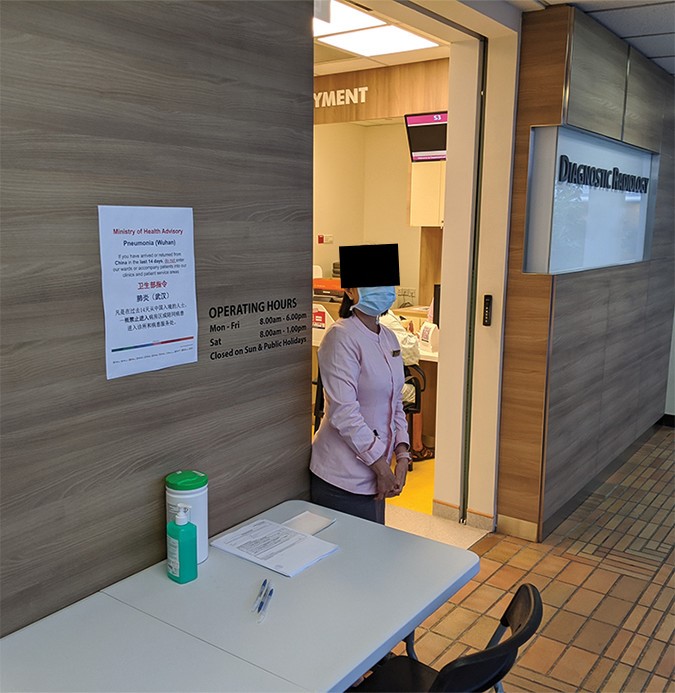
Fig. 1—Photograph shows screening station setup at radiology department entrance in early phase of outbreak, including staff member wearing mask. These smaller department-level screening stations were subsequently replaced by larger screening facilities at entrances to each building. Obscuring of facial features has been applied for privacy reasons for publication.
Share information so that all team members understand moment-to-moment changes in risks and resources needed to safely manage patients.
Personal protection equipment must be made available and properly donned and duffed.
Potentially infected patients must be identified and isolated.
Ideally, dedicated CT scanners will be identified and managed for high-risk patients.
Physical security and access control with proper signage must be assured (Fig. 1).
Alternate decentralized work areas should be identified.
Interventional radiology procedures should be modified for safety and efficiency.
Radiologists must rapidly report potential COVID-19 findings electronically and by telephone conversation, when appropriate.
So, what’s going on in Singapore today? As of April 14, 2020, there were just over 1,300 active cases of COVID-19 in the country. Of these, 1,287 patients were hospitalized but in stable condition, while 28 were listed as critical. Singapore recorded its 10th death from COVID-19 on April 14.
Similarly, Hosseiny et al. compared the clinical and imaging findings of COVID-19 with those of two previous coronavirus infections: SARS and Middle East respiratory syndrome (MERS). There are similarities, but there are differences, too. Clinical signs and symptoms of COVID-19 include fever, dyspnea, and dry cough. Complaints of sore throat and diarrhea are less common in most reported cases, though there is substantial variation in presentation other than fever. Typical COVID-19 findings on CT include multilobar ground-glass opacities (GGOs) often with consolidation. Normal CT, as seen in perhaps 15%–20% of scans, does not exclude SARS-CoV-2 infection. As expected, consolidation is an indicator for poor prognosis. Pulmonary fibrotic changes after recovery are less well-described.
ARRS is committed to providing all radiologists with open access to the latest imaging research on COVID-19 to help understand the imaging features associated with coronavirus.
In Wuhan, Hubei, China, Han and colleagues described early clinical and CT manifestations of COVID-19 pneumonia. Clinical manifestations in the 108 patients they studied were fever in 94 (87%), dry cough in 65 (60%), and fatigue in 42 (39%) patients. Laboratory findings included normal WBC count in 97 (90%), normal or reduced lymphocytes in 65 (60%), and high-sensitivity C-reactive protein elevation in 107 (99%) patients. CT distribution included one lobe in 38 (35%), two or three lobes in 24 (22%), and four or five lobes in 46 (43%) scans. Most lesions were peripheral (97 [90%]) and patchy (93 [86%]). GGOs were seen in 65 (60%) scans, with consolidation in 44 (41%) scans. The size of opacities varied from less than 1 cm (10 [9%]) to more than 3 cm (56 [52%]). Vascular thickening was noted in 86 (80%), the “crazy-paving” pattern was found in 43 (40%), air bronchograms were seen in 52 (48%), and the halo sign appeared in 69 (64%) CT scans.
Zhou and colleagues, also in Wuhan, described their findings in 62 patients with COVID-19 pneumonia. They emphasized GGOs and bronchial distortion as signs of COVID-19. Again, as of today, Wuhan seems to be doing well. China has lifted its 76-day lockdown, and the city is reemerging from the coronavirus crisis. From various news reports, you can see that the citizens of Wuhan are wearing protective masks—some of them better than the masks that we have in the United States.
Back in Hubei Province, Li and Xia from Tongji Hospital reported that, from their early experience, CT had a low rate of missed diagnosis of COVID-19 (3.9%, 2/51) and thus, “may be a standard method for the diagnosis of COVID-19 based on CT features.” The co-authors explained further: “Rapid diagnosis can lead to early control of potential transmission. With CT diagnosis of viral pneumonia, patients with suspected disease can be isolated and treated in time so that the management of patients will be optimized, especially for the hospitals or communities lacking nucleic acid testing kits.” They concluded, however, that “for the identification of specific viruses, CT is still limited,” also noting that “it is valuable for radiologists to recognize that the CT findings of COVID-19 overlap with the CT findings of diseases caused by other viruses.”
From Hunan, China, Zhao et al. reported on the relationship between chest CT findings and clinical conditions of COVID-19 pneumonia in a multicenter study of 101 patients retrospectively collected from four institutions. Most patients, 70%, were 21–50 years old, and 5% of the patients had family outbreaks. Fever was the onset symptom for 78% of patients. Fourteen patients in the emergency group were older than those in the nonemergency group. Most patients with COVID-19 pneumonia had GGOs (87 [86%]) or mixed GGOs and consolidation (65 [64%]), vascular enlargement (72 [71%]), and traction bronchiectasis (53 [52%]). Lesions were more likely peripheral (88 [87%]) and bilateral (83 [82%]) and lower lung predominant (55 [54%]) and multifocal (55 [54%]).
Salehi and colleagues published a nice systematic review of imaging findings in 919 patients with COVID-19. They concluded that although the majority of COVID-19 mortalities occur among patients with acute respiratory distress syndrome in the ICU, “in a patient population with low pretest probability of [SARS-CoV-2] infection, the typical imaging features should be interpreted with caution.”
One of the most unique papers AJR has published came from Wuhan. Liu et al. authored a preliminary analysis of the pregnancy and perinatal outcomes of women with COVID-19 pneumonia. Of the 15 pregnant women with chest CT-documented COVID-19, 11 had successful deliveries (10 cesarean, one vaginal) and four were still pregnant (three in the second trimester, one in the third) at the time of publication. Importantly, there were no abortions, neonatal asphyxias, neonatal deaths, stillbirths, or neonatal SARS-CoV-2 infections in any of the newborns. More recently, some papers have confirmed early-onset infection in neonates born to mothers with COVID-19, but mother-to-child transfer was not seen in this initial study of 15 patients.
Huang et al. in Wuhu, China analyzed 25 patients with RT-PCR-documented COVID-19. CT scores were rated 0–35 based on extent and intensity of lung involvement. Data were separated into two groups, based on time from symptom onset to diagnosis and treatment: group 1 was patients for whom this interval was less than or equal to 3 days and group 2 was those for whom the interval was greater than 3 days). CT scores were plotted against time, and after analyzing the resulting curves, the mean peak CT score was 10 and 16 for group 1 and 2, respectively, and the mean time to disease resolution was 6 and 13 days, respectively. The last CT scores were lower for group 1 than for group 2 (p = 0.025), which led to the conclusion that timely diagnosis and treatments are keys to providing a better prognosis for patients with COVID-19.
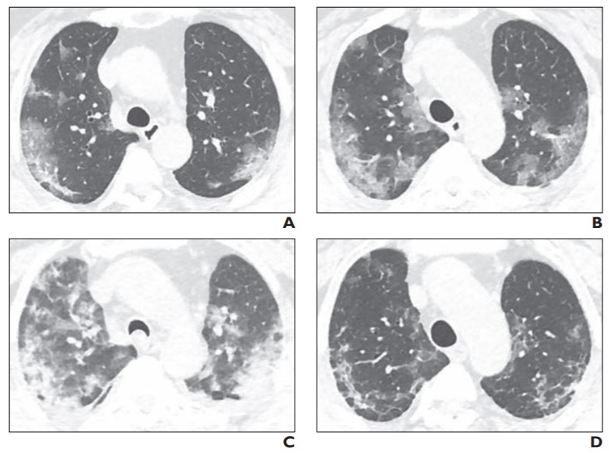
Fig. 2—59-year-old woman with coronavirus disease (COVID-19). A, Initial unenhanced axial chest CT image 4 days after admission shows multiple bilateral subsegmental peripheral patchy and ground-glass opacities with obscure boundaries and mainly subpleural distribution. Neither pleural effusion nor enlarged mediastinal lymph nodes are seen. B and C, Unenhanced axial CT images 6 (B) and 12 (C) days after admission and after initiation of treatment shows enlarged lesions; lesions of both lungs are diffuse and patchy compared with previous CT images (A). D, Unenhanced axial CT image 22 days after admission shows lesions in both lungs have absorbed gradually after treatment, and subpleural line can be seen.
On occasion, CT imaging showed asymptomatic opacities while RT-PCR testing was negative. As experiences with less-enriched COVID-19 cohorts were encountered, we learned that CT was considerably less efficient at detecting the many asymptomatic patients with COVID-19, especially compared with nucleic acid testing.
Logically, asymptomatic community members do not require RT-PCR testing unless there has been a known or potential exposure to COVID-19. CT is best reserved in planning therapy on selected patients with symptomatic COVID-19, or if doctors have reasonable suspicion that RT-PCR is falsely negative.
Of course, whereas the findings of CT lung opacities typical for COVID-19 may appear to be statistically reliable in the early stages of a pandemic, alternative diagnoses, including other infections and inflammatory conditions, cannot be readily excluded by image pattern alone.
Clinical Associate Professor, Trauma & Emergency Radiology NYU Langone Health, Bellevue Hospital
Eric A. Roberge
Assistant Professor of Radiology Uniformed Services University of the Health Sciences
Suzanne Chong
Associate Professor, Emergency Radiology Division, Radiology and Imaging Sciences Department Indiana University Health
Published April 20, 2020
Mark P. Bernstein’s “Mass Casualty Incidents: An Introduction for Imagers” was published in the Winter 2020 issue of ARRS’ InPractice magazine. Below, Bernstein et al. provide a primer for hospitals and health care systems responding to the disaster surge of COVID-19.
The coronavirus disease (COVID-19) pandemic has created a mass casualty disaster of staggering proportions. By April 2020, the novel coronavirus responsible for COVID-19 had forced many parts of the United States into crisis mode, while others race to prepare for the inevitable. In regions where the case numbers have not yet begun to climb, disaster planning teams have time to prepare for a crisis response and implement lessons learned from those who were impacted earlier. The goal is the greatest good for the greatest number of people, so hospitals and health care systems are turning the focus from individual health to population health in their disaster surge response to save as many lives as possible.
Mass casualty incidents (MCIs) can be man-made acts of violence, such as mass shootings, bioterrorism, or exploding bridges, or natural disasters in the form of earthquakes, tornados, tsunamis, and pandemics. Tragedies of intentional violence or infrastructure disasters create a sudden surge, demanding a rapid shift in a hospital’s daily routine, and are usually limited geographically—for example, the site of an active shooter or a train derailment. Natural disasters, however, cover much larger regions (i.e., the path of a tornado), whereas, by definition, pandemics know no boundaries.
One key variable in these disasters is time. Time, in most cases, determines our ability to prepare for and maintain a disaster response. In trauma MCIs, there is a window of time when patients arrive to local hospitals, which is often measured in minutes to hours. In the case of bioterrorism or pandemics, timelines are prolonged, measured in days to weeks. Regarding the ongoing COVID-19 pandemic, the window of time is indefinite and unknown. The disruption of a hospital’s daily routine for prolonged periods of time and the need for resources beyond those available, or worse, outstrips the supply chain, placing severe strain on the health care system. Our best tools to manage these challenges are preparation, planning, and practice.
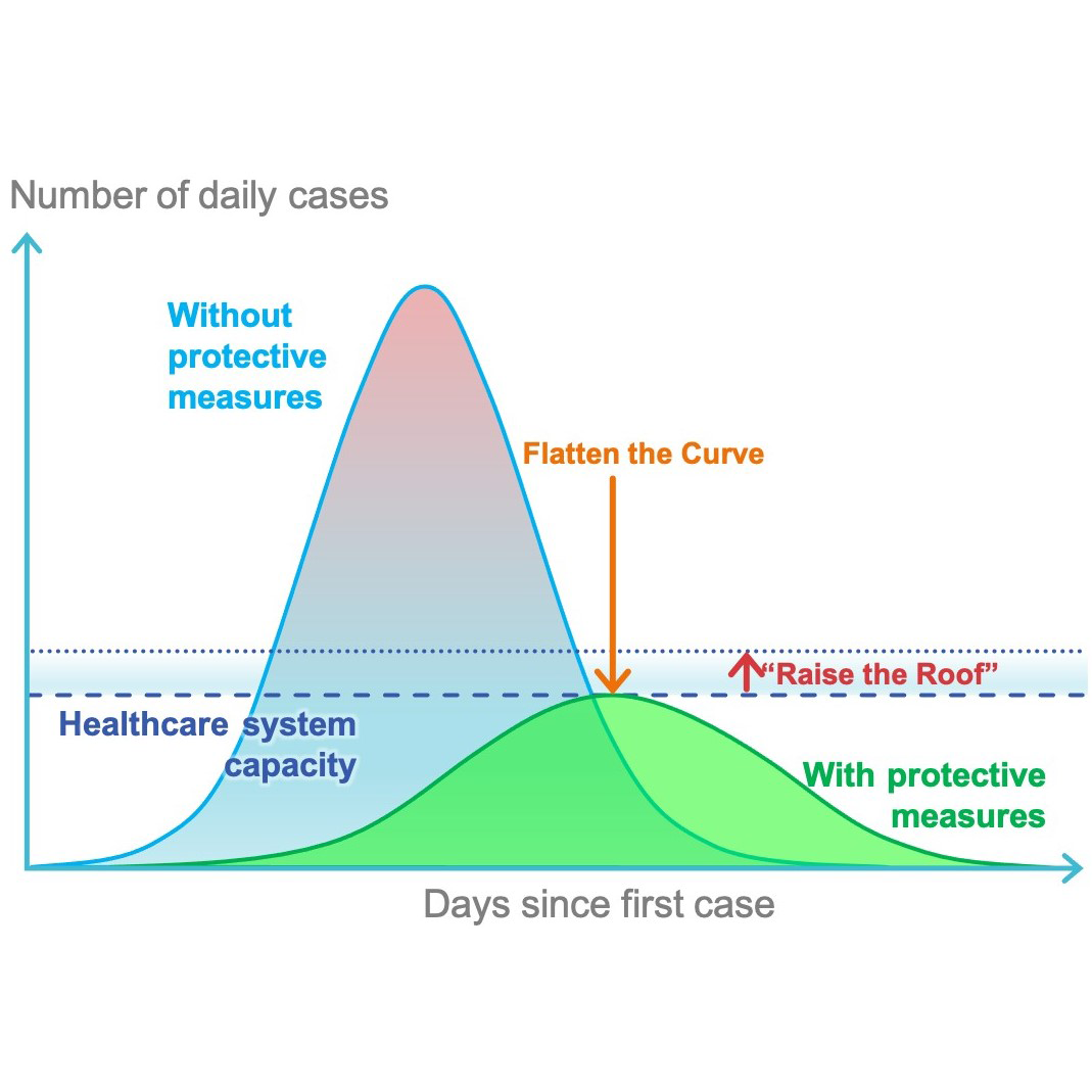
Preparation and planning take place from the federal and state levels to the community and local health care facility levels. Community planning should be coordinated with local governmental agencies, in accordance with state and federal disaster planning efforts, and integrated with local public health and emergency medical services. With respect to pandemics, community strategies must make every effort to “flatten the curve” in order to break the chain of transmission and slow the spread of infections. At the same time, hospital system strategies “raise the roof” of surge response by increasing health care system capacity (Fig. 1) through predesigned efforts focused on three factors: space, staff, and supplies. The hospital system is the backbone of these three elements.
Figure 1 – Community efforts to “flatten the curve” of coronavirus infections often intersect with health care system strategies to “raise the roof” for patient capacity (modified from Disaster Med Public Health Prep with permission from the Society for Disaster Medicine and Public Health).
Strategies for increasing health care system capacity will include conservation and substitution during a conventional response, adaptation and recycling during a contingency response, and, finally, reallocation of resources during a crisis response—essentially, withholding resources from one patient population to use them more effectively on another patient population. These “raise the roof” strategies involve nuanced ethical and legal considerations that must be addressed in advance, authorized by hospital leadership, and communicated clearly to frontline health care workers.
System
Ultimately, the hospital system component directs the response that determines the allocation of the three critical resources of space, staff, and stuff, which are based on supply and demand.
A robust hospital incident command system provides broad management for a multitude of issues, including: hospital controls (facility access, ventilation), communication (internal and external), community coordination (health care facilities, state and federal agencies, as well as utilities and supply chains), and continuity of emergency health care operation (vis-à-vis utility or other system failures). The hospital incident command should also determine and communicate which disaster response is being utilized. Disaster response can be described, in escalating intensity, as conventional, contingency, and crisis, dependent on surge severity and resource availability. The more severe the surge, the fewer the resources; the lower the hospital’s capacity to take care of victims, the more quickly the disaster response must shift into a higher mode (Fig. 2).
Space
Upon declaration of an MCI, efforts must be made to free up physical space for patients. The size and nature of the disaster will dictate the scope and speed necessary.
Figure 2 – As the hospital incident command system escalates the intensity of disaster response—from conventional to contingency to MCI—the minimum acceptable standard of care for patients is diminished (modified from Disaster Med Public Health Prep with permission from the Society for Disaster Medicine and Public Health).
The conventional response is for surges causing a 20% increase in patients beyond normal capacity. In this situation, all staffed beds are made available and filled. Elective procedures are postponed or cancelled, and patient discharge plans are activated to dedicate more space and empty beds to the surge.
A contingency response is used for surges that are twice a hospital’s capacity and demands more aggressive actions. As the numbers of patients greatly exceed the available hospital and critical care beds, hospital spaces designed for other purposes, including step-down units, observation units, and procedure suites, can be repurposed to recruit more space to bed patients. Transferring patients to other available facilities for ongoing, nonemergent care can be initiated.
A crisis situation completely overwhelms a health care facility. Patients fill hallways, and makeshift spaces, such as tents and offices, need to be devised. Erecting tent hospitals with intensive care units in city parks, converting convention centers into field hospitals, and docking of the United States Naval Ship (USNS) Comfort in Manhattan and USNS Mercy in Los Angeles are evidence that our nation is in crisis because of the COVID-19 pandemic.
Staff
As more space becomes available, achieving appropriate staffing and obtaining adequate supplies for the surge of patients is vital. The hospital incident command system should be convened for action as soon as a disaster is declared to urgently alert and mobilize necessary staff. The type of injuries that are expected (e.g., blunt trauma, penetrating trauma, or biological agent) will determine the type of staff best suited to respond. If staffing levels are insufficient, measures to increase staffing may be warranted, including expanding the scope of responsibilities, lengthening shifts, and enlarging patient-to-nurse ratios.
In a conventional response, trained and credentialed staff are able to care for patients with minor modifications, while maintaining usual standards of care.
The standard of care is challenged in a contingency response, as adequately trained staff must train and supervise off-service staff to safely provide care. Bringing in additional staff should be considered, and outside staff need to be given emergency privileges and credentialing.
A crisis response demands staff to perform clinical functions outside their usual domain. Aggressive staff recruitment and rapid training are necessary to meet the patient care demands and volume. During crisis mode, triage becomes necessary to ensure that acceptable care is provided for the largest number of people. Over- and under-triage can result in higher mortality rates.
Supplies (“Stuff”)
Supplies include medications, medical equipment, and personal protective equipment (PPE). Considerations must also be made for laboratory reagents, diagnostic testing, as well as for food, water, and linens.
The hospital system must be aware of onsite and offsite supply storage and availability through supply chains. The ability to adapt, reuse, and reallocate becomes necessary in both contingency and crisis situations.
In the current COVID-19 pandemic, we are witnessing contingency and crisis responses. Hospitals are experiencing severe shortages of ventilators and PPE, meaning patients may be deprived of life-saving care and health care providers are likely to be infected with dire, cascading ramifications.
Radiology Department Response
A departmental incident command team should be in place to implement a disaster management plan and engage in clear and consistent communication. The radiology department must have containment and mitigation strategies that ensure the safety of all staff and patients being imaged. For COVID-19, these measures include ensuring adequate PPE, especially for frontline technicians performing imaging studies, enforcing physical distancing, and limiting in-person interactions. Remote reading should be instituted, where possible. Decontamination protocols must be defined and executed. Nonemergent studies should be halted, including interventional procedures, to preserve PPE and limit exposure.
All real-time changes to address incident-specific issues should be frequently updated and communicated. Implementing these types of measures allows radiology departments to provide safe and appropriate care during surges and helps to ensure sustainable operations.
The lessons we learn from responses nationally and internationally should be incorporated into our hospital and departmental MCI and disaster planning process. Our ability to plan and prepare by focusing on system, space, staff, and stuff will make all the difference in the number of lives saved.
Suggested Reading
Christian MD, Devereaux AV, Dichter JR, Rubinson L, Kissoon N. Introduction and executive summary: care of the critically ill and injured during pandemics and disasters: CHEST consensus statement. Chest 2014; 146:8S–34S
Institute of Medicine (US) Committee on Guidance for Establishing Crisis Standards of Care for Use in Disaster Situations; Altevogt BM, Stroud C, Hanson SL, Hanfling D, Gostin LO, eds. Guidance for establishing crisis standards of care for use in disaster situations: a letter report. Washington, DC: The National Academies Press, 2009
Institute of Medicine (US) Committee on Guidance for Establishing Crisis Standards of Care for Use in Disaster Situations; Hanfling D, Altevogt BM, Viswanathan K, Gostin LO, eds. Crisis standards of care: a systems framework for catastrophic disaster response. Washington, DC: The National Academies Press, 2012
Institute of Medicine (US) Committee on Crisis Standards of Care: A Toolkit for Indicators and Triggers; Hanfling D, Hick J, Stroud C, eds. Crisis standards of care: a toolkit for indicators and triggers. Washington, DC: The National Academies Press (US), 2013
The views expressed are those of the authors and do not reflect the official policy of the Department of the Army, the Department of Defense, or the U.S. Government.
Associate Executive Director Diagnostic Radiology American Board of Radiology
Published April 10, 2020
Along with the Radiological Society of North America, American College of Radiology, American Radium Society, and American Medical Association Section on Radiology, the American Roentgen Ray Society (ARRS) co-sponsored the founding of the American Board of Radiology (ABR) in 1934. The mission of the ABR is to certify that our diplomates have demonstrated and maintained the requisite knowledge, skill, and understanding of their disciplines for the benefit of their patients.
Board certification serves as an important marker for the highest standard of care. It reflects the critical core values of compassion, patient-centeredness, and a commitment to life-long learning. Patients, physicians, medical physicists, health care providers, insurers, and quality organizations look for board certification as the best measure of a physician’s or medical physicist’s knowledge, experience, and skills to provide quality care within a given specialty.
Board certification and participation in a maintenance of certification (MOC) program has many benefits. It assures patients, privileging committees, payers, and regulators that the physician has successfully completed a training program and continues to expand his or her medical knowledge, which leads to improvements in their practice and patient safety. In 2007, the ABR instituted a requirement for practice quality improvement projects, which must be relevant to one’s practice, achievable, provide measurable results, and likely to improve quality. This remains a major component of the MOC program. Over the years, the number of qualified projects and participatory activities has been greatly expanded to reflect the integration of radiologists into the health care system.
Meet your MOC Requirements Leverage these member-exclusive benefits to meet your educational requirements with the ABR.
Since the founding of the ABR in 1934, the field of radiology has grown dramatically, and it became increasingly difficult to master the entire field. Thus, separate residency training programs were developed for diagnostic radiology and radiation oncology. (Most recently, a primary residency for interventional radiology has been approved.) Continued advances and the development of new imaging modalities resulted in many diagnostic radiologists restricting their practice domains to some extent. The ABR responded by providing subspecialty certification to reflect the importance of subspecialization. Subspecialty certification was offered for pediatric radiology and vascular and interventional radiology in 1994, for neuroradiology in 1995, and nuclear radiology in 1999. Given the speed with which these many advances in medical science changed the field of radiology, it became apparent that remote board certification was no longer pertinent. Something was needed to assure the public that physicians were keeping up with these new developments. The four subspecialty certificates offered by the ABR were timelimited from their inception, and the last lifetime primary certificates issued by the ABR were given in 2001. MOC would now be required to maintain ABR certification for all but lifetime certificate holders.
The four components of MOC are:
professionalism and professional standing
life-long learning and self-assessment
assessment of knowledge, judgment, and skills and
improvement in medical practice.
Originally, these requirements were met by maintaining an unrestricted state medical license in each state of practice, participating in continuing medical education that includes self-assessment, a cognitive exam, and participation in quality improvement projects. The ABR MOC requirements have been modified over the years, based on feedback from our diplomates.
Initially, a cognitive exam was required to satisfy Part 3 for MOC participants. However, this required radiologists to take time away from their practices and to pay for expenses to travel to a testing center. Furthermore, the cognitive assessment was required only every 10 years, an interval that many considered too long. An improved program was needed that would be meaningful, but not onerous for the diplomates.
The ABR Online Longitudinal Assessment (OLA) was introduced for diagnostic radiology in 2019, and for interventional radiology, radiation oncology, and medical physics in 2020. Each week, participating diplomates receive an email giving them the opportunity to answer one or two questions. Most diplomates are required to answer 52 questions a year. (Some with multiple certificates are required to answer more questions.) These questions were designed to test “walking around knowledge”—information diplomates should know “off the top of their head,” if asked by a colleague, resident, or patient. Furthermore, it is a learning experience, as the rationale for the correct answers and a reference is provided immediately.
Reaction to OLA has largely been positive. Many radiologists enjoy receiving two questions every week in their selected areas of practice. Since the “shelf life” of a question is four weeks, diplomates can elect to answer eight questions every four weeks, if they prefer “batching” the questions rather than answering two questions every week. Most radiologists have enjoyed participating in OLA, as it takes only a few minutes each week and does not require travel. Many continue to answer the weekly questions even after completing their yearly requirement of 52 items. More than 20,000 radiologists are now actively participating in MOC.
Questions for all ABR examinations are written by volunteers and reviewed by a subspecialty committee, before being submitted to be included in the cognitive assessment. The next step is the test assembly meetings, where all questions are again reviewed. Despite this rigorous process, an occasional problematic question may appear on an examination. These are picked up when ABR staff review the results of the exam. The ABR is fortunate to have two psychometricians and multiple experienced exam developers on staff, who review any potentially questionable item. Often, problematic questions are referred to a radiologist member of the Board of Trustees or the appropriate Committee Chair to participate in the decision whether to keep or remove the item from examination scoring.
Hear directly from ABR leadership as they provide an overview of the new ABR OLA program, which replaces the every ten-year MOC exam.
The ABR is a non-profit organization, which is highly dependent upon its many volunteers. The ABR has more than 900 diagnostic radiologists serving as volunteers on 68 different committees. Most of the paid office staff live in the Tucson, Arizona area and work at the ABR office building. Volunteers do much of their work electronically, but they do have periodic committee meetings in the Chicago testing center, near O’Hare airport, or at ABR headquarters in Tucson.
The volunteers contribute their time and expertise in writing questions, reviewing them for image quality and appropriateness as well as for constructing examinations. Before an examination is administered, another group of volunteers sets the passing standard (cutscore) using the Angoff Method. The Angoff Method is done by having a group of subject matter experts— many of whom are residency program directors—evaluate each item to estimate the proportion of minimally competent candidates who would correctly answer the item. The cutscore is the score that the panel estimates a minimally qualified candidate would receive. This is the legally defensible method used for many high-stakes examinations in the United States.
The goal of the ABR is to conduct examinations in which the candidates are comfortable and can do their best in demonstrating their knowledge. Thus, videos have been created to demonstrate the examination experience in both Chicago and Tucson. The ABR also communicates with their diplomates, candidates for certification, and the public through a variety of other means. The ABR has a booth at several of the larger radiology meetings to provide in-person answers and advice to attendees. The BEAM is the ABR’s newsletter that has recently increased from three to six issues a year. The ABR’s blog received more than 23,000 views last year. Additional communication efforts, which began in November 2018, include the social media outlets of Facebook, Twitter, Instagram, and LinkedIn.
The American Board of Radiology, along with the other 23 American Board of Medical Specialty member boards, strives to advance our field, improve patient care, and protect the public by assuring that our diplomates have acquired and maintained the requisite knowledge and skills to be effective practitioners. Board certification is an important marker of those attributes.
Associate Professor, Emergency Radiology University of Washington
Published April 23, 2020
Advancements in the accessibility, speed, and image quality of MDCT in the last 20 years have guaranteed that MDCT is the preferred imaging modality for evaluation of most conditions presenting in the emergency department, and this is particularly true for imaging after trauma. Vascular injuries, including those involving the thoracic and abdominal aorta, abdominal mesentery, pelvis, cervical vessels, and upper and lower extremities, are an uncommon but potentially lethal outcome of both penetrating and blunt trauma. Historically, diagnosis of vascular injuries relied on open exploration or conventional catheter-based angiography (CTA), both of which are invasive, time-consuming, and not broadly accessible. Today, however, the diagnosis or exclusion of many of these injuries is made on MDCT, oftentimes obviating the need for more invasive techniques. The timing and type of endovascular repair, particularly for aortic injuries, has also evolved.
Most deaths from traumatic aortic injuries still occur at the scene or before the patient reaches the hospital. However, those who reach the emergency department and undergo imaging can now be treated with a high rate of success. The majority of traumatic thoracic aortic injuries (TTAI) are found at the aortic isthmus. Injuries are found less commonly at the aortic root, ascending aorta, distal descending aorta, or at branch vessel origins, with multifocal injuries in up to 18% of patients. Although most aortic injuries are associated with high-energy trauma, there is no consensus as to the precise definition of “high-energy.” Furthermore, direct chest trauma or visible external signs of chest trauma are not necessary for the diagnosis. Therefore, liberal screening with chest CTA is encouraged in any patient with more than minimal deceleration injury.
Initial screening for mediastinal hematoma may be performed with a portable chest radiograph in many institutions. Signs suggesting mediastinal hematoma, and thus raising the possibility of a surgically relevant aortic injury, include right paratracheal stripe thickening, superior mediastinal widening, aortic arch enlargement or irregularity, opacification of the aortopulmonary window, rightward displacement of the trachea or enteric tube, inferior displacement of the left mainstem bronchus, obscuration of the descending aorta, widening of the paraspinal lines, or apical capping. These radiographic features are neither sensitive nor specific, and absence should not preclude chest CTA in high-risk trauma victims. Contrast-enhanced chest MDCT has very high sensitivity and a negative predictive value for acute aortic injuries, and it should be obtained in all at risk patients. Thoracic CTA with cardiac gating or ultrahigh pitch is the diagnostic study of choice, but nongated CTA is sufficient in most patients. Intimal flaps, intraluminal thrombus, intramural hematoma, irregular external aortic contour, focal luminal dilation or saccular outpouching (also known as pseudoaneurysm), or active extravasation are direct signs of aortic injury. Injuries can be graded according to the Society of Vascular Surgery (SVS) system or using a newer system of minimal/moderate/severe injuries that more directly guides management. Minimal aortic injuries have no external aortic contour deformity and intimal tear or intraluminal thrombus less than 10 mm in size (equivalent to SVS grade 1); these injuries do not require operative intervention and instead receive antiplatelet therapy for 4–6 weeks, with optional follow-up imaging. Moderate and severe TTAIs require surgical intervention.
Clinical Case-Based Review of Vascular and Interventional Imaging
Only about 5% of blunt aortic injuries involve the abdominal aorta. Sufficiently rare that many radiologists may never encounter one during their careers, this injury should be specifically sought when patients present with blunt abdominal trauma, such as a seatbelt sign or abdominal impact on the steering wheel, and have spinal fractures (particularly flexion-distraction injuries), duodenal or small bowel injuries, or pancreatic injuries. Isolated blunt abdominal aortic injuries (BAAI) are also rare. Two-thirds of BAAIs occur between the renal arteries and the aortic bifurcation, and up to one-quarter also have injuries involving the thoracic aorta. Abdominal CTA is the diagnostic study of choice, though venous-phase abdominal MDCT is sufficient for diagnosis and preintervention planning in most cases. In patients stable enough to be evaluated on MDCT, the most common appearance of BAAI is intimal flaps or intimal thrombi without external aortic contour deformity. Pseudoaneurysms of the abdominal aorta are only seen in 16% but require repair, either open or endovascular, depending on location. In the absence of external contour abnormality, BAAI can be managed nonoperatively with antiplatelet therapy and beta blockers. It is important to note that neither TTAI or BAAI can be excluded on a unenhanced CT because injuries, particularly intraluminal thrombi and intimal flaps, may not be accompanied by periaortic hematoma or stranding.
Injuries of the mesenteric vasculature are also uncommon in blunt trauma patients, more often resulting from penetrating trauma. Unfortunately, these are frequently lethal due to exsanguination, reflecting the difficulty in obtaining control of the proximal superior mesenteric artery (SMA), as well as back-bleeding from the valveless portomesenteric venous system. Though uncommon at initial laparotomy, bowel infarction and subsequent sepsis and multiple organ system failure are responsible for the bulk of delayed deaths from mesenteric vascular injury. Classification systems by the American Association for the Surgery of Trauma-Organ Injury Scale (AAST-OIS) and by Fullen et al. are both anatomy-based, reflecting the greater surgical difficulty and poorer outcomes associated with more proximal mesenteric arterial or venous injuries. Although immediate operative evaluation is appropriate in any patient with penetrating trauma to the peritoneum or with blunt trauma in extremis, patients with hemodynamic stability following blunt abdominal trauma can be imaged with contrast-enhanced MDCT. On MDCT, direct signs of surgically important mesenteric vascular injuries include mesenteric vascular beading, abrupt termination, or active extravasation. Intraperitoneal low- or intermediate-density free fluid is highly sensitive for either bowel or mesenteric injury, as is abnormal bowel wall thickening or enhancement, and surgical exploration is appropriate when any of these are found on MDCT. The absence of intraperitoneal free fluid has a high negative predictive value for surgically important mesenteric or bowel injury. Isolated mesenteric stranding or hematoma without active extravasation does not necessarily need surgical exploration, but these patients should be monitored carefully for delayed presentation of CT-occult bowel injury or mesenteric injury resulting in bowel ischemia.
Hemorrhage from pelvic ring injuries can be significant and life-threatening. Arterial hemorrhage accounts for 15–20% of pelvic bleeding, and low-pressure bleeding from venous structures or fractured edges of cancellous bone account for the remainder. These low-pressure bleeding sites are usually controlled by pelvic sheeting, external fixation, or internal pelvic packing, whereas arterial hemorrhage is amenable to endovascular control. Early triage to angiography may be considered for those patients with obturator ring fractures displaced at least 1 cm or pubic symphyseal diastasis of at least 1 cm, as these are independent predictors of major hemorrhage. If a patient with pelvic ring injuries is hemodynamically stable, multiphase MDCT can improve the sensitivity and specificity of detection of pelvic bleeding. Ideally, an arterial phase is obtained to identify arterial injury, as opposed to venous injury, and an additional phase differentiates active bleeding from pseudoaneurysm. Unenhanced MDCT or dual-energy CT with virtual unenhanced images may be necessary to identify bone fragments that mimic pseudoaneurysm or active extravasation. Absence of contrast extravasation on MDCT has a high negative predictive value for clinically significant pelvic bleeding. When conventional catheter-based pelvic angiography is performed, whether before or after MDCT, injection of the bilateral internal iliac veins and the bilateral external iliac veins should be performed.
Lower extremity CTA is the diagnostic study of choice for noninvasive evaluation of lower extremity vascular trauma. Any patient with hard signs of vascular trauma, including active hemorrhage, an expanding or pulsatile hematoma, a wound with bruit or thrill, a distal pulse deficit, or distal ischemic changes, should undergo CTA, unless emergency surgical intervention is necessary. Even those with lower extremity injuries, without hard signs of vascular injury, may still benefit from lower extremity CTA if the ankle-brachial index (ABI) is reduced below 0.9 (sensitivity 87–100%, specificity 80–100%). For those with ABI above 0.9, the likelihood of vascular injury requiring surgery is low, though these patients may still be observed with serial exams for 24–48 hours. One important protocol issue with lower extremity CTA on newer ultrafast scanners is that the scan may “outrun” the contrast bolus in the distal lower extremity, particularly in patients with lower cardiac output. For this reason, at my institution, our lower extremity CTA protocol includes the arterial phase from abdomen or pelvis through the toes, followed 7 seconds later by an immediate delayed (late arterial) phase from knees to toes. Inclusion of both lower extremities in the reconstructed FOV is helpful, even if the injury is unilateral, to provide internal comparison.
Upper extremity CTA is less commonly performed and is more variable in technique. If the upper extremity CTA is performed in isolation, the arm may be positioned above the patient’s head to improve image quality and reduce radiation dose, as long as the patient’s injuries permit such positioning, whereas if the CTA is performed concurrent with a chest CTA, the arm can be positioned at the patient’s side. Always consider contrast injection contralateral to the injured arm to avoid a nondiagnostic scan because of venous extravasation or extensive streak artifact. If both upper extremities require evaluation, a central line should be used for contrast injection. Furthermore, the suspected location of vascular injury may affect the scan range. Proximal injuries, such as those from scapulothoracic dissociation, may only require evaluation of the upper arm to the level of the elbow. If more distal evaluation of the forearm, hand, or fingers is required, some advocate a two-part CTA protocol with different energies and fields of view (100–120kV for aortic arch to elbow, small field of view and 80–100kV for elbow to fingertips, when elbow is positioned above the head).
CTA findings of vascular injury in the upper and lower extremities requiring intervention include vascular occlusion, dissection, extravasation, transection, pseudoaneurysm, and arteriovenous fistula. Differential considerations include preexisting peripheral arterial atherosclerotic disease, nonocclusive vascular spasm, extrinsic compression from adjacent bone fragments or compartment syndrome, or acute embolic occlusion, such as from proximal aortic injury.
Vascular trauma requires prompt recognition and appropriate treatment to prevent significant mortality or morbidity. Today, MDCT is by far the most common technique by which these injuries are diagnosed following trauma. Since vascular injuries are uncommon, many radiologists might not feel adept at imaging them, recognizing them, and characterizing them. It is imperative that an arterial phase MDCT protocol be developed for use in high-risk patients, that intravenous contrast be used in all cases, and that suspicious imaging findings are conveyed to the trauma team appropriately and urgently. These patients may benefit from referral to a level 1 trauma center for definitive treatment.
The opinions expressed in InPractice magazine are those of the author(s); they do not necessarily reflect the viewpoint or position of the editors, reviewers, or publisher.
Senior Faculty, Icahn School of Medicine Mount Sinai Hospital
Leonie Gordon
Vice Chair of Education Professor of Radiology and Nuclear Medicine Medical University of South Carolina
Don C. Yoo
Professor of Diagnostic Imaging, Clinician Educator, Warren Alpert Medical School of Brown University Director of Nuclear Medicine, Miriam Hospital
Esma Akin
Associate Professor of Radiology, Chief of Division of Nuclear Medicine George Washington University Medical Center
Katherine Zukotynski
Departments of Medicine and Radiology McMaster University
Published March 23, 2020
Theranostics is an exciting, emerging field where imaging and therapy are intimately tied together, as the same chemical molecule is used for imaging and therapy with a different radionuclide. Theranostics is a relatively new word, but radioiodine ablation with Iodine-131 (I-131) for imaging and treatment of thyroid cancer is one of the earliest examples of theranostics. Although radioactive iodine (RAI) has been used in clinical practice since the second world war, advances in imaging, therapeutic agents, and our understanding of the molecular basis of disease has slowly led to change. Developing a standardized approach to the management of thyroid cancer has been challenging and controversial. The American Thyroid Association (ATA) and the National Comprehensive Cancer Network (NCCN) guidelines for the management of thyroid cancer have undergone several iterations, some of which have been controversial, as conclusive data on the use of imaging and management strategies is often limited. Recently, however, discussion has led to the publication of the Martinique principles—setting the stage for increased interdisciplinary communication in an attempt to establish a set of recommendations based on the existing data and our wealth of accumulated experience.
Today, when a patient is diagnosed with thyroid cancer, they are assessed clinically and stratified as low, intermediate, or high risk. This is commonly done using the ATA or NCCN risk stratification system for recurrence, and the American Joint Committee on Cancer staging system predictions for long-term outcomes. Based on the risk stratification, a management plan is devised. Often, this includes pre-therapy imaging, therapy, and post-therapy imaging. There is a host of imaging that may be performed before and after therapy. Most commonly, ultrasound, CT, and planar imaging using RAI are performed with a gamma camera; however, SPECT and SPECT combined with CT may improve the sensitivity and specificity for the detection of disease compared with planar imaging alone. PET either with CT or MRI has a role for patients suspected of having thyroid cancer recurrence with rising thyroglobulin and negative diagnostic thyroid scans. There are several radioactive agents to choose from for imaging purposes. Although I-131 is typically less expensive and may be used for both imaging and therapy, Iodine-123 (I-123) has better imaging characteristics and is often used for diagnostic scans, especially in low- or intermediate-risk patients; however, I-123 cannot be used for therapy. Additionally, I-124 (although not routinely clinically used) and 18F-FDG may be helpful in certain situations, but they require access to PET.
ARRS Quick Bytes is a member-exclusive benefit that provides 20-minute videos on emerging topics in radiology for CME on the go.
There are several controversies regarding imaging and therapy of patients with thyroid cancer. For example, there is significant debate about the need for imaging before and after therapy using radioactive iodine vs other modalities, such as ultrasound and CT. Also, whereas RAI therapy has been a mainstay in thyroid cancer treatment for years, there have been recent changes in how this therapy is done. Historically, the amount of RAI to give for the purposes of therapy was based on disease extent, so those patients with distant metastases received a higher empiric amount of RAI than those with localized disease. Furthermore, pediatric patients were treated similarly to adults. Recently, however, we have tried to lower the amount of RAI administered based on the patient’s risk stratification and age to improve long term outcome and minimize radiation exposure, where possible. There is also a recognition that pediatric patients have some different clinical issues; thus, their approach to RAI has some key differences compared to adults.
While the use of and approach to theranostics in thyroid cancer is evolving, a few constants remain. Specifi cally, the experience and expertise of the multidisciplinary care team, as well as the desires of the patient, all need to be considered when making decisions about how to proceed. Imagers must recognize which information they can glean from their scans will best assist in determining the optimal course for treatment. When deciding on therapy, the amount and type of therapy to be given is based not only on pathology, but also on the medical team and patient’s wishes for short- and long-term follow up. Ultimately, as physicians who are intimately involved with both imaging and therapy, our insight can offer a lot to the overall care of the patient with thyroid cancer.
With technological innovation teeming and networks more globalized than ever before, teleradiology—an imaging practice heavily reliant upon both—would seem uniquely moored to rise with these two tides. As advancement often begets acquisition, and especially as radiology practices continue to amalgamate, some teleradiology experts are floating a seemingly counterintuitive notion: the teleradiology wave could be starting to crest.
To be sure, long-distance diagnosis has been dialed into imaging for some 30 years.
In late 1991, University of Kansas researcher Arch W. Templeton boasted in AJRthat more than 1,000 cases had been “digitized, transmitted, and printed on our teleradiology system.” Four years later for the journal, Douglas R. DeCorato detailed his after-dark developments, writing “all radiologic studies performed at Roosevelt Hospital between the hours of midnight and 8 A.M. were digitized and then transmitted over a T1 fiberoptic link to the radiology department of St. Luke’s Hospital, 4.8 kilometers away.”
Transmissions from individual institutions coming in loud and clear, by the summer of 2005, David B. Larson and colleagues had painted the first “comprehensive portrait of teleradiology in radiology practices.” Based upon a 66% response rate from the 970 practices that the American College of Radiology (ACR) surveyed in 1999, as Larson confirmed in AJR, “Seventy-one percent of multiradiologist practices had teleradiology systems in place, using them to interpret 5% of their studies. For solo practices, corresponding statistics were 30% and 14%.”
Flash forward a scant two years, when Todd L. Ebbert sought to capture and communicate just how big teleradiology had become. His 2007 AJR web exclusive had two distinct objectives: “to describe in detail the use of teleradiology in 2003 and to report on changes since 1999 in this rapidly evolving field.” Armed with the ACR’s Survey of Radiologists from 2003 (sent by mail, ironically), as well as its 1999 Survey of Practices, Ebbert et al. verified that 67% of radiology practices in the United States, “which included 78% of all U.S. radiologists,” had performed teleradiology.
Almost a decade’s worth of telemedicine would be performed until the first nationally representative approximation of telehealth practices across all medical specialties was published.
Now, nearly three decades removed from AJR’s initial frontline reporting, is the teleradiology revolution running out of steam? Well, the business answer at least is rather hazy.
Echoing Muroff’s sentiments, Elizabeth Krupinski, professor and vice chair for research in the department of radiology and imaging sciences at Emory University School of Medicine, indicated a swing allied with artificial intelligence, saying “I can see teleradiology changing as a byproduct of the overall industry shift.”
And already, less than a year later, as Muroff told Diagnostic Imaging this past September: “Teleradiology is a saturated, mature market that is no longer growing.” “If anything,” he compounded, “it’s shrinking somewhat because, as practices get larger, they have a greater capability of providing comprehensive call themselves.”
Corporate takeovers of independent radiology practices—what AuntMinnie flagged as one of 2019’s “biggest threats to radiology”—has taken on teleradiology, too. As reporters Brian Casey and Erik Ridley explained: “The rise of corporate radiology companies— which have oftentimes grown by acquiring smaller groups— is turning many radiologists from entrepreneurs into employees. Meanwhile, hospitals continue to expand by swallowing up outpatient centers that once operated independently.”
The bigger they are, the louder their call, indeed.
A newer survey from KPMG tapped 330 corporate, private equity, and investment banking executives in the life science industries for their two cents. And what did this “Big Four” accounting organization find? This year, the health care sector will endure even more absorption than it did in 2019.
Of course, suffering the slings and arrows of more and more mergers and acquisitions (M&A) doesn’t have to necessarily stymie teleradiology’s forward march.
In this past November’s issue of AJR, Michael A. Bruno and team pointed out that “in a fully integrated practice model a single group of subspecialist radiologists would provide care seamlessly at all practice sites, either on a rotational basis or by sharing cases through teleradiology or shared PACS systems, across the full spectrum of care.”
Ultimately, the truest test of tele-harmony writ large depends on who, how, and expressly where you ask.
In light of all the headline legislation, relentless coups, and exaggerated projections implying the demise of enterprise teleradiology, according to its most thorough clinical evaluation to date, the actual practice of teleradiology is very much alive and well.
Defining teleradiology as “the interpretation of medical imaging examinations at a separate facility from where said examination was performed,” Rosenkrantz and his colleagues solicited responses (appropriately enough, via email) from a random sample of 936 ACR members. While a clear majority, 731 respondents, designated their main work setting as non-teleradiology, 85.6% of that cohort indicated they had practiced teleradiology within the past 10 years. Furthermore, 25.4% stated teleradiology comprised a majority of their annual imaging volumes.
No longer the realm of nighthawks, a staggering 91.3% of respondents said that they had implemented teleradiology during normal business hours, while 44.5% to 79.6% said they had implemented teleradiology over evening, overnight, and weekend shifts.
In rural areas, 46.2% of American radiologists surveyed by Rosenkrantz reported performing teleradiology, and 37.2% reported performing teleradiology in critical access hospitals.
Helping working radiologists realize after-hours success and expand coverage for underserved patients, “despite historic concerns,” Rosenkrantz reassured, “teleradiology is widespread throughout modern radiology practice.”
Like most cutting-edge revolutions, efficacious telemedicine continues to spread.
A pilot study just published ahead-of-print in AJRby a team from Germany’s second-largest city and Europe’s third-largest port, Hamburg, christened the concept of maritime telemedicine with the inauguration of a PACS-centered service staffed 24/7 by specialized radiologists at a tertiary hospital on shore.
So, what do the trade machinations of tomorrow or the next administration’s regulations portend for teleradiology’s next wave, the clinical and the commercial?
Fresh out of stealth mode and buttressed by $16.5 million in Series A cash, the mission of this teleradiology company is decidedly asynchronous—assist radiologists in triaging head CT scans through machine learning.
Associate Professor of Clinical Radiology University of California, San Francisco
Published January 10, 2020
The latter half of 2019 saw the identification of an entirely new respiratory illness and introduction of a new diagnosis— e-cigarette, or vaping, product use associated lung injury (EVALI)—into the medical lexicon. Based on current understanding, EVALI is an acute or subacute respiratory illness that is often severe and in some cases fatal, purported to be a chemical pneumonitis resulting from inhalation of one or more toxic substances. As of November 5, the Centers for Disease Control (CDC) reports 2,051 cases of EVALI in the United States, including every state except Alaska, plus the District of Columbia, with 39 deaths in 24 states. The reported cases have affected patients as young as 13 and as old as 75, with males about twice as common as females. Nearly all of these patients have presented with acute or subacute respiratory symptoms, and not surprisingly, thoracic imaging (chest radiograph and CT) has become central to the diagnosis of these patients. Knowledge about these diseases is changing every day, and it is imperative that diagnostic radiologists have a general understanding of what vaping is, how it looks on imaging, and what we know so far.
What Is an E-cigarette, and What Is Vaping?
An e-cigarette is an electronic device that is designed to simulate traditional smoking. Instead of the combustion of tobacco (or, more recently, marijuana), e-cigarettes heat a substance (usually liquid, oil, or wax) to create a vapor that is inhaled, hence the term “vaping.” E-cigarettes were invented in 2003 and introduced in the U.S. around 2007.
Most devices have three main components: a chamber or cartridge that contains the substance to be heated and vaporized (also referred to as the e-liquid); an atomizer or heating element that vaporizes the substance in the cartridge, so that it can be inhaled; and a battery to power the heating source.
Like any piece of technology, these devices have evolved and are becoming increasingly sophisticated. Early devices tended to mimic tobacco cigarettes in shape, but now admittedly look outdated, compared to the most recent generations of products that are smaller, sleeker, and more easily concealed. Some bear more of a resemblance to USB thumb drives than smoking devices, and a few even have Bluetooth connectivity to track how much one vapes.
What Substances Do People Vape?
The substances that patients vape are almost limitless, and this variability is one of the main reasons it has been so difficult to pinpoint the recent surge of cases on one specific cause. A majority of recent cases have been associated with vaping tetrahydrocannabinol (THC) or other marijuana derivatives, and there is mounting evidence for vitamin E acetate as one of the main culprits. While a majority of patients with EVALI report vaping both THC and nicotine, some patients report vaping exclusively nicotine, so it is possibly not just the vitamin E acetate, a thickening agent in THC-containing vaping products, that is to blame.
Given the lack of regulation, there are many ways that the substances are stored, filled, and refilled, as well as many suppliers from where patients get their vaping substances. Some are refillable cartridges, whereas others are disposable pods; some people create their own “home brews” or buy products off the street that may not be sterile and may be adulterated. It is suspected that some of the aftermarket or “off-the-street” products may be more likely to cause injury, and the CDC advises against their use.
Nicotine is often mixed with flavoring agents or “vape juice,” and there are more than 15,000 different flavors. Adults may prefer more traditional flavors such as tobacco, mint, or menthol that try to mimic the taste of cigarette smoking. But other flavors more unabashedly appeal to teenagers and adolescents—many of whom were never smokers prior to vaping. A study in Pediatrics found that adolescents who vaped these nontraditional flavors (including fruit, candy, sweet or dessert, buttery, or other blends not including traditional flavors) were more likely to continue vaping at six months and take more puffs per occasion. The use of these flavors resulted in greater self-reported addiction and satisfaction in another study of young adults.
Why the Recent Rise in Vaping Cases?
Vaping is becoming more popular, particularly among adolescents and young adults; the variety of substances that can be consumed has expanded; and e-cigarette companies have increased the marketing of their products, just to name a few. But with the constant media coverage, everyone thinks about vaping as a cause of lung injury, and that has led to increased recognition by physicians, including radiologists.
The first case we suspected to be lung disease due to vaping was in 2014 in an adult male patient with ground-glass opacity (GGO) on CT, although this case could never be proven, as there wasn’t even a name for this disease then. The first confirmed case we saw was in 2017 in a female who was vaping THC to help her sleep. Case reports of EVALI date back to 2012, but our original article in AJR, “Imaging Findings of Vaping-Associated Lung Injury,” is the first to review and present all of the different imaging patterns that we have encountered so far. The varied appearances underscore the confusion and difficulty in these cases, arguing in support of a multifactorial cause.
What’s the Bare Minimum Radiologists Should Know About Vaping and EVALI?
using an e-cigarette or dabbing (i.e., heating concentrated cannabis oil or wax and inhaling the vapors) in 90 days prior to symptom onset
abnormalities on either chest radiograph or CT
negative infectious workup
no alternative plausible diagnosis (e.g., cardiac, rheumatalogic, or neoplastic)
A probable case of EVALI is similar—the one distinction that either an infection was detected by culture or polymerase chain reaction but not suspected of being the sole cause of lung injury, or minimum testing to exclude infection was not performed.
Imaging is part of the case definition, and as such, radiologists are critical to the diagnosis. The CDC definition verbatim is “pulmonary infiltrate, such as opacities, on plain film chest radiograph or ground-glass opacities on chest CT,” but to any radiologist, this sounds like a vague and generic explanation. One can review the many different patterns of lung injury in our AJR paper, but the one commonality from the cases we present, our review of the literature, and those cases we’ve encountered since is that these patients almost universally present with bilateral opacities that look like acute lung injury and/or organizing pneumonia. Cases may be diffuse, upper- or lower-lobe predominant.
In the appropriate clinical setting, it is arguable that chest radiograph should be sufficient for the diagnosis if bilateral opacities are present, although clinicians often order CT to evaluate for alternative causes, such as pulmonary embolism. Patients who present with acute illness may require ventilatory support. Unfortunately, some patients have died. Treatment with corticosteroids seems to be effective. While most patients completely heal, there is little data on the long-term appearance of survivors of EVALI.
Lung Pathology of EVALI
For most cases of EVALI, obtaining lung tissue is unnecessary for establishing the diagnosis, although some literature on pathology now exists. The largest series of cases where pathologic specimens were available was recently published and concluded that EVALI is “a form of airway-centered chemical pneumonitis from one or more toxic substances” in the aerosolized vapor. The presence of lipid-laden macrophages and positive oil-red-O stains has raised the possibility of exogenous lipoid pneumonia due to vitamin E acetate. Regardless of the underlying pathology, macroscopic fat has not been observed on CT imaging.
What Does the Future Hold for Vaping and EVALI?
The recent illnesses and deaths represent a grave tragedy and public health crisis with little precedent. However, it is important for physicians to not focus exclusively on the negative press. Lost in the daily media shuffle is the fact that for some patients, e-cigarettes may be an effective tool for smoking cessation. Recent data from a randomized control trial in the United Kingdom found that nicotine-containing e-cigarettes were almost twice as effective for smoking cessation at one year compared to other forms of nicotine replacement, but the abstinence rate was still only 18.0% (vs 9.9%). Patients in the e-cigarette group also experienced a greater reduction in cough and phlegm production. On the other hand, can patients stop vaping once they have started? At the one-year mark, 80% of the patients using e-cigarettes in this group were still using. Many health officials are worried that it is just replacing one habit (smoking) with a slightly less bad habit (vaping). Vaping might be safer than traditional cigarette use, but at this point, we just don’t know.
Hopefully, the agents responsible for cases of EVALI will be discovered, and cases of acute lung injury will subside, but what are the long-term effects of vaping? Vaping is a new practice that has been around for barely more than a decade, and it is certainly a worry that long-term vaping could lead to more chronic lung disease and fibrosis. We have seen some indication that vaping may progress to fibrosis, although this realm is largely unexplored and ripe for imaging research.
The opinions expressed in InPractice magazine are those of the author(s); they do not necessarily reflect the viewpoint or position of the editors, reviewers, or publisher.