
Tag: InPractice Articles

  • Imaging Advances Toward Autism Diagnosis

    Imaging Advances Toward Autism Diagnosis

    Logan Young
    Staff Writer

    Published March 21, 2020

    To be sure, radiology has come a long, long way. Only 10 years ago, the best medical imaging could do for children with autism spectrum disorder (ASD) was to identify key abnormalities in the brains of those already diagnosed—i.e., 1 in 59 children, according to today’s estimates from the Centers for Disease Control and Prevention’s (CDC) Autism and Developmental Disabilities Monitoring Network. A half-decade earlier, cortical gray-matter studies were discovering overall substantially thicker cortex for boys with autism, alongside similar findings in the temporal and parietal lobes, whereas diffusion tensor imaging was being used to illustrate disruption of white-matter tracts between regions implicated in impaired social cognition. Meanwhile, just as early functional MRI (fMRI) studies on ASD were exploring core symptom domains via activation patterns in response to mimesis, facial processing, theory of mind, semantic sentence comprehension, lexical semantic processing, and tasks involving variable imagery content, researchers were also looking to magnetic resonance spectroscopy (MRS) to assess models regarding excitation and inhibition ratios in ASD.

    Writing on MRS in the October 2004 issue of the Journal of Neuroscience, Matthew K. Belmonte from the Autism Research Centre at the University of Cambridge duly noted: “It has been said that people with autism suffer from a lack of ‘central coherence,’ the cognitive ability to bind together a jumble of separate features into a single, coherent object or concept. Ironically, the same can be said of the field of autism research, which all too often seems a fragmented tapestry stitched from differing analytical threads and theoretical patterns”.

    Fifteen years removed, while ASD remains very much an heterogeneous disorder of multifactorial etiology, evidencing an expansive range of symptoms and severities alike, radiology is in the process of reconciling so many image threads. True, bereft of a priori behavioral phenotyping (e.g., Autism Diagnostic Observation Schedule [ADOS], Social Responsiveness Scale, Kaufman Brief Intelligence Test, composite IQ score), right now, radiology alone still cannot definitively diagnose ASD in anyone, child or adult. There is good news, though. The radiology research paradigm is shifting—away from mere aberration identification to clinical diagnosis.

    The sands underneath it all first loosened in 2014, when University of Pittsburgh and Carnegie Mellon researchers utilized machine-learning algorithms to grade 34 young adults as either autistic or control with > 97% accuracy based upon fMRI neurocognitive markers for eight social interaction verbs: compliment, insult, adore, hate, hug, kick, encourage, and humiliate. Moving quickly, one year later, Virginia Tech Carilion Research Institute professor P. Read Montague synthesized nine years’ worth of previous trials to announce in Clinical Psychological Science that his team had developed an even more efficient technique to diagnose children with ASD in under two minutes: single-stimulus fMRI. Subjects were shown 15 images of themselves and 15 images of another child, matched according to age and gender, for four seconds per image in randomized order. Like the control adults in Montague’s earlier experiments with imaging for ASD, when viewing their own pictures, the control children had a high response in the middle cingulate cortex; by contrast, children with ASD showed an appreciably diminished reaction. Notably, Montague et al. could detect this disparity using one, solitary image.

    This May, much of Montague’s same colleagues, including principal investigator, Kenneth Kishida of the Wake Forest School of Medicine, made headlines for a Biological Psychology article demonstrating that a single stimulus and < 30 seconds of fMRI data were sufficient to differentiate ASD children from their typically developing (TD) peers. To test a hypothesis that responsiveness of the brain’s ventral medial prefrontal cortex (vmPFC) in children diagnosed with ASD is diminished for visual cues, denoting high-value social interaction, 40 participants (of which 12 had ASD and 28 were TD), aged 6–18 years old, were prompted to observe images of four faces and four objects, which were projected onto a screen and viewed through a mirror during fMRI scanning. With each image characterized as favorite, pleasant, neutral, or unpleasant, the favorite images depicted each of the participants’ self-selected favored face and object, and the remaining images were selected from the International Affective Picture System (IAPS) database. Each of the eight images was then displayed only once for five seconds during a block that repeated six times. Following the completion of 12- to 15-minute MRI scans, participants were shown the identical set of images on a computer screen, ranking them in order, from pleasant to unpleasant, with a self-assessing sliding scale. Results showed that the average response of vmPFC was significantly lower in the ASD cohort, compared to the TD cohort.

    “How the brain responded to these pictures is consistent with our hypothesis that the brains of children with autism do not encode the value of social exchange in the same way as typically developing children,” Kishida said in a prepared statement. “Based on our study,” he continued, “we envision a test for autism in which a child could simply get into a scanner, be shown a set of pictures, and within 30 seconds, have an objective measurement that indicates if their brain responds to social stimulus and non-social stimuli.”

    There are limitations here. Because these 40 children were permitted to specify favored objects and people, reasonably assuming that there were distinct visual differences between these non-IAPS images and that canonical cache, Kishida conceded the possibility that at least some of the reported response differential could simply be due to known vs. novel. Moreover, since ASD disproportionately affects male patients—i.e., four times more common among boys than girls, the CDC maintains—he acknowledged an optimal design could be updated to investigate the gender divide between the ASD and the TD children more thoroughly.

    “Based on our study, we envision a test for autism in which a child could simply get into a scanner, be shown a set of pictures, and within 30 seconds, have an objective measurement that indicates if their brain responds to social stimulus and non-social stimuli.”

    —Kenneth Kishida

    Another Wake Forest faculty member, Christopher T. Whitlow, has been presenting related research on ASD imaging since 2014. As his studies have surveyed patterns of joint variability in severely preterm infants, might we see an eventual diagnostic environment where Whitlow’s voxel-based morphometry informs Kishida and Montague’s single-stimulus exemplar to evidence brain dysfunction in patients younger than the age-six threshold?

    Although reproductive stoppage (i.e., the tendency for arrested propagation after diagnosis of an affected child) can lead to underestimates of sibling recurrence risk for ASD, with ascertainment biases and overreporting often pointing to its inflation, we should focus on the family first. In 2011, the multisite international network, Baby Siblings Research Consortium, conducted a prospective longitudinal study of 664 infants who had an older biological sibling with ASD, monitoring them from early life to 36 months, when they were classified as having or not having ASD—an ASD taxonomy requiring exceeding the ADOS cut-off, as well as an expert’s diagnosis. In total, 18.7% of infants developed ASD. Whereas infant age at enrollment, gender and functioning level of the infant’s older sibling, and other demographic circumstances did not predict ASD outcome, infant gender and the presence of > 1 older affected sibling were significant forecasters. Again, there was a nearly threefold risk escalation for male subjects and an additional twofold increase in risk if there was > 1 older affected sibling.

    Family history, meet deep learning. Recent findings published in Science Translational Medicine by University of North Carolina at Chapel Hill researchers revealed that when applied to functional connectivity MRI (fcMRI) data at six months of age in infants with high familial risk for ASD, a nested, cross-validated machine-learning algorithm predicted an ASD diagnosis with > 96% accuracy at 24 months. Citing several brain variances—both morphological and electro-physiological—members of his team had documented as early as six months in infants later diagnosed with ASD, “Given the complexity and heterogeneity of ASD,” lead author Robert W. Emerson surmised, “methods for the early detection of ASD using brain metrics will likely require information that is multivariate, complex, and developmentally sensitive.” Apropos, Emerson et al. employed an array of 230 regions of interest (ROI) previously defined across the entire brain to create functional connectivity matrices from the fMRI scans of 59 at-risk infants (11 diagnosed with ASD at 24 months, 48 who did not have ASD at 24 months) during natural sleep without sedation at their six-month visit. “Our logic was that these regions would be the most likely to contribute to the discrimination between groups in the 59 separate support vector machine models,” wrote Emerson. With data collection resulting in 26,335 usable ROI pairs exemplifying each infant’s whole-brain functional constitution by training MATLAB’s Statistics and Machine Learning Toolbox (Mathworks, Inc.) to ascertain the causal patterns of individual separation, the probability that infants with a positive classification truly had ASD (positive predictive value) at 24 months was 100% (95% CI, 62.9–100). Negative predictive value at 24 months was 96% (95% CI, 85.1–99.3).

    A first-of-its-kind study from November 2018 that leveraged the imaging archive of Geisinger Health System in Danville, Pennsylvania, takes us back to the future, examining early brain markers in ASD to further the promise of artificial intelligence for earlier detection. Renewing his dissertation research, Gajendra J. Katuwal and colleagues applied random forest ensemble learning to models trained on 687 brain features of Freesurfer v 5.3.0 (Martinos Center for Biomedical Imaging) to compare cortical and sub-cortical morphometric features for ASD vs. non-ASD classification. Their query of head MR images from Geisinger’s institutional tranche, after removing those with artifacts, motion, lesions, abnormally large ventricles, and neurodevelopmental disorders as identified by International Classification of Diseases code, yielded 112 non-ASD and 115 ASD subjects. Eschewing gender confounds, 20 non-ASD and 34 ASD scans of female subjects were excluded. Although total intracranial volume (TIV) of ASD measured 5.5% larger than the control, brain volumes of other ROI, when calculated as TIV percentage, measured smaller in ASD—partially due to larger (> 10%) ventricles in ASD. ASD’s larger TIV exhibited correlates with greater surface area and aggregate cortical folding, yet not with cortical thickness. ASD frontal and temporal white-matter tracts evidenced less image intensity, seemingly suggesting myelination deficit. Ultimately, Katuwal’s methodology was able to achieve 95% AUC for ASD vs. non-ASD classification using all brain features. When stochastic discrimination was discrete for each feature type, image intensity yielded the highest predictive power (95% AUC), followed by cortical folding index (69%), cortical and subcortical volume (69%), and surface area (68%).

    According to Katuwal, “the most important classification feature was white matter intensity surrounding the rostral middle frontal gyrus,” which measured lower (d = 0.77, p = 0.04) in ASD.

    Because medical technology also rises, medical imaging, itself, is sure to manifest a more prominent role over time among allied sciences with regards to forthcoming ASD diagnoses and concomitant, personalized care. To that end, in order to fully apprehend the neuroanatomical foundations of ASD, a comprehensive, multimodal surveillance of early brain alterations would seem to light the best forward path. Progress isn’t always a straight line, of course, so radiology has places yet to go, indeed.


    The opinions expressed in InPractice magazine are those of the author(s); they do not necessarily reflect the viewpoint or position of the editors, reviewers, or publisher.


  • White Horse, Yellow Journal: Berquist Bids Adieu to AJR

    White Horse, Yellow Journal: Berquist Bids Adieu to AJR

    avatar

    Logan K. Young
    Staff Writer

    Published March 21, 2020

    When Thomas H. Berquist logs off his iPad this summer, his 12-year tenure as the 12th editor in chief of the American Journal of Roentgenology (AJR) will capstone a period of unprecedented growth for the 113-year-old publication. Truly the end of an era, only two other men will have occupied the AJR’s chief chair longer than Berquist: Lawrence Reynolds, who picked up the mantle in 1930 and died in office 31 years later, and his immediate successor, Traian Leucutia, whose editorship (1961–75) lasted just two years longer than this lauded, yet humble Mayo Clinic radiologist.

    Articulating an expansive vision for radiology’s beloved “yellow journal” from his first days at the desk in late 2008, the ARRS Publications Committee, to whom Berquist has reported for some 150 issues of AJR, agrees that he’s fostered a unique editorial climate ever since—one of both exacting rigor and earnest diversity.

    “Dr. Berquist is one of the most inclusive leaders that I have ever had the pleasure to work with,” says Deborah Baumgarten, ARRS Publications Committee chair. “He solicits opinions and really listens to and considers what others have said. You feel like you matter to him.”

    Acknowledging achievement only if it’s data-borne, the internationally recognized author of 38 books on medical imaging tells InPractice he remains very much “a numbers guy.”

    Incidentally, despite ARRS’ announcement of Berquist’s retirement more than 15 months ago, submissions to the journal continue to pour in unabated. And although AJR’s acceptance rate “still hovers right around 20%,” Berquist does admit that the overall quality of the articles being submitted is likely as good as it has ever been.

    For once, there’s a causation implied by the correlation.

    Two years ago, Berquist himself took to these same pages to reassert AJR’s raison d’être in two words: “evidence-based articles”. Codifying the journal’s reporting guidelines for Original Research and Review articles “to assist authors in providing optimal consistent content,” he also detailed significant revisions to the Standards for Reporting Diagnostic Accuracy Studies (STARD) and Standards for Reporting Observation Studies in Epidemiology (STROBE) checklists, “in an effort to provide more imaging-friendly guidelines.”

    With humility and precision, Berquist now notes, “to date, there have been 128 STARD submissions and 33 STROBE submissions to AJR,” casually mentioning the “significant recruitment initiative” he’s spearheading to further improve these types of content enhancement, which he’s keen to note will soldier on even without him aboard.

    Circulation is up, too. With AJR enjoying record readership worldwide—especially unique views and clicks, online and mobile, at AJRonline.org—as the outgoing editor in chief wrote in his “Things We Learned Along the Way” editorial from November, “the online version is the journal of record.”

    It was Berquist’s predecessor, Robert Stanley, who introduced the notion of electronic article submission. Not surprisingly, his institution of web-based submissions yielded a marked increase in international authors submitting to AJR, sending Stanley and staff scrambling to enlist foreign-language reviewers. Sixteen years post-Stanley, Berquist recalls yet another telling audit.

    “Currently, there are 2,321 total AJR reviewers,” he says. “Eighty percent hail from the United States, and 20% are based internationally.”

    Asked how a more international distribution due to ever-increasing scholar globalization might alter the scope of AJR content to come, thus far, Berquist says he’s seen only one year, 2014, where foreign submissions outpaced submissions from the U.S. As always, Berquist’s bias tends toward scientific scrutiny, not identity politics, “particularly where a benign cultural difference could escalate to the level of significant medicolegal dilemma.”

    Ultimately, he’d much rather talk residents and reviewers than matters foreign and domestic. Regarding residents, Berquist is quick to credit Howard P. Forman for initiating the journal’s Trainee Reviewer program, pointing out the present group of “61 trainee reviewers and additional new reviewers who have mentors as they begin their reviewer role.”

    Naturally, Berquist has streamlined the onboarding process; it began two years ago at the Marriott Wardman Park Hotel in Washington, D.C.

    In 2018, alongside Cheryl S. Merrill, ARRS’ director of publications, Berquist debuted what would eventually become an essential component of the ARRS Annual Meeting that speaks volumes about the significance of scientific integrity—a two hour course the duo affectionately dubbed “Rock the Review: How to Get a Perfect Score.” A perfect score equals 4.0, sure, but what does said “rocking it” actually look like on the page?

    For that inquiry, Berquist settles in: “The review must include sophisticated, detailed comments to authors with line and page referencing to enhance the content and relevance of the work; concise, confidential comments to the editor; and the reviews must be completed in the allotted 14 days or earlier.”

    As for his active reviewers, again, Berquist knows their numbers by heart.

    “Ten percent of AJR reviewers have scores less than 3.0, and 40% have scores between 3.0–3.5,” he says. “Half of the reviewers for AJR, nearly 1,200, have a perfect 4.0,” Berquist half-beams, adding that each and every reviewer is evaluated at least yearly, “more frequently should their approach warrant it.” He reveals “any reviewer may request a review of themselves at any time,” too

    Resigned to the inherent difficulty of “consistent communication” with more than 2,300 reviewers across the globe, Berquist’s not bereft of procedure here either.

    “There are multiple data points available each day,” he says of his quality control, “including how many invitations a reviewer has received, accepted, and declined; how many times a reviewer has been uninvited for not responding; the last review accepted and completed and the last review declined; reviews in progress; and the mean reviewer score.”

    Lest you think he’s all scores and no play, there are prizes— albeit hard-won ones.

    Known for penning personal letters to stellar reviewers, Berquist also established “a Distinguished Reviewers category for individuals performing 10 or more reviews in a given year with score of 3.0 or higher.” Reviewers’ names are featured on the AJR masthead for the entirety of the following year, and their departmental chairs are notified of the distinction.

    For “above and beyond assistance,” states Berquist, “we initiated the Gold and Silver Distinguished Reviewer Achievement Awards in 2018 for reviewers with 100 or more reviews and 50–99 reviews, respectively.” These reviews must be scored at least 3.5, and Berquist remarks that 91 AJR reviewers have received 14 gold and 77 silver trophies during the Reviewer’s Luncheon at the ARRS Annual Meeting.

    “We now have Platinum and Diamond Distinguished Reviewer Achievement Awards for scores 3.5 or higher for 150– 199 reviews and 200 or more reviews, respectively,” he says with that muted smile returning.

    Acknowledging that the whole notion of peer review itself is in flux, Berquist’s not averse to the creep of new ideas. His interest in zeitgeist systems thinking, like Just Culture, has been abiding, and he confesses to “a certain anticipation” for an updated model of shared accountability.

    Never not teaching, the diagnostic radiologist is wont for a metaphor.

    “Peer review is a lot like the Supreme Court,” Berquist claims. “It’s by no means perfect, but it’s the best we have now.”

    For all reviewers, authors, and journal staff, time is always of the essence. Recalling an AJR authors’ survey “where 85% of respondents considered speed to publication extremely important,” once more, the numbers stay on Berquist’s side.

    “In 2013, the time to first decision was 37.6 days,” he says. Streamlined protocols in 2017, implemented by the journal’s inhouse staff, shrunk that time down to 25 days. For 2019, Berquist tallies “the average time to first decision is 18.8 days,” compared to 20 days at the same time the previous year.

    Prior to recent concerted efforts, he laments that the elapsed time from first decision to AJR publication measured a “protracted 147 days.” Understandably, Berquist happily reports that number has been cut almost in half.

    Asked who or what is most responsible for this optimized circle to publication, AJR’s chief editorial officer neither hesitates nor equivocates: “The journal staff deserve the credit—all of it.”

    Berquist balks at the term officer. His self-effacing streak matched only by his work ethic, this self-described “policeman with no gun” has doggedly pursued often competing commitments at AJR—everything from article enhancements, reviewer recognition, and production improvements to that ever-important journal impact factor.

    And at least until June, armed or not, the buck still stops with Berquist.

    “Right now, the impact score of AJR is 3.161,” he rattles off to the third decimal. “That’s the highest the score has ever been. But it’s not yet a 4, so in that regard,” he doesn’t pause, “I’ve failed.”

    Impact factor is a scientometric index, yes, but Berquist concedes there are more popular proxies, especially in pixels. It was under his administration that ARRS joined Twitter, posted a DOI on Instagram, and published an AJR video to YouTube. It was during Berquist’s watch that democratized platforms such as these breathed new, sometimes second lives to articles about magnetic eyelashes as MRI artifacts and his personal favorite, radiologic detection of inadvertently ingested wire bristles from a grill-cleaning brush.

    “It’s exciting, it’s all visibility, and we’re getting much better at it,” Berquist says of social media exposure. “We’re also exploring more and more things adjacent to it,” his subtle reference to AJR Podcasts, available cross-platform via iTunes and Google Play.

    At the end of the day, it’s not impact factor or Just Culture or homepage views that’s kept Berquist up at night these last 12 years.

    “For me, the key thing has always been scientific integrity,” he says. “In fact, it’s really only this. But in reality, there are times when I feel alone on this white horse, swinging at windmills.”

    Of course, the man of La Mancha never had to traverse this modern landscape of so many open-access journals—an “exponential proliferation” our Jacksonville, Florida physician cites as his chief concern for medical publishing moving forward.

    “There were about 80 radiology journals when I began at AJR,” Berquist remembers. Today, he inventories more than 800 open-access journals publishing medical imaging content on a regular basis. How could Berquist alone possibly guard against every duplicative breach?

    “I’m not sure we can anymore,” Berquist answers, invoking both pronouns.

    According to the editor in chief, AJR uses a huge database, Similarity Check (CrossRef), for manuscript evaluation, as well as “a 10% or greater duplication and singles source greater than or equal to 3% to help assess duplication more thoroughly.” Given that a typical year will end in more than 1,800 submissions to the cue, the journal’s two-factor safeguard invariably yields “a staggering number” of replicate queries.

    Noting that medical schools, residency programs, and fellowships lack a proper course in publishing ethics—Berquist’s biggest regret is not advocating harder for a nationwide curriculum—“only 1.8% of the submissions AJR receives contain less than 10% duplication,” he sighs. “We can’t keep kicking this can down the road.”

    With nuclear medicine becoming de rigueur in the 1960s, ARRS members of a certain vintage will recall how Traian Leucutia relented to rechristening AJR the American Journal of Roentgenology, Radium Therapy, and Nuclear Medicine. The late Melvin M. Figley not only changed it back in 1976, he kept the word “Roentgenology” in the title, maintaining its myriad historical associations. As radiology enters the third decade of the new millennium, does Berquist see an emerging modality or pressing topic that could necessitate another retitling of the journal?

    “Believe it or not, changing the name of AJR has come up,” he says. “Imaging, as a whole, is a lot of different things, and imaging is always evolving, so if the ABR’s requirements change accordingly, I could foresee the ARRS Executive Council maybe taking a vote.”

    Berquist has but one simple request for the next editor: “Whatever happens, I just hope we’ll keep on calling AJR the yellow journal.” Having championed the leading resource for practicing physicians and allied health professionals engaged in patient-centered medical imaging, let no one dare call what he’s done “yellow journalism,” though.


    The opinions expressed in InPractice magazine are those of the author(s); they do not necessarily reflect the viewpoint or position of the editors, reviewers, or publisher.

  • What’s Next for Us in Radiology?

    What’s Next for Us in Radiology?

    avatar

    Ruth Carlos

    2019-20 ARRS President

    In November 1982, AJR author Duncan Neuhauser wrote, “Just on the horizon are elaborate artificial intelligence diagnostic programs.” Back then, the price of gas averaged $1.22 a gallon and, for the first time ever, Time magazine’s “Man of the Year” was a literal object: the computer. Some 37 years ago now, as far back as I can tell, Neuhauser’s article (aptly titled “Careful Thinking”) was the first time those two words—“artificial intelligence”— appeared side-by-side in the yellow journal.

    Neuhauser’s event horizon ran long. Breaking Moore’s law, too, the words “artificial intelligence” wouldn’t reunite on the page in AJR for another five years, when Thomas Spackman and Kerry Bensman finally weighed in. Pointing out that radiology “has accepted computers and computer systems more completely than most other medical specialties have,” they also noted in the May 1987 issue that future PACS or DIMS (i.e., digital image management systems) “will require the novel application of expert systems and artificial intelligence, fields in which most radiologists have little experience.

    What a difference the decades make. Here at the dawn of 2020, Spackman and Bensman would be hard-pressed to find any board-certified radiologist without at least cursory exposure to artificial intelligence or working fluency with AI-adjacent algorithms like radiomics, predictive analytics, etc. For ARRS, AJR, or even our speciality at large, AI exposure and fluency are no longer the most pressing issues; access to the full suite of once and future AI technologies is.

    Both for today and for tomorrow, three distinct points of entry remain: coordination, location, and remuneration. Firstly, are our patients receiving convenient appointments for appropriate screening and diagnostics? Moreover, exactly where are these imaging facilities located, and can patients physically get to said facilities safe and sound? And, ultimately, will our patients still be able to afford whatever AI-assisted imaging care looks like in 2025 or 2030?

    Gone from our Earth forever are the so-called days of “unfettered wholesale imaging”. In its place, I hope, will stand a nononsense rubric of relevance versus reimbursement for whomever orders the clinical decision—be they man, woman, machine, or any mix thereof.

    One thing I do know for sure: In 2020 and beyond, radiologists will need to step out of the reading room and possess the complete value chain, from initial scheduling to clinician action. To quote Teddy Roosevelt—who occupied the White House when AJR was founded—“the credit belongs to the man who is actually in the arena…”


    The opinions expressed in InPractice magazine are those of the author(s); they do not necessarily reflect the viewpoint or position of the editors, reviewers, or publisher.

  • Mass Casualty Incidents: An Introduction for Imagers

    Mass Casualty Incidents: An Introduction for Imagers

    avatar

    Mark P. Bernstein
    Clinical Associate Professor, Trauma and Emergency Radiology
    NYU Langone Medical Center and Bellevue Hospital

    The events of the Columbine school shooting in 1999, the attacks on September 11, 2001, and the anthrax mailings the following week underscored the need for health care to be prepared to respond to acts of mass violence and bioterrorism.

    Many health care systems developed disaster preparedness plans, assuming that treatment would be delivered according to established standards of care with sufficient resources and facilities to serve their communities. However, with each subsequent mass casualty event it became apparent—at least in the immediate response, referred to as the “surge”—that resources were overwhelmed and delivery of health care to established standards was compromised. Consequently, health care systems needed to review and revise their disaster management plans with newly identified issues and renewed preparations.

    Mass casualty incidents (MCIs) are not defined by number of victims or severity of injuries, but rather by an imbalance of supply and demand. Therefore, the definition is fluid, dependent upon the demand for and availability of limited resources to provide optimal care for a population of casualties.

    The World Health Organization defines a mass casualty incident as “an event which generates more patients at one time than locally available resources can manage using routine procedures. It requires exceptional emergency arrangements and additional or extraordinary assistance.” The key message being that there is no threshold.

    Multiple Casualties vs Mass Casualties

    In daily operations of normal emergency care, there is an abundance of resources in relation to patient load. In this setting, health care follows our routine standard of care operations. When the emergency department experiences an influx of multiple patients in a short period of time, without overwhelming resources, this is simply a busy shift. In this multiple casualties scenario, although extra resources may be marshaled, there is no significant deviation from normal standard of care. In contrast, a mass casualty results from a rapid patient load that quickly overwhelms available resources with necessary changes to the delivery of care.

    MCIs may be natural, in the form of tornadoes, hurricanes, or floods; or they may be accidental, such as a building collapse or train crash; or they may be intentional, including mass shootings, riots, or explosive detonations. What MCIs share, however, is that these events are uncommon, unpredictable, and often occur without warning. Thus, responding to these events requires planning and practice.

    The Greatest Good for the Greatest Number

    The goal of health care in an MCI is optimizing outcomes for the greatest number of patients. Accordingly, changes in the usual standards of care are imperative to achieve this goal. Rather than doing everything possible to save every life, it will be necessary to allocate limited resources in a different manner, due to overwhelming demand. Those resources include operating rooms (ORs), interventional radiology (IR) suites, ventilation equipment, blood products, physical space in the emergency department, and imaging equipment—to name just a few.

    To that end, several considerations need to be addressed, including:

    • How should current standards of care be altered in response to an MCI to save as many lives as possible?
    • What is the minimal acceptable care?
    • What issues and principles should guide the planning of a medical response for an MCI?
    • What information, tools, and resources are available to address the needs of planners?
    • When and how are non-trauma centers integrated into the response and care for an MCI?

    Many disaster management plans do not provide guidance concerning altered standards of care necessary to respond to an MCI. Allocation of limited resources should be considered and planned for to ensure that access is both clinically sound and just.

    Triage: Red, Green, or Yellow?

    Triage is the act of sorting patients according to severity of injury, likelihood of survival, and availability of resources. It is a dynamic process, as resource accessibility changes (e.g., running out of ORs) and as patient condition changes (e.g., patient responds to fluid resuscitation and tourniquet application; conversely, a once stable “walking wounded” patient has suddenly decompensated). Moreover, triage needs to be flexible enough to respond to changes in MCI type and magnitude.

    The sorting process serves to identify those patients in need of immediate medical attention, tagged red; patients with minor injuries that can clearly wait (i.e., walking wounded), tagged green; and patients who are tagged neither red nor green. These yellow-tagged patients require urgent, though not immediate medical care, repeat physical examinations, and often benefit from imaging to improve triage accuracy.

    Human resources should also be considered, along with physical resources, to ensure a prolonged supply of qualified staff. Such considerations include staff transport into and out of the facility, nourishment, protection, adequate rest, and stress management.

    Avoiding the Bottleneck

    Multiple studies report CT and portable x-rays have created consistent bottlenecks during MCIs. Brunner J, Rocha TC, Chudgar AA, et al. The Boston Marathon bombing: after-action review of the Brigham and Women’s Hospital emergency radiology response. Radiology 2014; 273:78–87

    Campion EM, Juillard C, Knudson MM, et al. Reconsidering the resources needed for multiple casualty events: lessons learned from the crash of Asiana airlines flight 214. JAMA Surg 2016; 151:512–517

    Mueck FG, Wirth K, Muggenthaler M, et al. Radiological mass casualty incident (MCI) workflow analysis: single-centre data of a mid-scale exercise. Br J Radiol 2016; 89:20150918

    Dick EA, Ballard M, Walker HA, et al. Bomb blast imaging: bringing order to chaos. Clin Radiol 2018; 73:509–516
    To prevent the radiology bottleneck, imaging should be integrated into the MCI protocol.

    The role of imaging is to improve triage accuracy: identify life threatening injuries to determine who is most in need of critical resources, including the OR, IR suite, or other life-saving measures. Detailed diagnosis at this stage to identify each and every rib fracture is not the mission in an MCI. Keep in mind that if the purpose of casualties coming to a hospital is to access such lifesaving resources, then a process modifier, such as imaging, should not be the rate-limiting step forming a bottleneck.

    Essential radiological tasks are threefold: first, identify surgical and interventional cases; second, communicate critical results; and third, reduce over-triage to the OR.

    During the surge, imaging should be limited to yellow-tagged and select red-tagged patients (those awaiting OR to prioritize).

    Radiography during the surge phase of an MCI should be limited to portable chest x-rays to prevent misuse of non-emergent radiographs while other patients are waiting. No other radiographs should be allowed, until clearance from the senior triage physician.

    CT scanning during the surge should be limited to high-priority hemodynamically stable patients and those responding to resuscitation.

    Consider limiting scan protocols to a single whole-body CT (WBCT) to eliminate variation for optimal efficiency and greatest throughput. Imaging in an MCI is a departure from daily practice. CT is a limited-resource triage modifier and should always be viewed as such to prevent a bottleneck.

    Imaging strategies include: a dedicated radiologist stationed at the CT console for immediate review; use of a paper form for critical imaging results; “no frills” WBCT protocols to eliminate immediate post-processing of multiplanar reformations, in favor of volume reading on a dedicated CT workstation, where possible; and consideration of thicker image slices, if scanner processing and/or hospital network is slow. Remember that spinal precautions can be maintained until after the surge, and reconstructions can be performed later, as necessary.

    It is important to recognize that although CT usage during the MCI surge will be selective and may be altogether avoided, CT volumes will predictably increase post-surge. Post-surge imaging in an MCI may take up to 72 hours to complete; ensure staff are available beyond the initial phase.

    Integration Is Preparation

    Mass casualty events are increasing in frequency, creating stress on the hospital system as a whole, including the radiology department. Because dealing with an MCI presents a departure from routine standard of care, radiology must be incorporated into the hospital’s overall disaster management plan. Considering and understanding the issues the radiology department faces, as well as the role radiologists play in planning for these incidents, is vital for saving lives and improving outcomes.

    The question is no longer if, but rather when, your department will become involved in some capacity. The need to be prepared is self-evident, and history has shown this requirement applies to all practice types, yet the integration of imaging into the MCI response remains a relatively novel concept—and can seem like an overwhelming one.

    When planning and preparing for hospital-based medical care during disasters and mass casualties, radiologists must act as subject matter experts on the crucial role imaging plays. Proper integration can help develop a ready and resilient response that optimizes efficient and effective care while conserving vital resources.


    The opinions expressed in InPractice magazine are those of the author(s); they do not necessarily reflect the viewpoint or position of the editors, reviewers, or publisher.

  • Interactive Multimedia Radiology Reporting

    Interactive Multimedia Radiology Reporting

    avatar

    Cree Gaskin
    Professor and Chief, Musculoskeletal Imaging and Intervention; Vice Chair, Clinical Operations and Informatics; Associate Chief Medical Information Officer University of Virginia Health System

    Read anything on the internet today and you can expect to find enriched content typical of digital communication—pictures that help tell the story, text formatting that calls attention to key information, and hyperlinks that connect us to additional content with just a click.  Such features improve the experience of the reader, resulting in faster and clearer communication.

    How about your radiology report? Does it do that? Probably not. Even though it’s digital, it’s likely a static plain-text-only document, just like an old newspaper. It’s surprising really, especially when you consider all the existing digital advancements within our field and the importance of the report itself to our specialty. The report is the primary means by which we, the diagnostic radiologists, deliver clinical care. And yet the formatting of our reports remains archaic.

    Fortunately, times are changing. Collaborations between vendors and radiologists have led to recent advancements in technology that support interactive multimedia reporting, or the ability to create imaging reports with enriched content and better connections to the images.

    The idea of a multimedia report is not new. Authors in the mid-1990’s described preliminary systems for multimedia radiological communications, but over time neither proved practical or impactful enough for widespread adoption. In recent years, several independent groups of authors reported that adding images to reports would add value, following the old adage that “a picture is worth a thousand words.”

    “The report is the primary means by which we, the diagnostic radiologists, deliver clinical care. And yet the formatting of our reports remains archaic.”

    —Cree Gaskin

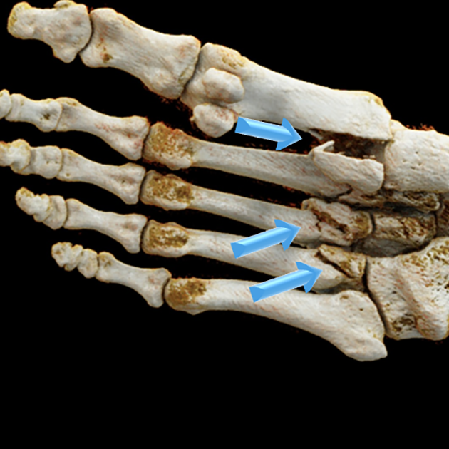
    It is easy to see how adding key images to the report can help us communicate better, especially when summarizing a complex study such as a CT scan (Fig. 1).  But there are practical challenges, both technical and cultural, to implementing this simple idea.

    Radiologists understand the potential value of a multimedia report for their referring colleagues. However, even if they had the available technology, they simply do not have time for an extra step, unless it is quick and easy. Some vendors offer the ability to add images to reports, but that can be cumbersome. Without tight integration, having separate applications from two different vendors can complicate the process of importing images from the viewer (i.e. PACS) into the reporting system.

    One solution is the hybridization of reporting and viewing applications into one. Because the systems are combined, the reporting system is more naturally aware of what is in the viewer, thus facilitating the import of key images. At the University of Virginia Health System (UVaHS), this approach (Vue PACS with Vue Reporting, Carestream Health) allows us to add images into our clinical reports [1]. The process is as follows: important images are optimized in the viewer, marked as “key” by a mouse click, and then a voice command inserts the images into the report (Fig. 2). The process only takes seconds to complete.

    The concept of the reporting system being “aware” of what is in the viewer can be applied in additional ways. Radiologists routinely compare the study they are interpreting to relevant prior studies and dictate identifying details into their report. With the reporting system alert to prior studies opened in the PACS, a voice command can automatically insert details of these studies into the report. This can save time and reduce transcription error.

    An even more important advancement is the ability to place hyperlinks within the report. This dynamic addition powers a variety of interactive elements through a URL to enhance the experience of those viewing the report, whether to a referring provider, downstream radiologist, or patient.

    Consider the example where a radiologist annotates an image in the PACS, either by drawing an arrow pointing to a subtle finding or measuring a small lesion. A voice command inserts hyperlinked text into the report, carrying context from the last annotation made in the viewer. From the radiologist’s perspective, measurements, identifying series, and image numbers are automatically inserted into the report without dictating these details, again saving time and reducing transcription error. From the referring provider’s perspective, important findings stand out in the report as colored hyperlinked text that can provide immediate access to relevant images (Fig. 2). This could accelerate report and image review time, as well as improve report clarity.

    At UVaHS, we have found this multimedia reporting approach to be enough of a departure from traditional plain-text-only reporting that it requires more than just access to the technology; it necessitates brief training and months or more for cultural adoption. Nevertheless, our radiologists now commonly create interactive multimedia reports for complex imaging studies like CT, PET/CT, and MRI. This elective change in care delivery indicates that our radiologists find value in the result.

    Hyperlinks can further enhance reports by connecting to a variety of additional content beyond key imaging findings. Conceptually, once hyperlinks are supported, any content available via URL activation could be leveraged. For example, links within the report could be enabled to launch: secure sites to facilitate patient- and study-context email communication; a report grading system for providers or patients to contribute feedback; a webpage to share radiologist’s credentials; or a webpage with patient-friendly content to help the patient learn useful information about the examination.

    Beyond the technological advancement needed to create interactive multimedia reports and the hurdle of cultural adoption by radiologists, another barrier to report evolution is the ability to distribute and view the advanced reports. It is routine for an electronic health record (EHR) to receive, archive, and display plain-text-only reports; however, the system may not be designed to handle more contemporary document formats, like RTF and PDF, to convey enriched content.

    For the last couple of years at UVaHS, we have worked around this problemSH to enable our referring providers to access our advanced reports through the EHR in two ways. One is through a link to a PDF copy of the report stored in a document management system (OnBase, Hyland). The other is through a link to a lite digital viewer (Vue Motion, Carestream Health) that displays both the interactive multimedia report and all scrolling images from the study.

    Recently, our EHR vendor (Epic Systems Corporation) completed development that should support interactive multimedia report content over an interface message in RTF format. We are collaborating with our vendors to test enriched report content directly within the EHR to increase viewing ease for clinicians.

    We would also like for our patients to be able to see these advanced reports. If patients had an interactive report that allowed viewers to click on various findings and direct them to display the relevant images, patients may become more engaged with their imaging results and could develop a better understanding of their conditions. We are getting closer to this reality. As with many health care systems, our patients already can access their imaging results via a patient portal to the EHR. As a next step, we are working with our vendors to connect this patient portal to a patient-facing lite viewer that displays both radiology images and the interactive multimedia report.

    In many aspects of daily life, digital communications are routinely enhanced beyond plain text with images, text formatting, and interactive elements. It seems natural to extend these improvements in communication to radiology reporting. We have already achieved technical success in doing so, and we have observed solid, sustained adoption by our radiologists. This is just the beginning. There are still challenges in distributing advanced reports, and referring providers are not even expecting to see them. This will change. In the future, interactive multimedia radiology reporting will likely become standard. Referring clinicians, and some discriminating patients, will come to expect enhanced reports. Radiologists will get used to creating them. Eventually, we will drop the words “interactive multimedia” and simply call them “radiology reporting.”


    The opinions expressed in InPractice magazine are those of the author(s); they do not necessarily reflect the viewpoint or position of the editors, reviewers, or publisher.

  • State of the Art Imaging for Chronic Liver Disease

    State of the Art Imaging for Chronic Liver Disease

    avatar

    Khaled M. Elsayes
    Professor of Diagnostic Radiology
    University of Texas, MD Anderson Cancer Center and McGovern Medical School

    Chronic liver disease is an increasingly common and important disorder, now afflicting more than 25% of the world’s population. Regardless of its underlying cause, chronic liver disease can progress to cirrhosis, development of liver cancer, and liver-related death. Recent advances in imaging knowledge and technology have elevated the role of radiology in the diagnosis and management of patients with or at risk for chronic liver disease across its entire spectrum and course.

    Noninvasive Monitoring of Chronic Liver Disease

    With a rising epidemic of obesity and metabolic syndrome in the United States, nonalcoholic fatty liver disease and its progressive form, nonalcoholic steatohepatitis, are increasingly encountered in practice. Additionally, overconsumption of alcohol and viral hepatitis remain common causes of chronic liver disease. Irrespective of etiology, chronic liver disease represents a spectrum of inflammation, injury, fibrosis, and eventually, cirrhosis. Although in the past, cirrhosis was considered an irreversible injury, recent medical advances provide opportunities to halt and even reverse fibrosis. Likewise, identification and modulation of risk factors, like fat and iron deposition in the liver, are essential to managing patients at risk for or with chronic liver disease. Pathological assessment remains a reference standard, but risks and cost associated with biopsy reduce the benefit in the setting of longitudinal monitoring. Radiologists play an essential role in providing noninvasive quantitative information to direct management.

    State of the art imaging options for assessing chronic liver disease focus on three key elements: liver fat, iron, and fibrosis. Liver fat can be identified by ultrasound, CT, and MRI. The most accurate, accessible, and precise method for monitoring patients with chronic liver disease is MRI-proton density fat fraction (PDFF). MRI-PDFF takes advantage of the fact that owing to differences in molecular structures, fat and water protons experience different magnetic fields and precess at different rates. This offset in frequency (“chemical shift”) is the basis for in- and opposed-phase dual echo imaging with its familiar appearance, including “India ink” etching at fat-water interfaces on opposed-phase images, due to signal cancellation when water and fat are precessing directly opposite each other. MRI-PDFF expands on this concept and is estimated by acquiring images at multiple echo times, selected to optimize separation of fat and water signals, and by taking into account several confounders that otherwise introduce errors into fat quantification. Iron is one of the most important confounders and is also an important factor contributing to chronic liver disease. Iron is ferromagnetic and causes signal decay (T2*), due to disruption of local magnetic fields. The decay of signal over time (R2*) is directly proportional to iron content over a wide pathophysiological spectrum, allowing us to estimate R2* values and convert them to liver iron concentrations. Hence, MRI-PDFF quantifies both fat and iron.

    The most validated and clinically used method for estimating liver fibrosis is elastography. Elastography can be done with ultrasound or MRI. Elastography is an imaging technique that quantifies the stiffness of tissue, or resistance to deformation following application of external pressure. Imaging methods estimate stiffness by generating shear waves in the liver and measuring their propagation. Ultrasound methods differ based on how they generate shear waves and whether they produce gray-scale images (point shear wave elastography and 2D elastography) or not (vibration controlled transient elastography). Ultrasound methods measure shear wave speed, which can be converted into tissue stiffness values. The speed measurements may differ between manufacturers and etiologies of chronic liver disease, which challenges establishment of universal thresholds for stages of fibrosis. Magnetic resonance elastography (MRE) utilizes a standard system for shear wave generation and measurement across all vendors and platforms; as a result, the tissue stiffness values obtained from MRE are thought to be more reproducible.

    Standardizing Diagnosis of Liver Cancer in Patients with Chronic Liver Disease

    Hepatocellular carcinoma (HCC) can be confidently diagnosed based on imaging, in contradistinction to most malignancies that require tissue examination for their diagnosis. The noninvasive diagnosis of HCC is justified by the high positive predictive value of CT and MRI for this purpose when stringent criteria are applied in high-risk patients (i.e., high pre-test probability). Filippone A, Blakeborough A, Breuer J, et al. Enhancement of liver parenchyma after injection of hepatocyte-specific MRI contrast media: a comparison of gadoxetic acid and gadobenate dimeglumine. J Magn Reson Imaging 2010; 31:356–364

    Roberts LR, Sirlin CB, Zaiem F, et al. Imaging for the diagnosis of hepatocellular carcinoma: A systematic review and meta-analysis. Hepatology 2018; 67:401–21

    Hanna RF, Miloushev VZ, Tang A, et al. Comparative 13-year meta-analysis of the sensitivity and positive predictive value of ultrasound, CT, and MRI for detecting hepatocellular carcinoma. Abdom Radiol (NY) 2016; 41:71–90 Additionally, cross-sectional imaging assesses local spread and distant metastases.

    Current standards in the noninvasive diagnosis of HCC follow the guidelines of the American Association for the Study of Liver Disease (AASLD), Organ Procurement and Transplantation Network (OPTN), and Liver Imaging Reporting and Data System (LI-RADS). Duncan JK, Ma N, Vreugdenburg TD, Cameron AL, Maddern G. Gadoxetic acid-enhanced MRI for the characterization of hepatocellular carcinoma: a systematic review and meta-analysis. J Magn Reson Imaging 2017; 45:281–290

    Guo J, Seo Y, Ren S, et al. Diagnostic performance of contrast-enhanced multidetector computed tomography and gadoxetic acid disodium-enhanced magnetic resonance imaging in detecting hepatocellular carcinoma: direct comparison and a meta-analysis. Abdom Radiol (NY) 2016; 41:1960–1972

    Liu X, Jiang H, Chen J, Zhou Y, Huang Z, Song B. Gadoxetic acid disodiumenhanced magnetic resonance imaging outperformed multidetector computed tomography in diagnosing small hepatocellular carcinoma: a meta-analysis. Liver Transpl 2017; 23:1505–1518
    These guidelines agree on certain imaging features that should be present in an observation to provide the required high positive predictive value for HCC, such as a maximum diameter of at least 10 mm and characteristic dynamic enhancement characteristics discussed further below. Distinct differences used to exist between the three guidelines in the categorization of hepatic lesions, until the release of the latest LI-RADS guidelines for CT and MRI in 2018. The latest release comprised minor modifications to LI-RADS version 2017 to facilitate its integration into the AASLD clinical practice guidelines in August 2018. Matsui O, Kobayashi S, Sanada J, et al. Hepatocelluar nodules in liver cirrhosis: hemodynamic evaluation (angiography-assisted CT) with special reference to multi-step hepatocarcinogenesis. Abdom Imaging 2011; 36:264–272 LI-RADS and AASLD now have identical criteria for definite HCC, and the OPTN criteria are nearly identical to LI-RADS and AASLD.

    LI-RADS is a comprehensive system that provides standards for terminology, technique, interpretation, and reporting of liver imaging. It has been developed by a multi-disciplinary and increasingly international team of diagnostic and interventional radiologists, hepatobiliary surgeons, hepatologists, and hepatopatholgists, alongside support from the American College of Radiology. Choi YS, Rhee H, Choi JY, et al. Histological characteristics of small hepatocellular carcinomas showing atypical enhancement patterns on gadoxetic acid enhanced MR imaging. J Magn Reson Imaging 2013; 37:1384–1391 Since its first release in 2011, LI-RADS has been updated periodically, with the latest update in 2018. Kitao A, Matsui O, Yoneda N, et al. The uptake transporter OATP8 expression decreases during multistep hepatocarcinogenesis: correlation with gadoxetic acid enhanced MR imaging. Eur Radiol 2011; 21:2056–2066

    Kim BR, Lee JM, Lee DH, et al. Diagnostic performance of gadoxetic acid-enhanced liver MR imaging versus multidetector CT in the detection of dysplastic nodules and early hepatocellular carcinoma. Radiology 2017; 285:134–146

    Nakamura S, Nouso K, Kobayashi Y, et al. The diagnosis of hypovascular hepatic lesions showing hypo-intensity in the hepatobiliary phase of Gd-EOBDTPA-enhanced MR imaging in high-risk patients for hepatocellular carcinoma. Acta Med Okayama 2013; 67:239–244

    LI-RADS assigns a diagnostic category code for each observation to communicate the likelihood of being benign or being HCC, ranging from LR-1 (definitely benign) to LR-5 (definitely HCC). The LR-5 category has a reported specificity of 95% for HCC. In addition to the previous five categories, LI-RADS also provides three other categories—LR-NC (not categorizable), LR-TIV (tumor in vein), and LR-M (probably or definitely malignant, not necessarily HCC)—with certain criteria for each category.

    The imaging diagnosis of HCC in LI-RADS is based on the presence or absence of five major imaging features and a number of ancillary features (AFs). Major features include nonrim arterial phase hyperenhancement (APHE), nonperipheral “washout” appearance, enhancing “capsule” appearance, size, and threshold growth.

    The AFs are divided into three groups: AFs that favor malignancy in general, AFs that favor HCC in particular, and AFs that favor benignity. A preliminary LI-RADS category is assigned based on the present major features, then the AFs are used at the interpreter’s discretion to adjust the preliminary category.

    In summary, the latest advances in imaging of HCC allow for a confident noninvasive diagnosis of this malignancy and comprehensive assessment of other lesions and pseudolesions depicted by imaging.

    Improving Sensitivity for Liver Cancer Diagnosis with HBAs

    HBAs are gadolinium-based intravenous MR contrast agents that permit hepatobiliary phase (HBP) imaging, in addition to conventional dynamic post-contrast phases. Gadoxetate disodium is the most commonly utilized HBA, due to high hepatobiliary excretion and convenient HBP timing of 10–30 minutes.

    Gadoxetate offers several advantages for patients with cirrhosis. Of all available modalities, gadoxetate-enhanced MRI has the highest overall per-lesion sensitivity (86%) and positive predictive value (94%) for diagnosis of HCC, as well as the highest sensitivity (84–96%) for detection of ≤ 2 cm HCCs. Unlike APHE, reduced gadoxetate uptake is an early event in hepatocarcinogenesis: up to 38% of early HCCs may be seen only on the HBP, and 82% of high-grade dysplastic nodules (DN) and 76% of early HCCs are hypointense on the HBP.

    HBP hypointense nodules without APHE are unique to HBA MRI. If sampled histologically, 74% of such nodules are HCCs, and 10% are DN, although these numbers may be inflated by selection bias; if followed, 16–43% progress to hypervascular HCC within 24 months. Nakamura S, Nouso K, Kobayashi Y, et al. The diagnosis of hypovascular hepatic lesions showing hypo-intensity in the hepatobiliary phase of Gd-EOBDTPA-enhanced MR imaging in high-risk patients for hepatocellular carcinoma. Acta Med Okayama 2013; 67:239–244

    Cho YK, Kim JW, Kim MY, Cho HJ. Non-hypervascular hypointense nodules on hepatocyte phase gadoxetic acid-enhanced MR images: transformation of MR hepatobiliary hypointense nodules into hypervascular hepatocellular carcinomas. Gut and Liver 2018; 12:79–85

    Saitoh T, Sato S, Yazaki T, et al. Progression of hepatic hypovascular nodules with hypointensity in the hepatobiliary phase of Gd-EOB-DTPA-enhanced MRI in hepatocellular carcinoma cases. Intern Med 2018; 57:165–171

    Yang HJ, Song JS, Choi EJ, Choi H, Yang JD, Moon WS. Hypovascular hypointense nodules in hepatobiliary phase without T2 hyperintensity: longterm outcomes and added value of DWI in predicting hypervascular transformation. Clin Imaging 2018; 50:123–129

    Hwang JA, Kang TW, Kim YK, et al. Association between non-hypervascular hypointense nodules on gadoxetic acid-enhanced MRI and liver stiffness or hepatocellular carcinoma. Eur J Radiol 2017; 95:362–369

    Briani C, Di Pietropaolo M, Marignani M, et al. Non-hypervascular hypointense nodules at gadoxetic acid MRI: hepatocellular carcinoma risk assessment with emphasis on the role of diffusion-weighted imaging. J Gastrointest Cancer 2018; 49:302–310

    Kim YS, Song JS, Lee HK, Han YM. Hypovascular hypointense nodules on hepatobiliary phase without T2 hyperintensity on gadoxetic acid-enhanced MR images in patients with chronic liver disease: long-term outcomes and risk factors for hypervascular transformation. Eur Radiol 2016; 26:3728–3736

    Rosenkrantz AB, Pinnamaneni N, Kierans AS, Ream JM. Hypovascular hepatic nodules at gadoxetic acid-enhanced MRI: whole-lesion hepatobiliary phase histogram metrics for prediction of progression to arterial-enhancing hepatocellular carcinoma. Abdom Radiol (NY) 2016; 41:63–70
    In patients who undergo resection for early-stage HCC, the presence of HBP hypointense nodules predicts high HCC recurrence risk and lower overall survival. Toyoda H, Kumada T, Tada T, Sone Y, Maeda A, Kaneoka Y. Nonhypervascular hypointense nodules on Gd-EOB-DTPA-enhanced MRI as a predictor of outcomes for early-stage HCC. Hepatol Int 2015; 9:84–92 Intermediate to long-term recurrence-free survival may be improved, if these nodules are treated concomitantly at the time of HCC resection. Matsuda M, Ichikawa T, Amemiya H, et al. Preoperative gadoxetic acid-enhanced MRI and simultaneous treatment of early hepatocellular carcinoma prolonged recurrence-free survival of progressed hepatocellular carcinoma patients after hepatic resection. HPB Surg 2014; 2014:641685

    HBP hypointense nodules without APHE are also markers of increased HCC risk elsewhere in the liver: the cumulative three year rate of HCC elsewhere in the liver is 22%, compared to 6% in patients with no such nodules. Komatsu N, Motosugi U, Maekawa S, et al. Hepatocellular carcinoma risk assessment using gadoxetic acid-enhanced hepatocyte phase magnetic resonance imaging. Hepatol Res 2014; 44:1339–1346 Presence of these nodules in patients with early HCC is associated with decreased recurrence-free survival and higher intrahepatic recurrence rates following resection or ablation. Inoue M, Ogasawara S, Chiba T, et al. Presence of non-hypervascular hypointense nodules on gadolinium-ethoxybenzyl-diethylenetriamine pentaacetic acid-enhanced magnetic resonance imaging in patients with hepatocellular carcinoma. J Gastroenterol Hepatol 2017; 32:908–915

    Lee DH, Lee JM, Lee JY, et al. Non-hypervascular hepatobiliary phase hypointense nodules on gadoxetic acid-enhanced MRI: risk of HCC recurrence after radiofrequency ablation. J Hepatol 2015; 62:1122–1130

    Song KD, Rhim H, Lee MW, Kim YS, Kang TW. Intrahepatic distant recurrence after radiofrequency ablation for hepatocellular carcinoma: precursor nodules on pre-procedural gadoxetic acid-enhanced liver magnetic resonance imaging. Acta Radiol 2017; 58:778–785

    The degree of gadoxetate uptake may predict tumor differentiation: poorly-differentiated HCCs are more frequently HBP hypointense (98%), compared with well- or moderately-differentiated HCCs (86%). Erra P, Puglia M, Ragozzino A, et al. Appearance of hepatocellular carcinoma on gadoxetic acid-enhanced hepatobiliary phase MR imaging: a systematic review. La Radiol Med 2015; 120:1002–1011 Up to 15% of HCCs may be iso- or hyperintense on the HBP, and such HCCs have more favorable outcomes, including improved recurrence-free and overall survival. Kim JY, Kim MJ, Kim KA, Jeong HT, Park YN. Hyperintense HCC on hepatobiliary phase images of gadoxetic acid-enhanced MRI: correlation with clinical and pathological features. Eur J Radiol 2012; 81:3877–3882

    Kitao A, Matsui O, Yoneda N, et al. Hypervascular hepatocellular carcinoma: correlation between biologic features and signal intensity on gadoxetic acid-enhanced MR images. Radiology 2012; 265:780–789

    Kitao A, Zen Y, Matsui O, et al. Hepatocellular carcinoma: signal intensity at gadoxetic acid-enhanced MR Imaging–correlation with molecular transporters and histopathologic features. Radiology 2010; 256:817–826

    Use of gadoxetate in patients with cirrhosis is associated with several important pitfalls. Smaller contrast dose and volume affect timing of the arterial phase (AP) and may lead to reduced peak enhancement of HCC in the AP. Fujinaga Y, Ohya A, Matsushita T, Kurozumi M, Ueda K, Kitou Y, et al. Effect of hepatobiliary uptake of Gd-EOB-DTPA on the hepatic venous phase of dynamic magnetic resonance imaging on a 3.0-T apparatus: comparison between Gd-EOB-DTPA and Gd-DTPA. Jpn J Radiol 2011; 29:695–700

    Tirkes T, Mehta P, Aisen AM, Lall C, Akisik F. Comparison of dynamic phase enhancement of hepatocellular carcinoma using gadoxetate disodium vs gadobenate dimeglumine. J Comput Assist Tomogr 2015; 39:479–482
    Furthermore, gadoxetate is associated with higher incidence (5–22%) of transient severe motion, which occurs at or around the time of the late AP and leads to image degradation. Davenport MS, Viglianti BL, Al-Hawary MM, et al. Comparison of acute transient dyspnea after intravenous administration of gadoxetate disodium and gadobenate dimeglumine: effect on arterial phase image quality. Radiology 2013; 266:452–461

    Davenport MS, Bashir MR, Pietryga JA, Weber JT, Khalatbari S, Hussain HK. Dose-toxicity relationship of gadoxetate disodium and transient severe respiratory motion artifact. AJR 2014; 203:796–802

    Kim SY, Park SH, Wu EH, et al. Transient respiratory motion artifact during arterial phase MRI with gadoxetate disodium: risk factor analyses. AJR 2015; 204:1220–1227
    Poor quality of the AP may affect depiction of APHE, a feature that is required for noninvasive HCC diagnosis. Chernyak V, Fowler KJ, Kamaya A, et al. Liver imaging reporting and data system (LI-RADS) version 2018: imaging of hepatocellular carcinoma in at-risk patients. Radiology 2018; 289:816–830

    Portal venous phase (PVP) “washout” appearance in combination with APHE allows for nearly 100% specificity of HCC diagnosis. Marrero JA, Hussain HK, Nghiem HV, Umar R, Fontana RJ, Lok AS. Improving the prediction of hepatocellular carcinoma in cirrhotic patients with an arterially-enhancing liver mass. Liver Transpl 2005; 11:281–289 Parenchymal uptake of gadoxetate starting as early as the PVP results in observations potentially appearing relatively hypointense to the parenchyma, due to lower uptake of gadoxetate rather than true “washout.” As a result, hypointensity in the transitional phase (TP) is not equivalent to hypointensity during the PVP or delayed phases with extracellular agents: if hypointensity in the TP is considered “washout,” the specificity for HCC decreases from 98–100% to 86–95%. Choi SH, Lee SS, Kim SY, Park SH, Park SH, Kim KM, et al. Intrahepatic cholangiocarcinoma in patients with cirrhosis: differentiation from hepatocellular carcinoma by using gadoxetic acid-enhanced MR imaging and dynamic CT. Radiology 2017; 282:771–781 Therefore, LI-RADS restricts assessment of “washout” with gadoxetate to the PVP. Santillan C, Fowler K, Kono Y, Chernyak V. LI-RADS major features: CT, MRI with extracellular agents, and MRI with hepatobiliary agents. Abdom Radiol (NY) 2018; 43:75–81 Another effect of the early parenchymal enhancement with gadoxetate is the potential to obscure enhancement of the “capsule.”

    In patients with decompensated cirrhosis, diminished parenchymal uptake of gadoxetate results in less enhancement during the TP and the HBP. Motosugi U, Ichikawa T, Sou H, et al. Liver parenchymal enhancement of hepatocyte-phase images in Gd-EOB-DTPA-enhanced MR imaging: which biological markers of the liver function affect the enhancement? J Magn Reson Imaging 2009; 30:1042–1046 As a result, conspicuity of HCC in the HBP is decreased in patients with poor hepatic function. Kim JY, Lee SS, Byun JH, et al. Biologic factors affecting HCC conspicuity in hepatobiliary phase imaging with liver-specific contrast agents. AJR 2013; 201:322–331 Additionally, interpretation of HBP intensity of liver observations—particularly if iso- or hyperintense to the background— may be unreliable in the setting of suboptimal HBP enhancement.

    Although HBP hypointensity improves detection of HCC and high-grade DN, TP and HBP hypointensity are not specific to HCC, as any lesion without functional hepatocytes (e.g., cysts, hemangiomas, non-HCC malignancies, etc.) will appear hypointense in the HBP. Joo I, Lee JM, Lee DH, Jeon JH, Han JK, Choi BI. Noninvasive diagnosis of hepatocellular carcinoma on gadoxetic acid-enhanced MRI: can hypointensity on the hepatobiliary phase be used as an alternative to washout? Eur Radiol 2015; 25:2859–68

    In conclusion, use of gadoxetate in patients with cirrhosis offers certain advantages—particularly higher sensitivity for HCC, if liver function is preserved and AP quality is adequate—but radiologists should be aware of the various pitfalls of gadoxetate to optimize patient selection and image interpretation.

    Acknowledgements
    Kathryn Fowler, Claude Sirlin, and Victoria Chernyak also contributed to this article.


    The opinions expressed in InPractice magazine are those of the author(s); they do not necessarily reflect the viewpoint or position of the editors, reviewers, or publisher.

  • Gender Affirmation Imaging Revisited

    Gender Affirmation Imaging Revisited

    avatar

    Florence X. Doo
    Resident Physician Diagnostic Radiology
    Icahn School of Medicine at Mount Sinai West

    Alexander S. Somwaru
    Assistant Professor of Diagnostic Radiology
    Icahn School of Medicine at Mount Sinai West

    How often does gender or sex come up in your daily practice? In a publication or article you read? In your daily interactions with your family or colleagues?

    Based on your answers to the above questions, suffice it to say that although gender is arguably a core piece of each individual’s identity, it variably affects one’s daily lived experience. For example, do you experience your gender differently in different contexts—such as when you were eight years old vs as a 45-year-old, or in different settings like a black-tie event? Just from these instances, it is clear that many factors influence our experience of gender, including language, societal norms, cultural background, etc. A recent article in the Journal of the American College of Radiology (JACR) excellently described basic gender terminology and definitions, emphasizing the need for greater gender inclusivity in radiology.

    Whereas radiologists are not expected to be gender studies experts, there are several key aspects of gender that radiologists should be aware of in daily practice.

    1. Be able to clearly and accurately describe imaging findings of physical anatomy, including gender affirmation therapy and surgeries, which we described in our original article in AJR, “Gender Affirmation Surgery: A Primer on Imaging Correlates for the Radiologist”. Gender affirmation therapy is multidisciplinary by nature; as radiologists, we are part of the team providing necessary care for individuals experiencing gender incongruence.
    2. Consider adjusting protocols for relevant anatomy: training staff to allow self-insertion of transvaginal probes; utilizing an endovaginal, rather than transrectal, approach for prostate cancer screening in transwomen who have undergone gender affirmation surgery; and correctly evaluating atrophied structures secondary to hormone therapy.
    3. Study developing educational cases and share research in transgender health and imaging. Our colleagues (Maglione, Margolies, Jaffer, et al.) published one of the first descriptions of imaging findings of breast cancer in transgender women in AJR, and others have proposed adapting breast cancer screening guidelines for transgender women. More investigations with outcomes-based research are needed to improve understanding of transgender healthcare, including developing screening guidelines and protocols.
    4. Collaborate with counselors, pediatricians, endocrinologists, general practitioners, surgeons (breast, plastic, maxillofacial, gynecologic, general, etc.), and allied health professionals who specialize in gender affirmation therapy, as there are treatment options (i.e., hormonal therapy) and ongoing advancements that may impact imaging interpretation.
    5. Be aware of your local facility and institution’s Human Rights Commission “Healthcare Equality Index” score, and how you may either maintain or improve that score. Our own institution, Mount Sinai Health System, is fortunate to have a 100/100 score and a strong multidisciplinary team at the Center for Transgender Medicine and Surgery.

      What is the experience of LGBTQ+ patients in your facility? Are all staff trained? Two separate studies in 2015 and 2017 found that almost a quarter of transgender people said they avoided doctors or health care for fear of being discriminated against. The 2017 study, which detailed LGBTQ+ American experiences, showed that 33% had one or more negative experiences with a health care provider.
    6. Assess workplace and educational climates for colleagues and trainees, who may face invisible or systemic barriers, even if not addressed explicitly in the scoring system. A 2015 study of medical students showed that approximately 43% of sexual and gender minorities concealed their identities, due to fear of discrimination. Do you consider your workplace gender diverse? Have you personally advocated for gender inclusivity, or defended someone from discriminatory attitudes, remarks, or behaviors? How are issues reported and addressed individually and systemically?
    7. Encourage your professional society and journal publications to be aware of sex and gender terminology. Many journals, including AJR, require sex to be labelled on image captions. This posed an interesting dilemma, particularly in our recent article, as “sex assigned at birth” (SAAB) and “anatomical sex” are not necessarily equivalent, and anatomical sex may change. And, of course, these labels do not necessarily match the patient’s gender identity or chosen pronouns. These guidelines should be contextualized when publishing articles on transgender imaging.
    Data Sources: Nature Reviews Endocrinology & Hormone Research in Pediatrics

    8. Examine your local, state, and national policies regarding gender rights and safety protections. As described in a recent call to action published in JACR, the Association of University Radiologists moved its annual meeting location, in part, due to California’s Assembly Bill 1887, which prohibited California state employees (including those at medical centers) from state-funded travel to states that have discriminatory laws in place. Other radiology organizations should be encouraged to follow this example. Only half of the United States has state or local laws protecting LGBTQ+ workers. Is your state one of them?

    9. Survey identification (ID) laws, as well as how difficult it may be to correct identification to reflect gender identity. The National Center for Transgender Equality’s 2015 study also revealed that 32% of transgender individuals who have shown an ID with a name or gender that did not match their gender presentation were verbally harassed, denied benefits or services, asked to leave, or assaulted. What are the requirements for ID in your institution, and how are staff trained to respond?

    Imaging is simply one, albeit vital, facet in the overall care that we provide to our patients. The landscape of gender and sex, both inside and outside of radiology, continues to evolve, as we improve our individual understanding and address systemic flaws. All of us—ourselves, our healthcare providers, our patients, and our society—benefit by respectfully recognizing our individual identities. Let us be allies for one another.


    The opinions expressed in InPractice magazine are those of the author(s); they do not necessarily reflect the viewpoint or position of the editors, reviewers, or publisher.
  • In-the-Moment Aggressions

    In-the-Moment Aggressions

    avatar

    Ruth C. Carlos
    2019–2020 ARRS President

    Here’s a word that you still won’t find in Stedman’s: microaggression. A short, often casual exchange that, regardless of intent, can send a disparaging message to a certain individual because of group membership, there’s nothing particularly new about it. Lest you think this word was forged in a social media storm, in fact, it was a tenured professor of education and psychiatry at Harvard Medical School who first used it. Coined in 1970 by Chester M. Pierce, the first African-American full professor at Massachusetts General Hospital, to combine the subtle dismissals and outright injustice non-black Americans inflict upon African Americans, today’s wider term applies to any routine marginalization based on race, gender, sexuality, age, ability, or socioeconomic status. As Meridith J. Englander and Susan K. O’Horo observed in last October’s AJR Journal Club, microaggression has “entered our everyday vernacular”.

    Need the word in a sentence, with some real-life examples? When an attending physician tells an international student that he or she is doing exceptionally well on rounds, “especially considering their background.” The subordinate or colleague who keeps hearing that they’re “just too pretty” or “far too handsome” to be stuck in a dark reading room.

    “Think of microaggressions like mosquito bites,” wrote Carolynn M. DeBenedectis, lead author of “Microaggression in Radiology,” the September commentary published in the Journal of the American College of Radiology. “A single or occasional mosquito bite is annoying for a second but can be ignored, but when the mosquito bites are unrelenting and in large numbers, they can be damaging.” Left untreated, these bites of bias— again, unconscious or not—can lead to diminished self-confidence, poorer self-image, as well as serious mental health conditions like anxiety and depression.

    Responding to the August commentary in JAMA Pediatrics, the New York Times’ headline wondered, “Is it possible to train doctors without hurting anyone’s feelings?”. Of course, as JAMA’s own title duly noted, “Avoiding the eggshells is not the answer”. We practice medicine, and sooner rather than later, all practicing diagnostic radiologists will have to render a difficult face-in-PACS decision and engage in a potentially problematic discussion face-to-face. For academic and mentor radiologists alike, it’s our duty to point out the mistakes of others—shedding light on each artifact and every pitfall.

    Acknowledging that a person’s gender, ethnicity, or circumstances are real-world facts, how should we respond as the “other?” Moreover, what does a true ally do in these most fraught moments? To start, always consider the source, where context remains key. Being too brusque helps neither party, especially if the microaggressor is closely connected to the microaggrieved. “Keep the initial conversation short, and schedule a time to talk about it later to give the other person time to think things over,” suggests NiCole T. Buchanan, an associate professor of psychology who leads workshops on microaggressions at Michigan State University.

    At the same time, passive-aggressiveness can hurt everyone, beyond the microaggressor and the microaggrieved even. As leadership consultant Mario Rodriguez noted during this year’s Association of Medical Imaging Management annual meeting, following a simple, three-step process often helps to diffuse conflicts in a pointedly, albeit respectful manner. According to Rodriguez, “you should describe the problem simply, explain clearly how it makes you feel, and, then, outline the changes you’d like to see”.

    As Judy Melinek wrote, “social justice is good medicine”. Together, we can be both radiologist and advocate.


    The opinions expressed in InPractice magazine are those of the author(s); they do not necessarily reflect the viewpoint or position of the editors, reviewers, or publisher.

  • Leadership to Equitable Radiology

    Leadership to Equitable Radiology

    avatar

    Ruth C. Carlos
    2019-2020 ARRS Scholar

    According to the January issue of AJR, women occupy only 14% of available leadership positions in academic nuclear medicine departments, both at American and Canadian institutions. A recent Emory University assessment of gender disparity in labor divisions found that female general, abdominal, and musculoskeletal radiologists read fewer advanced imaging modalities like CT and MRI. Perhaps more lopsided is the male-to-female ratio, itself—approximately three men for every one woman among radiologists and radiology residents, as per ACR’s annual industry survey. Furthermore, according to the October edition of AJR, just 8% of all interventional radiologists in the United States are women.

    Like much of the news, health care headlines such as these can leave us asking ourselves, “How much progress have we made?” Dredging this sea of conspicuous data points and trends on gender inequality in medical imaging, men and women alike question, “What does ‘progress’ even look like?”

    One hundred years removed from the ratification of the Nineteenth Amendment to the U.S. Constitution that confirmed my right to vote, as the 119th ARRS president, I am proud to join the august ranks of Kay Vydareny, Theresa McLeod, Ella Kazarooni, and Melissa Rosado de Christenson—four previous female presidents who have served this oldest radiology society in North America. And now, for the first time ever, six major radiology organizations are all led by women—International Society for Magnetic Resonance in Medicine, Radiologic Society of North America, Society of Interventional Radiology, American Society of Neuroradiology, American College of Radiology, and, of course, the American Roentgen Ray Society.

    In the wake of this boom of female leadership in our specialty, why is it that more women continue to enter other specialties such as pediatrics and gynecology, while radiology remains a male-dominated field? The proportion of women in our specialty has remained static at 20–25%.

    Might this radiological gender gap present an opportunity for “failing up” in radiology, as described in my previous column for InPractice? Conversely, are there intrinsic differences in the practice of radiology compared to, say, pediatrics that will preferentially steer women toward non-radiology specialties? Although I can’t say that 20–25% is the right or wrong proportion, we must collectively work to reduce those barriers that we can control, such as increasing role-modeling, mentorship, and sponsorship and decreasing implicit and explicit bias.

    “And now, for the first time ever, six major radiology organizations are all led by women— International Society for Magnetic Resonance in Medicine, Radiologic Society of North America, Society of Interventional Radiology, American Society of Neuroradiology, American College of Radiology, and, of course, the American Roentgen Ray Society.”

    Ruth C. Carlos

    As an equal opportunity mentee, I have had the privilege of being supported and mentored by a wide variety of individuals such as Melissa Rosado de Christenson, who inspired my career-long affiliation with the ARRS, JACR Editor-in-Chief Emeritus Bruce Hillman, who shared his knowledge and love of scholarly publishing, and John Fennessy and Ruth Ramsey, to whom I owe considering radiology as a career. All those in a position to lead have a responsibility for closing the gender gap. This gap can be as obvious as the lagging proportion of women in radiology or as subtle as “manels,” all-male speaker panels. I applaud Francis S. Collins, director of the National Institutes of Health, for his resolution to no longer speak on all-male panels. “Breaking up the subtle (and sometimes not so subtle) bias that is preventing women and other groups underrepresented in science from achieving their rightful place in scientific leadership must begin at the top,” read his memo, “Time to End the Manel Tradition”.

    A radiologist’s gender is a fact, not a quota. Nevertheless, consistent, thoughtful attention to increasing diversity not just in gender but in all the axes of diversity benefits us all.


    The opinions expressed in InPractice magazine are those of the author(s); they do not necessarily reflect the viewpoint or position of the editors, reviewers, or publisher.
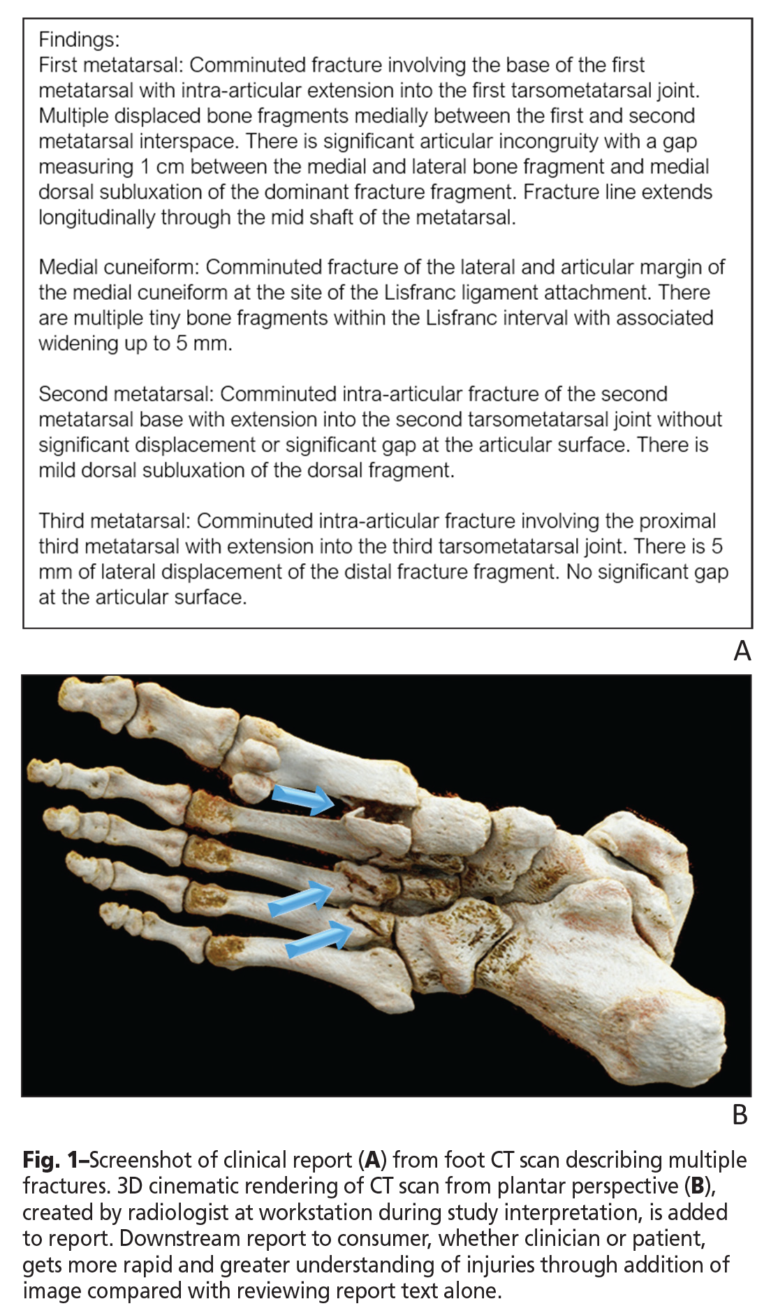
  • Moving from Peer Review to Peer Learning

    Moving from Peer Review to Peer Learning

    avatar

    Nadja Kadom
    Department of Radiology and Imaging Sciences Emory University; Pediatric Imaging Assistant Editor, AJR
    Cindy Lee
    Department of Radiology NYU Langone Health

    Competency of radiologists is an important factor in the delivery of high-quality patient care. To meet maintenance of certification (MOC) and Joint Commission requirements for ongoing professional performance evaluation (OPPE), radiologists are participating in peer review, a system that uses accuracy of interpretation as a surrogate marker for competency. A widely used example of such a system is RADPEER, a web-based product that was developed by the American College of Radiology (ACR).

    Benefits of peer review as a means of assessing radiologist competency include: Availability of commercial products; accreditation that is accepted by the Joint Commission; credit towards American Board of Radiology MOC part 4; and familiarity with established peer review systems. Existing peer review systems are generally set up to create as little additional workload as possible, which fosters compliance.

    There are, however, several issues regarding peer review systems, including great variability in how peer review data are collected. Some programs select cases randomly, whereas others allow physicians to select cases, which could introduce bias or result in case selections that are quick to review. There is also variability in how peer review is executed (i.e., the number of cases to review and the frequency of peer review).

    Systems that select cases for peer review—and that are not integrated in the daily exam reading workflow—carry a risk that participants will wait until the deadline and then hastily review a large number of cases, calling into question the quality of such reviews. Most peer review participants are never formally instructed on how to use the scoring system, and lack of examples for each scoring category can result in scoring inconsistencies Peer review participants prefer anonymous review, although in most practices, both the reviewer and the reviewed can identify each other. Friendly and unfriendly relationships may influence the scoring of agreement. There is often a stated peer review goal, such as achieving a less than 5–10% disagreement rate, which can create bias and result in under-reporting. Regarding the use of peer review data, as a ground rule, data should be collected and reported in a fashion that does not invite medicolegal action or repercussions at the local level. Allowing peer review data to be reviewed by other radiologists or officials at higher levels of the organization incites fear in participants that could lead to adverse effects on relationships within the department or the organization. Participants may need transparency regarding local and state policies and medicolegal safeguards in order to trust and honestly use the review system. Whereas some radiology practices have even used peer review data to terminate radiologists’ contracts, instead, peer review should serve to coach and judge at the same time. Moreover, there is no evidence to suggest peer review is an appropriate tool for identifying radiologists who are inadequate performers.

    It is not clear whether or not participation in peer review leads to performance improvement. This may be especially true for systems that do not provide feedback on reviewed cases, nor discuss relevant cases in a group setting. It should be noted that a disagreement does not equal an error or low-quality patient care. A discrepancy could simply be a difference of opinion on how a finding should be interpreted and reported. Peer review can underestimate the number, as well as the severity, of errors. Any discussion around such disagreement may be of little value in the absence of clinical follow-up or tissue-proven diagnoses. In traditional peer review, there can be a significant gap between the time the report was rendered for patent care and the time the report was reviewed and an error was identified, delaying any changes to patient care.

    To improve patient outcomes, we need to move from quality assurance to quality improvement. Our goal should be to reduce diagnostic errors, which contribute to 10% of patient deaths and 6–17% of adverse events in hospitals.

    Radiologists should be able to learn from their mistakes—an essential component of improving patient safety. Disclosing and discussing errors for learning and improvement purposes requires a so-called Just Culture that acknowledges human error, avoids blame, promotes fair accountability, and focuses on fixing system deficiencies. Just Culture often goes hand-in-hand with Safety Culture, which is found in high-reliability organizations, where error rates are very low. This culture entails, among other traits, embracing every opportunity to learn from mistakes and fix systems and processes for error prevention.

    Several sources are available for providing timely feedback. For example, during the read-out, a radiologist may find an error after reviewing the prior study. Consultation with a referring clinician and second review of studies for multidisciplinary conferences can reveal errors, comparison of pathology or surgical reports with imaging results can uncover errors, and complaints to radiology leadership or incident reporting systems can disclose errors. Radiologists may decide which system for feedback they prefer, be it a collegial email, a templated email, or if this information should be routed through third parties, such as section chiefs or quality officers. Many automated systems are available that facilitate peer feedback as part of the daily workflow rather than a separate activity, such as integration with PACS, dictation systems, or other apparatuses that automatically send surgical and pathology results to the radiologist who made the diagnosis. Each method has different ramifications regarding medico-legal discoverability, which may be an important consideration for practicing radiologists.

    Recommendations for best practices to raise learning opportunities to the group level include establishing a small committee to select cases with the highest learning potential, removal of all identifying case information and anonymity of the radiologist who interpreted a case, recording of peer learning conferences to enable asynchronous participation, avoiding blame or finding fault, encouraging discussion of pitfalls, mimics, and strategies for error prevention, and even providing relevant scientific references. Compliance with ongoing OPPE can be achieved in ways other than peer review discrepancy rates, such as recording participation in peer learning conferences, case submissions, or improvement initiatives that were completed as a result of peer case discussions.

    In 2019, the ARRS Performance Improvement Subcommittee decided to tackle the topic of transitioning from peer review to peer learning. The subcommittee assumed that most peer review programs focus on error detection, numerical scoring, and radiologist-specific error rates, with questionable effectiveness regarding learning and systemic improvement. The subcommittee created a 21-question, multiple-choice survey, and this survey was emailed to 17,695 ARRS members; 742 (4.2%) responded. Most respondents were in private practice (51.7%, 283/547) with a size of 11–50 radiologists (50.5%) and in an urban setting (61.6%). Significant diversity was noted in several aspects regarding peer review systems, including use of RADPEER (44.3%), selection of cases by commercial software (36.2%) versus manually (31.2%), and varying numbers of cases mandated for review per month (range < 10 to > 21). Interestingly, > 60% of respondents noted using peer review for group education. A great need for turning peer review into peer learning exists, as almost half (44.5%) of respondents reported being dissatisfied with their current process, stemming from insufficient learning (> 70%) and a sense of inaccurate performance representation (57.1%). Most respondents found the following peer review methods feasible in daily practice: incidental observation (65%), focused practice review (52.9%), professional auditing (45.8%), and blinded double reading (35.4%). Overall, it seems that a majority of practicing radiologists have already migrated toward peer learning systems and consider a variety of workflow-integrated review methods practicable. Establishing a peer learning system may require investments in staff and time, but as evidence continues to mount that peer learning—as opposed to traditional peer review—results in improved practice, better patient outcomes, and higher radiologist satisfaction, these investments appear to be justified.


    The opinions expressed in InPractice magazine are those of the author(s); they do not necessarily reflect the viewpoint or position of the editors, reviewers, or publisher.
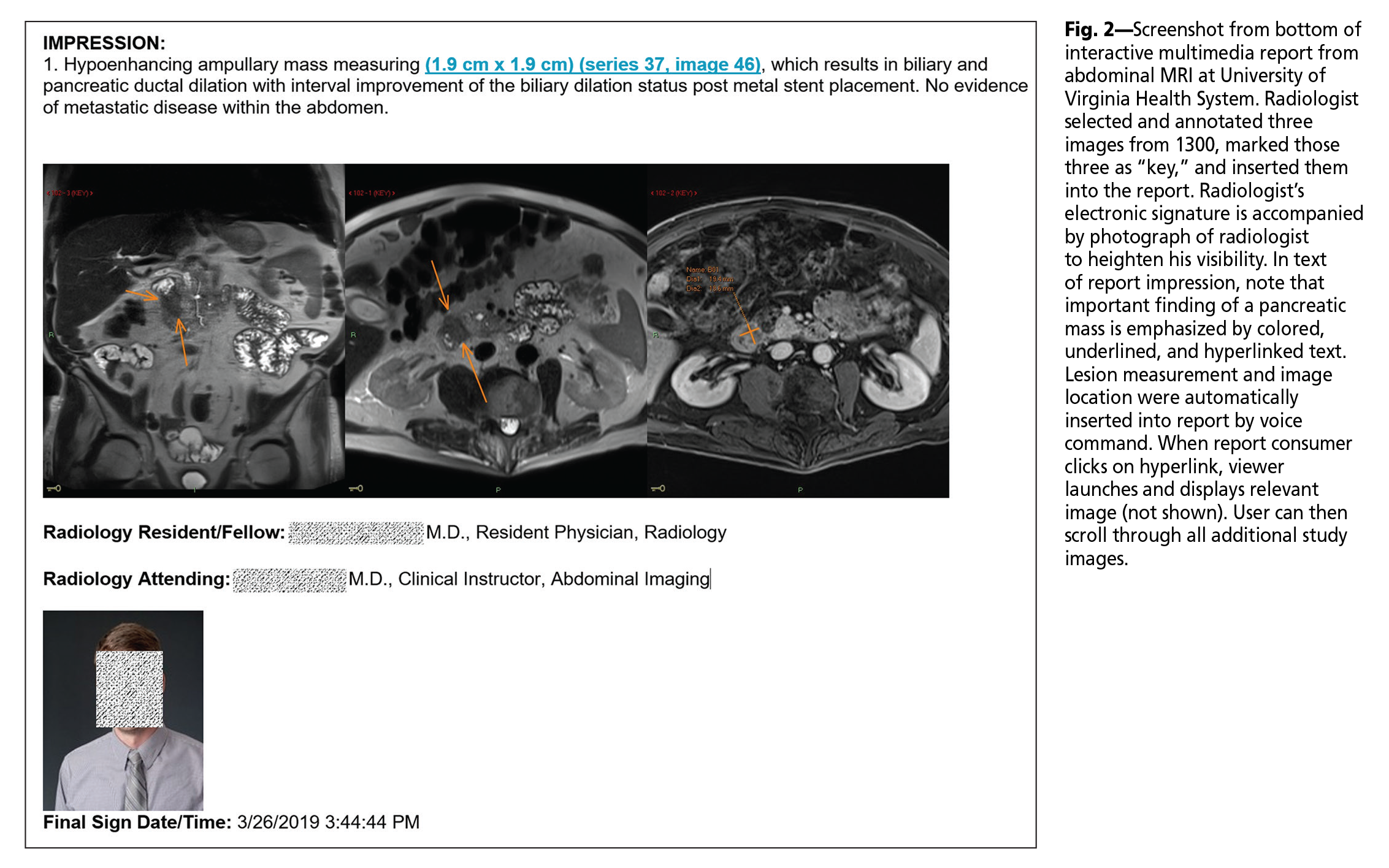
  • Vascular Imaging: Crossing Body Territories and Modalities

    Vascular Imaging: Crossing Body Territories and Modalities

    Imaging has become so engrained and valuable to the practice of medicine that sub-specialization has become necessary for radiology practices to manage the tremendous body of knowledge. This concentration has allowed us to practice diagnostic radiology at a high level to meet the needs of the many medical and surgical specialties. Clinical practices have shifted dramatically from utilizing imaging not only for surveillance and initial diagnosis, but also for surgical planning and long-term management of disease, which also requires greater understanding of the specific therapies in each field. Several medical and surgical specialties now rely on noninvasive vascular imaging to manage disease and for preoperative planning and postoperative observation.

    Although the current silos of subspecialization have been largely beneficial for radiology, some distinct areas of imaging cover the radiological spectrum and do not conform to our present subspecialty definitions. Vascular imaging is one such discipline, which has become somewhat orphaned because of its pervasive nature and tendency to cross body territories and imaging modalities.

    Clinical features of diseases with vascular etiologies frequently overlap with non-vascular diseases. For example, multiple diseases may present with post-prandial abdominal pain—though only a small fraction may end up benefiting from a course of steroids for vasculitis. A clinician’s preliminary diagnosis may be mesenteric ischemia, but the final judgment determined by MRI/MR angiography may be pancreatic cancer, or vice versa. An ulcerated plaque with a dangling thrombus may appear on a thoracic CT angiogram on one examination but disappear on the next—coinciding with a stroke, a bout of ischemic colitis, or a pulseless extremity. Rheumatologists, cardiologists, and surgeons do not terminate their attention to patients at the end of a body territory, though radiologists have largely broken down along territorial lines to improve our efficiency.

    As our specialty becomes increasingly subspecialized, clinical diagnosis has become progressively distributed among a larger number of physicians. This structural change has created a certain peril of failing to “connect the dots” and fading expertise for disciplines that “just don’t fit” into the existing framework of subspecialties. Vascular imaging champions are required, whether ordered into dedicated service lines or attached to conventional subspecialty silos. Complicating matters, indications for imaging examinations can be rather varied, including specific attention on the vessels themselves, (atherosclerotic disease to vasculitis) or diseases of the end-organs (stroke, myocardial infarct, mesenteric ischemia).

    Over that last 30 years, our field has witnessed tremendous advances in imaging technologies that have promptly changed the practice of cardiovascular imaging, shifting markedly from invasive catheter angiography to noninvasive imaging. Radiologist practices, for the most part, have seen noninvasive vascular imaging far outpace invasive imaging, especially as these techniques have improved in their reliability and effectiveness. Although imaging has recently been scrutinized as a cost center, noninvasive diagnosis is strikingly cost-effective, particularly compared to invasive angiography and surgical procedures undertaken without the benefit of the road map that imaging provides.

    These technologies continue to rapidly evolve with dramatic changes even in the last five years, which persist in shaping our clinical practices. However, the non-invasive technologies have not translated into all radiology practices with equal vigor. This non-adoption is due, in part, to the pervasive nature of vascular disease that impacts so many radiological subspecialties, as well as the roles that several imaging modalities play. Ultrasound, CT, and MRI each have complementary functions and strengths, such that screening, definitive diagnosis, and disease management often require more than one. It is difficult to find any subspecialty or individual radiologist with mastery of all these modalities, much less the ability to translate their latest advancements into practice.

    One prevailing question is how to achieve high-level vascular imaging across the wide range of clinical radiology. Ultimately, developing new service lines is exceedingly challenging, especially in the modern era where the pressures of relative value unit-based productivity dominate our practices. The activation barrier is high. Nevertheless, I believe that pursuit of diagnostic excellence is an endeavor worth committing a career to. Our patients count on it. No other specialties are prepared to carry this torch with imaging.

    I also believe radiologists possess a unique skill set and are well-positioned to be champions of multimodality imaging diagnosis. Many of us have been trained as master diagnosticians—to recognize and analyze disease, exploiting the strengths of and overcoming the weaknesses in individual imaging modalities. No, artificial intelligence doesn’t stand a chance without us.

    The opinions expressed in InPractice magazine are those of the author(s); they do not necessarily reflect the viewpoint or position of the editors, reviewers, or publisher.
  • Expansion of Rectal Cancer Staging MR

    Expansion of Rectal Cancer Staging MR

    avatar

    Marc J. Gollub
    Professor of Radiology Memorial Sloan Kettering Cancer Center

    Mukesh G. Harisinghani
    Professor of Radiology Massachusetts General Hospital; Genitourinary Imaging Section Editor, AJR

    Rectal cancer comprises approximately one-third of all cases of colorectal cancer—the third leading cause of cancer death in the United States. In the last two decades, the role of imaging has become crucial to patient care. Until recently, staging of rectal cancer patients was mostly done by surgeons using three office procedures: digital rectal exam, endorectal ultrasound (ERUS), and endoscopy. A more recent shift away from ERUS towards pelvic MRI has now positioned MR imaging front and center for staging rectal tumors. We have noted this growing percentage at our own institution, Memorial Sloan Kettering Cancer Center.

    Owing to direct, point-of-contact proximity and the intrinsic high-resolution, ERUS is superior to MRI in staging lower T-category tumors (T1 and T2); however, ERUS is limited by its restricted FOV and the lack of visualization of the radial (circumferential) tumor margin and the proximal margin. As a result, the mesorectal fascia (i.e., the intended circumferential resection margin for an ideally performed standard total mesorectal excision surgery), pelvic sidewall lymph nodes, upper pelvic lymph nodes, and high rectal tumors cannot be reliably imaged with ERUS. Pelvic MRI can overcome these limitations, thus allowing accurate surgical staging by providing critical information needed for successful surgery, as well as for initial treatment planning, which is needed by medical and radiation oncologists.

    If one combines this current shift to pelvic MR imaging with the relative lower incidence of rectal cancer compared with prostate and gynecologic cancers, as reported by Cancer Facts and Figures 2019 (e.g., rectum: 44,180 projected cases; prostate: 174,650 projected cases; 109,000 combined gynecologic projected cases), the relative lack of exposure and inexperience by general and even abdominal-specialized radiologists can be understood. Because of this increased referral pattern to pelvic MRI, and particularly in view of the latest trends indicating a disturbing increased incidence of rectal cancer among persons 54 years and younger, there is a heightened interest and determination among radiologists to become competent in rectal cancer staging. Currently, the complexity of pelvic MRI acquisition and interpretation, combined with the lack of radiologists with relevant expertise, puts some patients at risk for inaccurate diagnosis and suboptimal outcomes.

    This interactive, case-based course at the 2020 ARRS Annual Meeting will highlight pearls and pitfalls in using rectal MR for staging and follow-up evaluation.

    Although locally advanced rectal cancer is common, achieving cure requires complex tri-modality care with major morbidity. Tri-modality therapy consisting of radiation, surgery, and chemotherapy is the standard of care for locally advanced rectal cancer and achieves cure for the majority of patients. These treatments improve local control and induce tumor downstaging in approximately 50–60% of patients and complete pathological response in 15–38%. Radiation is used to decrease local recurrence rates and increase the potential for rectal sphincter-sparing surgery, but radiation causes significant long-term toxicity, including fecal incontinence, impotence, and vaginal stenosis. High rates of distant recurrence and poor postoperative chemotherapy adherence have led to increasing use of chemotherapy preoperatively. Recent clinical trials have challenged the tri-modality paradigm—testing whether high cure rates can be achieved with two instead of three modalities, including only chemoradiotherapy and systemic chemotherapy (so-called non-operative management or “watch and wait”) or only systemic chemotherapy and surgery-avoiding radiation. From these varying and significant quality-of-life altering treatment options, it can be seen that the staging of rectal cancer is complex.

    Fortunately, performance of pelvic MRI and knowledge of pelvic anatomy is familiar to many body radiologists from their experience with prostate and gynecologic cancers, so minimal protocol variation is needed to perform good studies. Retraining to look at the intestinal tract presents perhaps a greater challenge but is not insurmountable. Standards for rectal MRI performance are well-described by both American and European societies, and extensive training in the form of hands-on workshops is underway worldwide. Although there are remaining challenges to achieving uniform rectal MRI protocol performance around the world, larger and more specialized centers and practices perform satisfactory studies most of the time. A greater administrative challenge now faces academic and large-volume medical practices: task assignment.

    Who should read rectal MRI? Few centers have the luxury to super-subspecialize, meaning that only a small group of radiologists reads studies according to their interest and expertise. Furthermore, departments vary in their administration of division of labor according to their leadership’s preference, local practice patterns, work volume, and available faculty. A recent survey we performed—which included centers represented by an expert panel of radiologists from the Society of Abdominal Radiology, responsible for publishing topical guidelines— revealed great variation not only in volume, but also in resources and responsibilities of those radiologists charged with officially interpreting rectal MRI (Table 1).

    Table 1.

    Recognizing a need for expertise among radiologists, surgeons, and pathologists for this important cancer subtype, the Committee on Cancer—a quality program of the American College of Surgeons, working with the College of American Pathologists and American College of Radiology (ACR)—has created the first version of their standards, entitled National Accreditation Program for Rectal Cancer (NAPRC). The objective of NAPRC is to establish centers of accredited expertise to promote safe, standard, and excellent quality of care for patients with rectal cancer. The authors are among a group of radiologists who have created the teaching module required of radiologists on the ACR website and who continue to work with surgeons to update requirements to accommodate the varying practice patterns that exist in already recognized centers of medical excellence.

    Similar to staging for other primary cancers, rectal cancer staging by MRI is well-suited to a reporting style called “structured reporting” or even “synoptic reports.” Initially used by surgeons and pathologists to allow searchable fields for data-mining and quality assurance, such report templates should now be used by radiologists to ensure not only standardized communication of important staging features that could otherwise be lost in a long free-text report, but also as a teaching tool and quality indicator for which features are critical to evaluate and report. This is especially useful for practices where a great number of interpreting radiologists are also responsible for everything from pediatric bone age radiographs to whole body MRI studies for myeloma. Rather than trying to memorize what is important for each tumor or type of imaging study, synoptic reports with pre-populated pick lists guarantee a higher-quality report. As for other types of radiologic exams, the incremental benefit of these structured reports is well documented in the literature.

    Pelvic MRI has also begun to be used for restaging rectal cancer after preoperative therapy. This is because several options now exist for further treatment, where previously these were not considered. A good response to chemoradiotherapy will likely result in planned surgery and the need for confirmation of margins and any lymph nodes that may not be in the standard surgical field. An excellent response, particularly for a low tumor requiring removal of the anal sphincter and thus a permanent colostomy, may now prompt a “watch and wait” strategy of prolonged follow-up, anticipating a complete response rate potential of about 25% on average. A poor response, if not noted clinically mid-treatment, can be detected at MRI and can suggest the need for pre- or postoperative intensification of therapy. Restaging MRI is less widely practiced, due to poor overall sensitivity to detection of residual tumor. Fortunately, the ability of MRI to use functional information, such as diffusion weighted imaging, allows increased sensitivity with minimally decreased specificity, according to a widely cited meta-analysis. However, unlike solid organ tumors, the method of quantitating response is very challenging, cannot use established methods such as Response Evaluation Criteria in Solid Tumors and continues to undergo extensive investigation. Metrics and nomograms are desperately needed to standardize the treatment approach on the one hand and allow more personalized treatment—according to individual patient co-morbidities and lifestyle needs—on the other hand. Beyond the scope of this essay, the reader is referred to reviews on the topic of response assessment. Additionally, the importance of standard imaging features on rectal MRI, including T and N categories and the mesorectal fascia, may be shifting as newer imaging markers with emerging prognostic importance are discovered, including extramural venous invasion, tumor deposits (so-called “N1c”), and lateral pelvic side wall lymph nodes. As such, there is an ever-changing, dynamic, and academically demanding body of knowledge that accompanies the field of rectal MRI; much remains to be learned about how to best stage rectal cancer, including the potential to use machine learning to identify response.


    The opinions expressed in InPractice magazine are those of the author(s); they do not necessarily reflect the viewpoint or position of the editors, reviewers, or publisher.