A benign-looking liver lesion turned out to be a hepatic artery pseudoaneurysm—all thanks to color Doppler.
The Big Picture
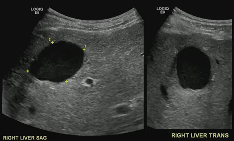
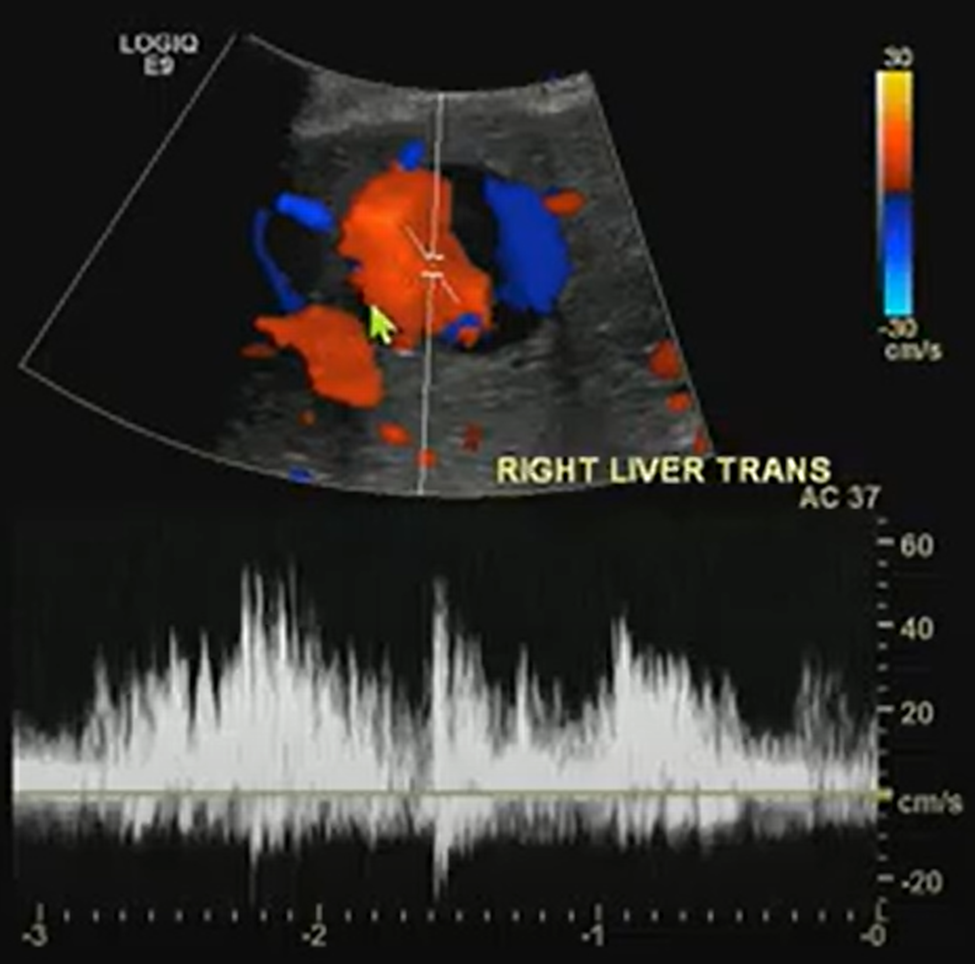
What looks like a simple hypoechoic cyst on ultrasound may hide a critical vascular pathology. Color Doppler is essential for distinguishing cystic lesions from vascular anomalies like pseudoaneurysms.
Key Takeaways
Always Doppler: Even cyst-like structures require Doppler evaluation to rule out vascular causes.
The Pepsi Sign: Swirling vascular flow within a lesion may signal a pseudoaneurysm.
High stakes: Hepatic artery pseudoaneurysms can mimic benign lesions but require urgent recognition and intervention.
Next steps: Interventional radiology embolization can be lifesaving.
Challenges Ahead
Differentiating pseudoaneurysms from other vascular or cystic lesions remains tricky.
Missing Doppler evaluation risks misdiagnosis and delayed treatment.
Awareness of teaching signs like the “Pepsi sign” is uneven among trainees.
Bottom Line
Never skip Doppler. The “Pepsi sign” may be the clue that transforms a benign-looking lesion into a critical vascular diagnosis.
When it comes to the cervical spine, cord integrity matters most. Even mild changes can spell trouble if the cord is compromised.
The Big Picture
Cervical canal stenosis isn’t just about the degree of narrowing; it’s about whether the spinal cord, itself, is at risk, too. Even without measurable stenosis, cord flattening can cause myelopathy. Understanding Dr. Lea Alhilali’s fishbowl analogy helps clarify how to distinguish mild, moderate, and severe cases.
Key Takeaways
Cord first: Regardless of canal narrowing, deformity or signal changes in the cord point to a higher risk of myelopathy.
Not just static: Static imaging may underestimate the impact; dynamic forces, repetitive microtrauma, or microischemia may drive symptoms.
Dr. Alhilali’s fishbowl analogy . . .
Mild stenosis:
Either ventral or dorsal CSF is effaced, but the cord still has room to “swim.”
Moderate stenosis:
Both ventral and dorsal CSF are lost, restricting cord movement.
Severe stenosis:
No CSF remains—cord is compressed, “fish” crushed.
Challenges Ahead
Why cord flattening causes myelopathy without stenosis remains unclear, and mechanisms are still debated.
Dynamic assessment may offer better insight than static MRI but isn’t standardized.
Management depends on correlating imaging with clinical findings, which are often nuanced.
Bottom Line
Think of the cervical cord like a fish in a bowl: it needs space to move. Once the CSF “water” is gone, the cord, as well as the patient, suffers. Classifying stenosis by available space—not merely narrowing—sharpens diagnostic accuracy and clinical relevance.