
Dual-energy CT (DECT) is becoming a must-use tool in musculoskeletal (MSK) imaging—not just for hardware, but for crystals, marrow, and trauma. Orthopedic imaging often suffers from two problems: metal streak and ambiguous density. But as the ARRS Online Course “Practical Dual-Energy CT Throughout the Body” duly notes, DECT tackles both by separating materials and controlling photon energy, giving rads clearer views of bone, soft tissue, and implant interfaces.
Where Does DECT Make the Biggest Difference?
- Metal Artifact Reduction (MAR) That Actually Works
High-keV VMIs (110–150 keV) reduce photon starvation and scatter from:
- Spine instrumentation
- Hip and knee arthroplasties
- Fracture fixation hardware
- Shoulder anchors
Result: cleaner cortices, more visible fractures, and better evaluation of infection or loosening.
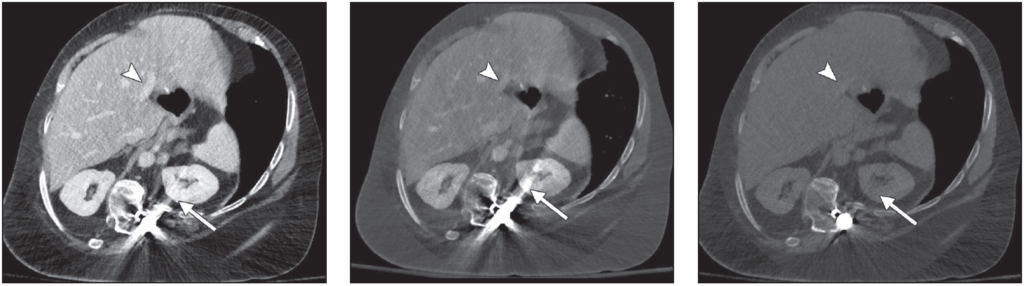
Portal venous phase abdominal CT images obtained after spinal reconstruction surgery. Left, Normal blended image shows considerable amount of metal artifact (arrow) overlaying left kidney and good contrast in portal veins (arrowhead). Center, Virtual monoenergetic image of same slice shown in Left, obtained at 50 keV, shows high portal venous contrast (arrowhead) and high amounts of metal artifact (arrow).Right, Virtual monoenergetic image of same slice shown in Left, obtained at 150 keV, reveals decreasing amounts of metal artifact (arrow) and loss of portal venous contrast (arrowhead).
2. Crystal Imaging: Knowing Exactly What You’re Looking At
Material decomposition differentiates uric acid from calcium, which helps:
- Confirm gout even in unusual locations (spine, tendons, postoperative joints)
- Distinguish gout from infection or tumor
- Map tophus burden for treatment decisions
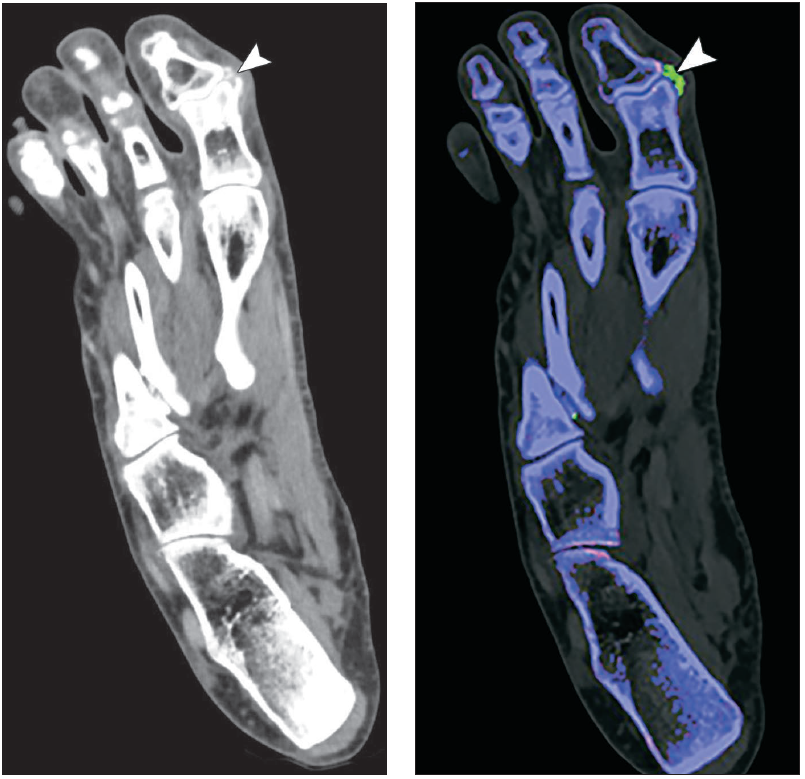
Patient with suspected tophus of first interphalangeal joint caused by gout. Left, Mixed CT image that is equivalent to single-energy scan acquired at 120 kVp shows tophus (arrowhead). Right, Material decomposition image applied to highlight urate crystals (green area indicated by arrowhead) confirms that lesion seen on regular CT scan is tophus caused by gout.
3. Bone Marrow Edema Detection
DECT water-specific reconstructions reveal bone marrow edema in trauma, stress injuries, and arthritis (especially helpful when MRI is unavailable or contraindicated).
Why Are MSK Rads Adopting DECT?
- Saves nondiagnostic postoperative studies
- Improves fracture conspicuity
- Reduces MRI dependence
- Helps differentiate infection, inflammation, and crystal deposition
- Speeds up decision-making for orthopedic surgeons
Bottom Line: MSK DECT is no longer just “nice to have.” When hardware, crystals, or marrow ambiguity stand in the way, DECT gives you answers a conventional CT simply can’t.
Leave a Reply