For radiologists especially, artificial intelligence (AI) is no longer just over the horizon; it’s in the reading room, right now. This practical immediacy is precisely the premise behind the 2026 ARRS Annual Meeting Categorical Course, Clinical Artificial Intelligence in Radiology. Presented live and virtually from the David L. Lawrence Convention Center in Pittsburgh, PA, this two-day ARRS Categorical Course continues our 125-year-old legacy of forward-looking education by arming radiologists with a robust understanding of how AI is reshaping the specialty.
Dr. Shandong Wu | Cat Course Codirector
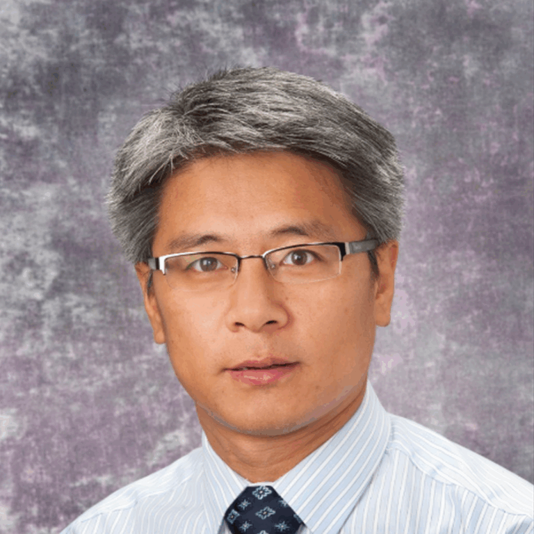
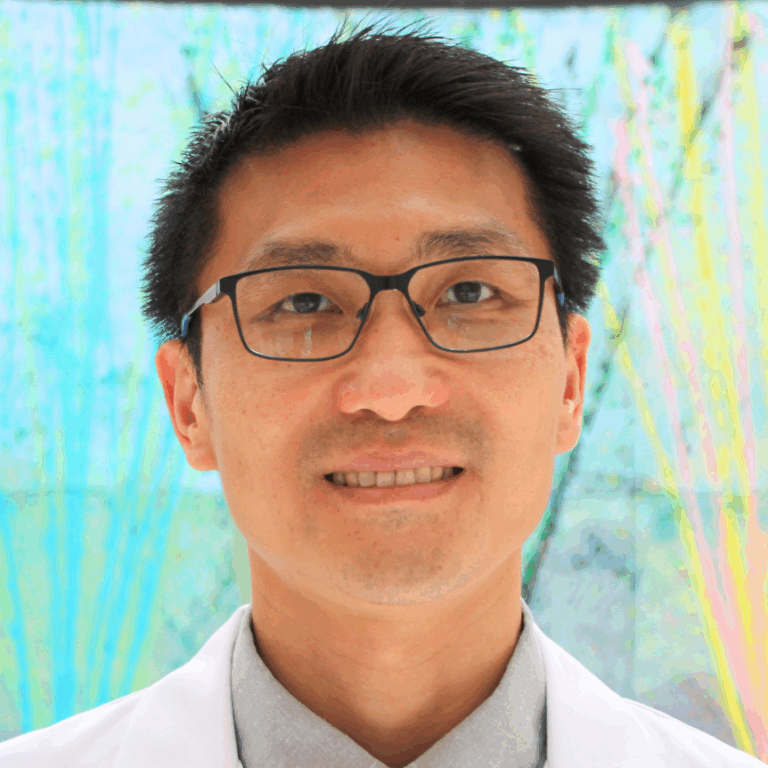
Clinical Artificial Intelligence in Radiology brings together more than 20 distinguished faculty from leading institutions across the globe, all led by Shandong Wu, PhD, founding director of the University of Pittsburgh’s Center for AI Innovation in Medical Imaging, a cross-campus initiative including more than 130 researcher and clinician members. Also a professor of radiology, biomedical informatics, bioengineering, intelligent systems, clinical and translational science, one of Dr. Wu’s ARRS Cat Course codirectors is an abdominal radiologist and director of diagnostic AI at the University of Washington, Yee Seng Ng, MD.
Dr. Shandong Wu | Cat Course Codirector
Alongside codirector and 2024 AJR Lee F. Rogers International Fellow in Radiology Journalism Hyun Soo Ko, MD (Peter MacCallum Cancer Centre, Australia), the trio is curating a curriculum of more than two dozen lectures and panels across seven thematic sections, giving registrants a comprehensive view of AI’s current and future roles in everyday practice.
SUN, APRIL 12—From Concept to Clinic: Building AI Literacy
Day one of Clinical Artificial Intelligence in Radiology kicks off with “Getting to Know AI,” a primer tailored for all levels of experience. Tessa Cook, MD, PhD (University of Pennsylvania), provides an overview of radiological progress in AI, while Dr. Ko demystifies essential concepts, such as machine learning, deep learning, radiomics, as well as generative and agentic AI.
Dr. Linda Moy | Vice Chair of AI, NYU Radiology
Up next, inaugural vice chair of AI at New York University’s radiology department, Linda Moy, MD, will provide an invaluable look into leveraging AI to improve workflow efficacy and effectiveness alike. Dr. Wu himself closes the Cat Course’s first session. The leader of Pittsburgh’s Intelligent Computing for Clinical Imaging lab will explore and explain how AI is enhancing imaging interpretation for computational insights—from screening and triage to diagnosis and prediction.
Clinical Implementation: From Regulation to Real-World Deployment
Section two of Clinical Artificial Intelligence in Radiology, “AI Clinical Implementation,” addresses legal, regulatory, and operational frameworks essential for radiologists seeking to implement or evaluate AI tools in practice. Didactic highlights will include guidance on U.S. Food and Drug Administration (FDA) regulations and performance monitoring by Melissa Davis, MD, MBA (Yale), as well as insights into distinguishing high-quality AI models from market hype.
In a uniquely insightful presentation, Julian Rivera, JD (University of Pittsburgh), will tackle the legion of legal considerations accompanying AI adoption: liabilities, ethical perspectives on signing contracts, collaborative business modes with AI companies, etc. Dr. Cook returns to share her expertise on evaluating local versus commercial solutions when measuring ROI, while a panel moderated by Dr. Moy will outline best practices and common pitfalls.
Beyond the Pixel: Multimodality and Multidimensional AI
The promise of any good AI expands significantly when paired with non-image data. The “Going Beyond Images to Multimodality” session explores emerging applications that leverage large language models, vision-language models, and foundation models. Presenters Heather Whitney, PhD (University of Chicago), and Lifeng Yu, PhD (Mayo Clinic), will delve into data curation, federated learning, and the physics of AI model performance. With Christian Bluethgen, MD (University Hospital of Zurich), having assessed multimodal data methodologies in his presentation, a panel discussion on tackling technical challenges to find opportunities rounds out day one of this ARRS Cat Course.
MON, APRIL 13—Practical Impact Across Subspecialties
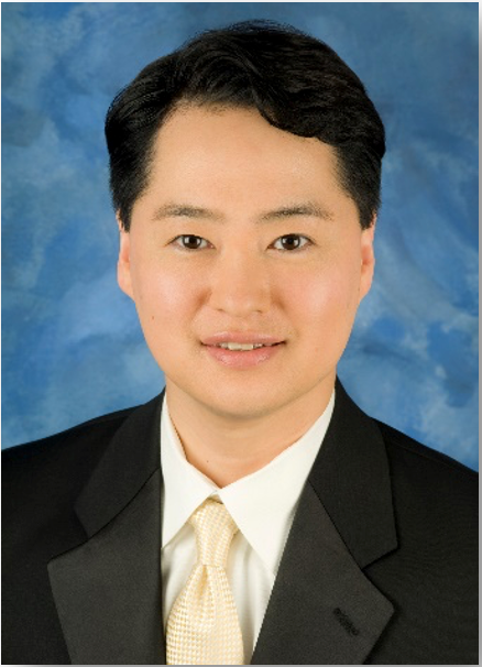
AI’s reach across subspecialties is the focus on Monday. Presenters including Constance Lehman, MD, PhD (Harvard), Ali Guermazi, MD, PhD (Boston University), and 2022 ARRS Gold Medalist Edward Y. Lee, MD, MPH (Harvard) will detail AI tools in breast, musculoskeletal, pediatric imaging, respectively. Dr. Ng’s highly anticipated survey of AI and abdominal imaging will be followed by a lecture from neuroradiologist Paulo De Aguiar Kuriki, MD (UT Southwestern).
Dr. Edward Y. Lee | ARRS Gold Medalist
That’s not all either. Real-world cardiothoracic, interventional, and nuclear medicine cases will further demonstrate how AI is already reflowing imaging workloads, improving diagnostic accuracy, and personalizing care across organ systems and patient populations.
Shaping Tomorrow: Research, Education, and Ethical Engagement
Day two of Clinical Artificial Intelligence in Radiology continues with “AI Research and Education,” including a model development demonstration by Dooman Arefan, PhD (University of Pittsburgh), and an exploration of MD–PhD collaboration opportunities from Dr. Wu. Justin Peacock, MD, PhD (Uniformed Services University), will discuss educational roadmaps and training resources, addressing a key concern for attendees seeking to build or deepen their AI competencies.
This 2026 ARRS Annual Meeting Categorial Course concludes with “Humanity and AI,” a thought-provoking session covering radiologist–AI collaboration, fairness and bias, and imaging’s ever-evolving role in AI-powered services. Florence Doo, MD (University of Maryland) will help us find a foothold in our present human–AI ecosystem, followed by a warning for all the disparities AI run amok could actually exacerbate care of Judy Gichoya, MD, MS (Emory). Eduardo Mortani-Barbosa, MD, MBA (University of Pennsylvania), will then detail specific skill sets that AI-forward radiologists will need to hone in their practices and in their communities. Finally, ARRS Scholar and Gold Medalist and editor of Radiology: Artificial Intelligence Charles E. Kahn, MD (University of Pennsylvania), joins to facilitate a panel discussion on action items and what to do next.
Dr. Charles E. Kahn | Editor, Radiology: Artificial Intelligence
With each live lecture accompanied by an e-book chapter, Clinical Artificial Intelligence in Radiology will provide strategic context and tactical guidance for imagers of each practice type and at every level of training.
And as Dr. Wu tells InPractice, “AI in radiology is not just a technical shift—it’s a cultural one. This ARRS Categorical Course is about empowering radiologists to shape that future, not just react to it.”
With content spanning conceptual foundations to the most practical of pearls, the curriculum curated by Wu, Ng, Ko and colleagues this April is poised to be an essential learning experience for working radiologists looking to engage with AI at the frontlines of medical imaging care.
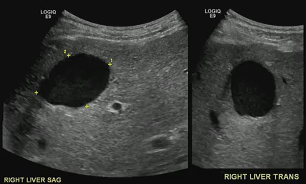
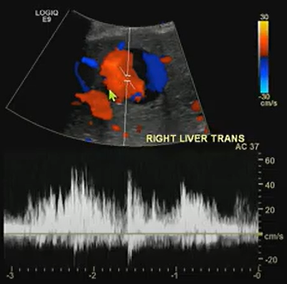
A benign-looking liver lesion turned out to be a hepatic artery pseudoaneurysm—all thanks to color Doppler.
The Big Picture
What looks like a simple hypoechoic cyst on ultrasound may hide a critical vascular pathology. In this ARRS Annual Meeting case from Kristin Rebik, DO, color Doppler proved essential for distinguishing cystic lesions from vascular anomalies like pseudoaneurysms.
Key Takeaways
Always Doppler: Even cyst-like structures require Doppler evaluation to rule out vascular causes.
Pepsi Sign: Swirling vascular flow within a lesion may signal a pseudoaneurysm.
High stakes: Hepatic artery pseudoaneurysms can mimic benign lesions but require urgent recognition and intervention.
Next steps: Interventional radiology embolization can be lifesaving.
Challenges Ahead
Differentiating pseudoaneurysms from other vascular or cystic lesions remains tricky.
Missing Doppler evaluation risks misdiagnosis and delayed treatment.
Awareness of teaching signs like the “Pepsi sign” is uneven among trainees.
Bottom Line
Never skip Doppler. The “Pepsi sign” may be the clue that transforms a benign-looking lesion into a critical vascular diagnosis.
An intersocietal panel of experts in CT convened by the American Association of Physicists in Medicine (AAPM)—with representation from clinical practice, academia, and industry input from Siemens Healthineers and Canon—examined a new performance measure in the quality-based payment programs of the Centers for Medicare & Medicaid Services (CMS). Publishing their findings in the American Journal of Roentgenology [1], the panel identified 20 important issues and ambiguities with the new measure, which became effective this year.
Collectively, these issues reflect unclear definitions, opaque methodologies, technical and legal barriers, and potential misalignment with clinical realities—posing significant obstacles to consistent, equitable, and scientifically valid implementation across diverse care settings.
Ambiguity surrounds where reporting is required versus optional and exactly which adult study types qualify, compounded by difficulties in consistent inpatient versus outpatient categorization. Terminology inconsistencies and unclear mapping of studies to dose and image quality categories add to the confusion. Meanwhile, patient size assessment methodology and calculation of size-adjusted dose diverge from established standards, while noise measurement lacks a recognized protocol. Criteria for excluding studies and handling combination studies remain undefined.
Then, there are the tech queries: is HL7 EHR connectivity mandatory, are alternative data transfer mechanisms even feasible, what potential IT burdens and/or security liabilities will radiology practices have to shoulder? Also, performance expectations for compliance thresholds are unspecified, as are methods for comparing diverse protocols under a single set of thresholds. Identical thresholds across different categories raise additional questions.
“Transparency and stakeholder engagement are essential for effective quality initiatives in medicine,” said Mahadevappa Mahesh, MS, PhD, president of AAPM.
Dr. Mahesh | President, AAPM
“We wrote this paper to call attention to issues and ambiguities with the CMS measure, and we look forward to working with CMS to address these issues and continue the culture of quality and safety that has developed in CT imaging over the past two decades.”
Balancing Image Quality and Patient Safety
One of the benefits to patients that will come from “The New CMS Measure of Excessive Radiation Dose or Inadequate Image Quality in CT: Issues and Ambiguities—Perspectives from an AAPM-Commissioned Panel” in AJR is that the expertise of the entire imaging community will be used to develop quality improvement initiatives that will keep radiation doses as low as possible while maintaining the quality of medically essential CT imaging. From physicians and physicists to technologists, regulators, and business leaders, “we’re confident that we can get this right by working together,” said Dr. Mahesh.
Technology Has Already Lowered Doses
A lifesaving technology used to diagnose disease and guide treatment, CT is the first-line imaging technique in many cases, especially in emergency departments and cancer centers. Concerns have been raised about the increased utilization of CT in medicine because the modality uses ionizing radiation, which at very high doses is known to increase a patient’s risk for developing cancer. However, at the low doses of radiation utilized in medical imaging, including in CT, the risk is extremely small—perhaps negligible.
Over the past two decades, imaging and allied health professionals have collectively worked to reduce CT doses. New scanner technologies have played a starring role in decreasing doses, including features that automatically measure the size of the patient and adjust the radiation dose to the right value. This is especially important for children, who require lower doses than adults due to their smaller size.
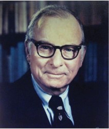
Dr. McCollough | Prior President, AAPM
“Some authors multiply the very small potential risk of a CT scan by the millions of patients who receive one and predict that we will see an increase in cancer,” said Cynthia McCollough, PhD, past president of AAPM.
“This can lead to alarmist stories and patients who really need a CT refusing to get one. Further, at the low doses we are talking about, it is debated whether the risk is even real. CT has been around for over 50 years and the predicted increases in cancer just aren’t being seen.”
Editorials Stress Ticking Clock, Call for Clarity
In her accompanying AJR editorial, Stephanie Leon, PhD, of the University of Florida in Gainesville, noted that “quality-based payment programs will be impacted starting in January 2027,” which means that imaging has two years and counting to figure all of this out [2].
CMS Quality Reporting Program
CMS Payment System
Reporting Requirement
Timeline
Hospital IQR Program
HIPPS
Optional. Hospitals are required to report three eCQMs self-selected from a list and three eCQMs mandated by CMS. The measure will be available on the self-selection list and thus its reporting is optional.
Reporting will begin in CY 2025; CY 2025 results will impact FY 2027 payments.
Hospital OQR Program
HOPPS
Required. Once the measure is fully implemented, hospitals will be required to report the measure.
Reporting will be voluntary in CY 2025 and mandatory in CY 2027; CY 2027 performance will impact CY 2029 payments.
MIPSᵃ
MPFS
Optional. Participants are required to report six MIPS quality measures, including at least one outcome measure, that are self-selected from a list (possibly a specialty-defined measure set depending on the reporting mechanism). If more than six measures are available, then reporting the measure is optional.
Reporting will begin in CY 2025; CY 2025 results will impact FY 2027 payments.
Another AJR editorial written by Kishore Rajendran, PhD, of the Mayo Clinic in Rochester, MN, and chair of the working group on the physics of quantitative imaging at AAPM, called for improved transparency, too. “A nonproprietary, community-based approach is imperative to ensure full transparency, achieve consensus among CT stakeholders, and provide reliable clinical diagnoses at the lowest radiation dose possible,” wrote Dr. Rajendran [3].
Watch as AJR senior author Ehsan Samei, PhD, and first author Jered R. Wells, PhD, call for a fundamental shift toward open-source, open-access, consensus-based, and community-owned strategies and resources to ensure quality and safety of CT: YouTube.com/@AJR_Radiology
References:
Wells JR, Christianson O, Gress D, et al. The new CMS measure of excessive radiation dose or inadequate image quality in CT: issues and ambiguities—perspectives from an AAPM-commissioned panel. AJR 2025 May. doi: 10.2214/AJR.24.32458
Leon, SM. CMS measure on CT dose and image quality: good intentions, but not quite ready for prime time. AJR 2025 May. doi: 10.2214/AJR.25.32908.
Rajendran K. Transparency and stakeholder engagement as cornerstones for effective quality initiatives in medical imaging. AJR May. doi: 10.2214/AJR.25.32859
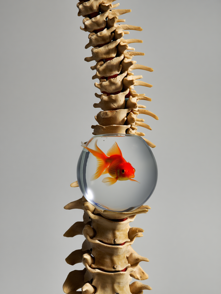
When it comes to the cervical spine, cord integrity matters most. Even mild changes can spell trouble if the cord is compromised.
Big Picture: Cervical canal stenosis isn’t just about the degree of narrowing; it’s about whether the spinal cord, itself, is at risk, too. Even without measurable stenosis, cord flattening can cause myelopathy. Understanding Dr. Lea Alhilali’s fishbowl analogy from the ARRS Neuroradiology Longitudinal Course helps clarify how to distinguish mild, moderate, and severe cases.
Key Takeaways:
Cord first: Regardless of canal narrowing, deformity or signal changes in the cord point to a higher risk of myelopathy.
Not just static: Static imaging may underestimate the impact; dynamic forces, repetitive microtrauma, or microischemia may drive symptoms.
“Fishy” Analogies…
Mild stenosis: Either ventral or dorsal CSF is effaced, but the cord still has room to “swim.”
Moderate stenosis: Both ventral and dorsal CSF are lost, restricting cord movement.
Severe stenosis: No CSF remains—cord is compressed, “fish” crushed.
Challenges Ahead
Why cord flattening causes myelopathy without stenosis remains unclear, and mechanisms are still debated.
Dynamic assessment may offer better insight than static MRI but isn’t standardized.
Management depends on correlating imaging with clinical findings, which are often nuanced.
Bottom Line: Think of the cervical cord like a fish in a bowl: it needs space to move. Once the CSF “water” is gone, the cord, as well as the patient, suffers. Classifying stenosis by available space—not merely narrowing—sharpens diagnostic accuracy and clinical relevance.
Reiterating, the house of radiology’s influence in shaping our nation’s health care policy writ large, the American Medical Association (AMA) House of Delegates (HOD) advanced several measures with significant implications for American Roentgen Ray Society (ARRS) members during its own annual meeting in Chicago this June.
In short: expect DICOM mandates to simplify imaging transfers, elevated oversight of AI, and more rigorous validation for CT-based calcium scoring [1].
Finally, Federally Interoperable DICOM
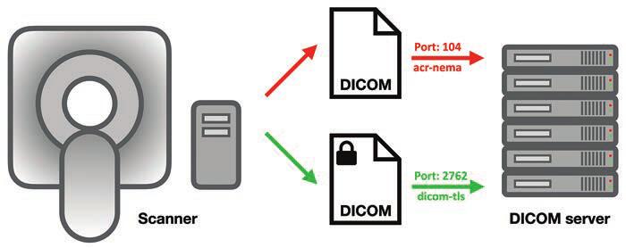
The HOD passed a pivotal resolution calling on AMA advocacy for federal health IT interoperability standards to include the DICOM format, a critical “missing link” that delegates have long championed. Despite over two decades of EHR development and federal mandates, DICOM has been excluded from formal interoperability frameworks. As a result, radiological images frequently cannot travel seamlessly through EHR systems, frustrating patients and providers alike. For one example, patients arriving for mammograms at new facilities are often dumbfounded that previous studies cannot be accessed digitally from elsewhere. The absence of interoperable imaging standards contributes to delayed care, redundant exams, unnecessary radiation exposure, and burdens for patients. And the security risks are legion (Fig. 1).
Fig. 1—Schematic shows DICOM server, computers that can exchange and store DICOM objects. Server offers DICOM service, which is software that can send and receive DICOM messages, running via specific computer ports (i.e., communications channels). Secured DICOM service is known as dicom-tls (port 2762), which uses transport layer security for negotiations, authentication, and encryption. A service that cannot be queried by hackers because it uses strong authentication mechanisms, this service sends and receives encrypted DICOM messages that cannot be read by hackers either. However, this is only true for manufacturers that have chosen to implement its strong authentication and encryption features. Arrows show direction of data transmission.
Spearheaded by neurology and orthopedic associations, this resolution urges inclusion of DICOM in the U.S. Core Data for Interoperability (USCDI) and seeks regulatory action requiring EHR and imaging archive vendors to support secure, efficient exchange of DICOM data. Testimony also highlighted policy fissures stemming from the Health Information Technology for Economic and Clinical Health (HITECH) Act of 2009, which exempted radiologists—alas, not viewed as patient-facing—from certain data-sharing requirements, thereby keeping imaging outside USCDI [2]. A significant win for medical imagers across the country, delegates did have to amend the resolution to get it passed, but this is meaningful progress toward closing the interoperability gap that hampers timely, coordinated, and secure care.
More Oversight and Transparency for AI
With AI digging even deeper into the specialty—at last count, over 75% of the more than 1,000 algorithms cleared by the Food and Drug Administration target radiology [3]—the HOD continued sounding the alarm on the “black box” nature of so many machine-learning, deep-learning, and radiomic systems. Resolution 519, though not adopted thanks to too much overlap with AMA’s existing AI policies, successfully highlighted acute issues of explainability, advocating for evidence-based, transparent AI within a deliberately structured framework [4]. Aligning with AMA’s stance that the physician’s expertise remains central to clinical decision-making, everyone in Chicago agreed that today’s radiologists must be able to comprehend and articulate how generative AI, agentic AI, or some future proprietary amalgam of the two arrives at any given verdict. All too often, seemingly slight updates to vendor hardware, scanning protocol, or patient demographics end up altering algorithmic performance, further underscoring the need for responsible vetting and robust monitoring of AI.
No LDCT for Coronary Calcium, Yet
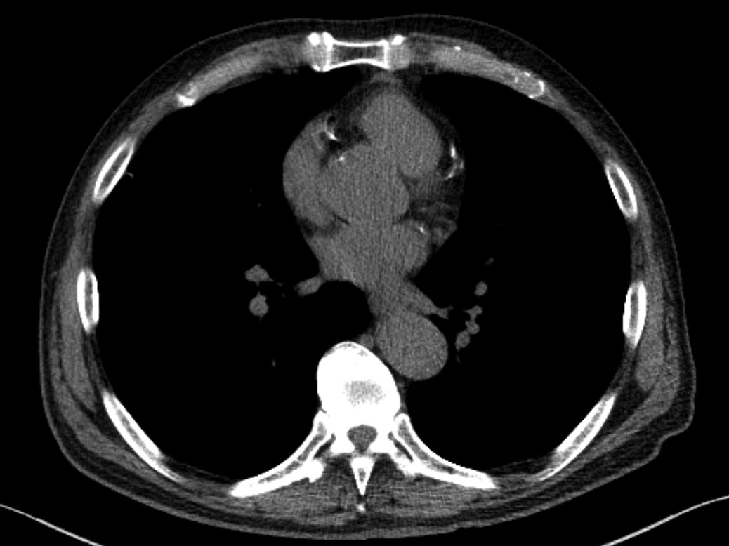
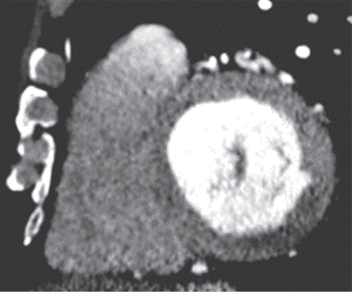
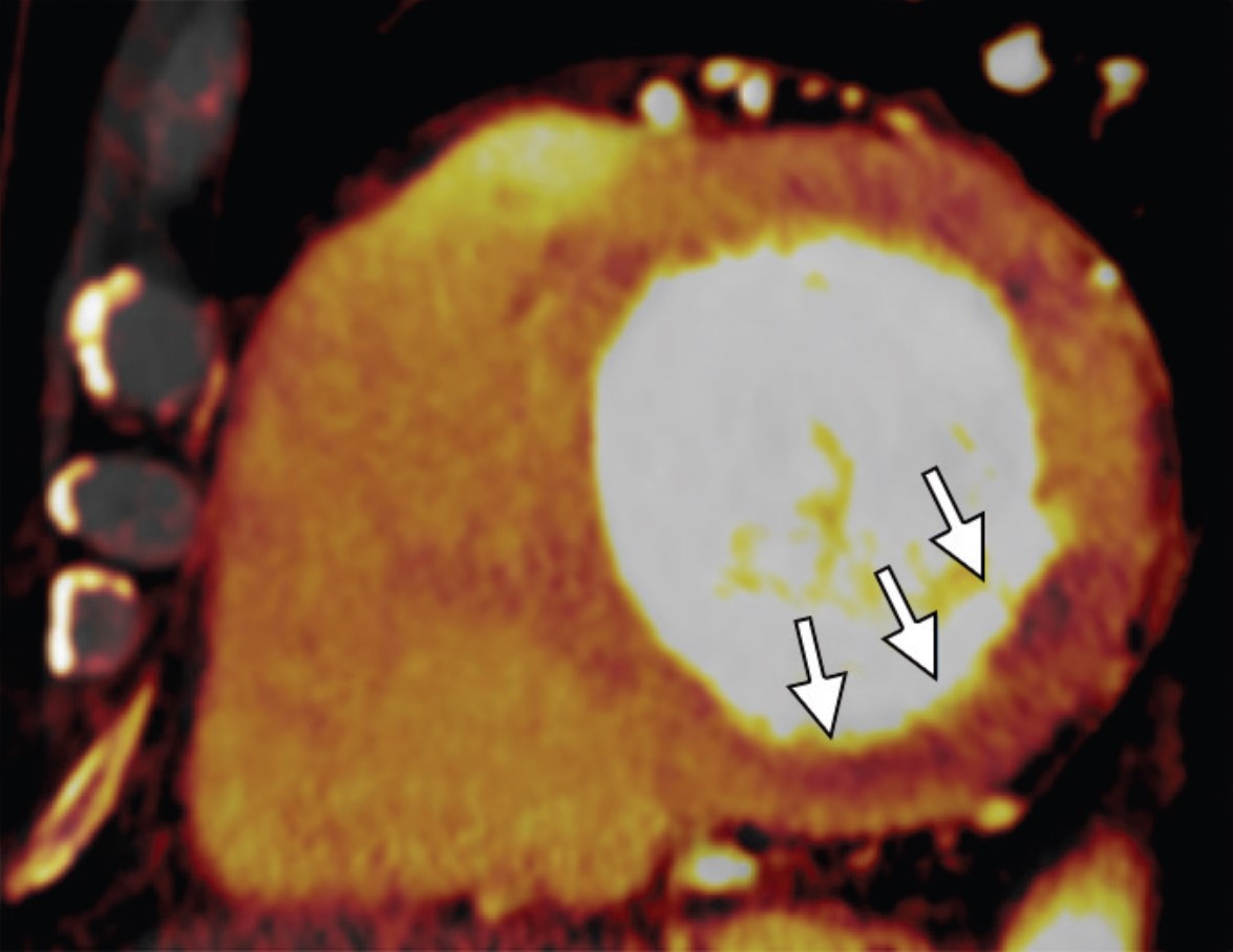
AMA also tabled a resolution regarding expanded promotion and usage of low-dose CT (LDCT) to screen both lung cancer and coronary artery disease via coronary calcium scoring. Emphasizing the modality’s value as a public health tool for high-risk individuals, particularly those with pack-year history of smoking, LDCT delivers far less radiation than standard CT and can detect small lung nodules early. Indeed, crucial research from the National Lung Screening Trial shows it can reduce lung cancer mortality by up to 20% [5].
And yet, uptake is still cripplingly low; fewer than 6% of eligible patients receive LDCT screening. To buttress the resolution’s goals, the American College of Radiology is launching complementary efforts, including expanding its early lung cancer registry to capture incidental findings from routine CTs, not just formal screening exams [6]. Such distinction will deepen insights into nodule detection and follow-up.
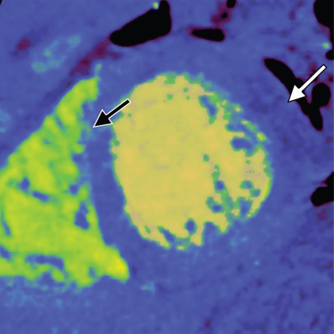
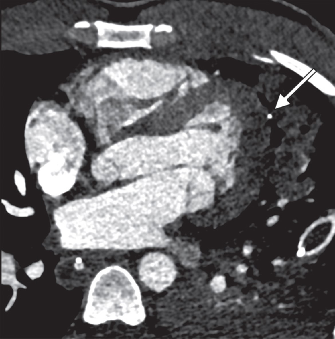
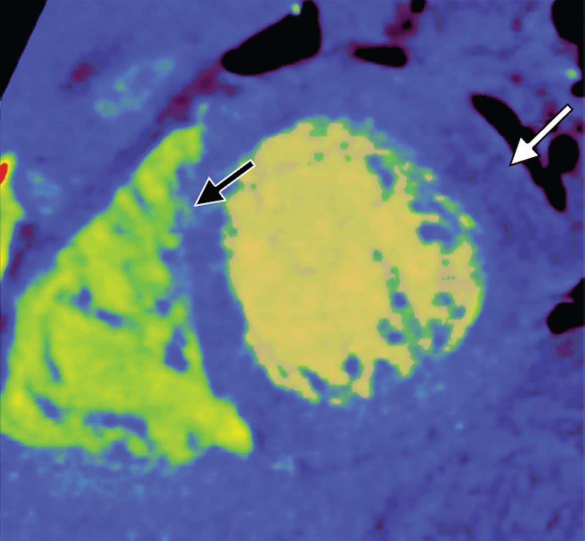
Fig. 2—73-year-old patient who underwent lung cancer screening by LDCT of the chest. Axial CT image shows coronary artery calcification (CAC). CAC was assessed as severe by consensus visual analysis.
More broadly, many hospitals have begun offering low- or no-cost LDCT screenings as an entry point for preventive care. Apropos, this resolution specifically solicits a coordinated national effort of public awareness campaigns and provider education to ensure affordable, widespread access to this potentially life-saving tool.
For further details about the 2025 Annual Meeting of the HOD, click here.
The American Roentgen Ray Society (ARRS) is pleased to announce Francis Baffour of Mayo Clinic in Rochester, MN, as the 2024 Melvin M. Figley Fellow in Radiology Journalism. ARRS also recognizes Hyun Soo Ko of the Peter MacCallum Cancer Centre and Epworth Medical Imaging in Melbourne, Australia, as the 2024 Lee F. Rogers International Fellow in Radiology Journalism.
Supported by The Roentgen Fund® and named for two distinguished Editors Emeriti of ARRS’ own American Journal of Roentgenology (AJR), the Melvin Figley and Lee Rogers Fellowships offer practicing radiologists an unparalleled opportunity to learn the tenets of medical publishing via “the yellow journal”—the world’s longest continuously published radiology journal. Through hands-on experience with ARRS staff and AJR personnel—as well as personal apprenticeship with AJR’s 13th Editor of Chief, Andrew B. Rosenkrantz—Drs. Baffour and Ko will receive expert instruction in scientific writing and communication, manuscript preparation and editing, peer review processes, journalism ethics, and both print production and digital publication.
Founded in 1907, AJR is one of the specialty’s leading peer-reviewed journals, publishing clinically oriented content across all imaging subspecialties and modalities relevant to radiologists’ daily practice. Overall, “the yellow journal” garnered 35,480 citations in 2022, ranking AJR fourth among all radiology journals.
Since 1990, The Roentgen Fund has granted millions of dollars to hundreds of imaging professionals for both research pursuits and professional development. Today, through six vital scholarship and fellowship programs, the generosity of The Roentgen Fund’s donors is channeled to every corner of the globe—establishing dual foundations in innovation and leadership for a true diversity of radiology’s next generation.
Francis Baffour practices as a diagnostic radiologist with expertise in advanced MRI and CT techniques for musculoskeletal imaging. His clinical and research interests align with his goal of identifying novel applications for advanced imaging technologies, then rapidly translating these discoveries into practical patient care. As associate medical director of the CT Clinical Innovation Center in Mayo Clinic Rochester’s department of radiology, he supports the mission of facilitating high-impact imaging innovations with direct effect on patients, such as radiation dose reduction techniques, novel CT technologies, and quantitative assessment of disease activity. Dr. Baffour earned his M.D. from the Albert Einstein College of Medicine in New York City and a B.A. in biochemistry from Vassar College in Poughkeepsie, NY.
A German and Australian board-certified radiologist with over 20 years of international experience, currently, Hyun Soo Ko practices at Peter MacCallum Cancer Centre and Epworth Medical Imaging in Melbourne, Australia, holding affiliations with the University of Melbourne and University of Bonn in Germany. Her expertise is diverse, with specialized fellowships in breast imaging (Melbourne), imaging research (Toronto), and pediatric radiology (Heidelberg, Germany). Dedicated to advancing translational medicine, her research focuses on AI and radiomics to discover predictive imaging biomarkers. She is an active member of the Royal Australian and New Zealand College of Radiologists (RANZCR) Advisory Committee on Artificial Intelligence and the German Roentgen Society (DRG) Working Party Methodology and Research. As a clinician researcher, Dr. Ko remains committed to mentoring and supporting emerging radiologists, firmly believing multidisciplinarity and inclusiveness are key elements for equitable progress and impact.
From May 5–9, 2024, Drs. Baffour and Ko will attend the 2024 ARRS Annual Meeting in Boston, MA, where they will co-present the AJR Year in Review Sunday Session and participate in the Editor’s Forum.
More about the Figley and Rogers Fellowships
Melvin M. Figley (1920-2010) assumed the editorial mantle of AJR with the January 1976 issue. Hiring a full-time professional staff and, for the first time, a managing editor, he appointed associate editors to facilitate the peer review process. Publication accelerated, and the quality of illustrations improved, leading to the AJR Pictorial Essay. Dr. Figley was also central to the partnership between ARRS and the American Society of Neuroradiology, including the founding of the American Journal of Neuroradiology in 1980. After more than a decade of dedicated service to “the yellow journal,” Dr. Figley retired in 1985.
Lee F. Rogers, the distinguished musculoskeletal radiologist and longtime chair of imaging at Northwestern University Medical School, was named AJR’s chief editorial officer in 1996. Highlights from Rogers’ tenure at the journal included more contemporary design, introducing of a medicolegal column, authored by Dr. Leonard Berlin, and his popular editor’s notebook. Both the quality and the variety of papers published in AJR remained quite high, as the information revolution propelled “the yellow journal” into new formats, such as CD-ROM, and frontiers, like the internet.
Five prior Melvin M. Figley Fellows of The Roentgen Fund® will also present during the 124th ARRS Annual Meeting in Boston, MA:
In radiology, whether you work in a large or small private practice, remotely by yourself, an academic department in a medical center, or part of a mega radiology practice, there has been a palpable shortage of radiologists. This shortage is fueled by a trend toward exclusive subspecialization with declining numbers of radiologists who can handle general work, ever-increasing expectations for service to our patients, referring doctors, hospitals, and health care systems. We have been stretched thinner. There is a desire by radiologists to have more flexible work hours or, simply stated, to work less hours overall compared to years past. There is a concern about what role artificial intelligence and machine learning will play; will we be displaced? Reimbursement has been decreasing relative to inflation and compared with other specialties. As a result of these realities and others, there is clear evidence of burnout among radiologists, similar to health care workers in other specialties. On top of that, sometimes, we find that the leaders in our organizations may be distant, or too corporate, or suffer from “toxic positivity,” which may be worse than “toxic negativity.”
There has been a steady headwind for years, but it now feels like a gale force wind. And a lot of this feels out of our control. So, goodness, how do we manage all of this? Hold on, let’s take a breath. One strategy that we can embrace and control is to develop a culture of teams within our workplaces. In fact, I have titled this series “The Teamwork Imperative” because we must establish teamwork as a core value within the radiology workforce. I believe that if we foster a culture of teams, we can mitigate and shield ourselves from some of these headwinds.
Let me be clear. Here, when I say teams, I am specifically not referring to the “macro teams” that many of us find ourselves in. For example, at Duke Health, it is said that the 30,000-plus employees are my “teammates.” That very well may be true. But no, I am referring to your local and focal team. I am referring to the individuals that you rely on daily or weekly to deliver your work product. It’s the folks you huddle with. And the teams develop where you huddle. If you are in training, I am referring to your team of co-residents, your chief residents, maybe your program director or program coordinator who you lean on. If you are in a private practice, I am referring to those that you share physical space with, or perhaps switch call with, or the individuals you show difficult cases to, or the referring docs you have developed close relationships with, and who rely on you to deliver care. In an academic environment like mine, it might be the members of your subspecialty division. If done well, the division pulls together as a team to deliver care, service, teaching, and research.
Those divisions that have a culture of team are far more effective than those who are unable to act as a team. If you are lucky enough to have these local and focal teams (and these often form and evolve organically), many challenges at work open up and become more manageable and attainable. The clouds begin to lift. Specifically, your deliverables, whatever they may be, are far more easily and effectively achieved if you have your team and approach your work from the perspective of that team.
Work becomes more efficient and fulfilling and, frankly, more fun. The work becomes more manageable, with more aspects under your control. You become more engaged. And that then becomes an antidote to burnout. Teams, therefore, contribute to retention.
Coaches discuss this all the time. I’ll borrow here from Mike Krzyzewski, the legendary Duke University men’s basketball coach. “Coach K” famously talked about the five keys to an effective team, likening them to the fingers on a hand. Each finger is individual and can stand alone, but when the five fingers of communication, trust, responsibility, caring, and productivity come together into a fist, the fist proves to be much stronger than the sum of the individual fingers.
Communication
Yes, of course, communication is about sharing news back and forth, accurately and honestly, but more importantly, communication is to be able to probe, to be able to respectfully question, and to be able to expect honest answers from your teammates. Sometimes, the questions aren’t easy, and the answers may not be easy either. To foster an effective team requires the ability and the safety of pointing out the opportunities—those ones are easy. More importantly, it is to have safety in pointing out deficiencies—those are more difficult. It is critical for teammates to be able to receive and internalize the information coming from within the group, whether it is a kudo, an observation, or a deficiency or a criticism.
You have to talk to each other. Actually talk. And in a world of remote work and texting, we don’t talk enough. Maybe the talking occurs in a partner meeting, defined clinical case conference, resident meeting, or division or department meeting. Maybe it is your team taking a coffee break or going for a midday walk to achieve “step” goals. Hopefully, the team dynamics are such that one can tap a teammate on the shoulder and engage in an effective and safe conversation.
Communication needs to be practiced. That is why I think standing, regular, in-person meetings, even if the agenda is light, are very important. The opportunity to come together regularly promotes the importance and expectation of communication. And it is habit forming. You get better at it.
Trust
Trust means telling the truth, and telling it sooner rather than later; knowing that within a team, that can be hard. It can be hard because so many of us struggle with confrontation and conflict and try to avoid them. But consider it this way, from the perspective of the iconic University of Tennessee women’s basketball coach, Pat Summitt. She said, “The absolute heart of loyalty is to value those people who tell you the truth, not just those people who tell you what you want to hear. In fact, you should value them the most, because they have paid you the compliment of leveling with you, assuming you can handle it.”
Honest and fair difficult conversations almost always produce results. If you can get through the first thirty seconds of a difficult conversation, often the clouds lift and a very productive conversation follows. For me, I need to write down the key first few sentences for that opening thirty seconds and the rest flows. If difficult conversations don’t produce results, you have learned something.
Collective Responsibility
A coach might say, “We win together, we lose together, there is no “blame game.” In the workplace of radiology, the collective responsibility is the pulling together to deliver on our various missions. Everyone does not play the same role. One partner may be a fast and efficient reader, that’s great. Her partner is slower because he spends a lot more time consulting and discussing with referring docs. Both these individuals are important to the team. Or in an academic environment, one may be more focused and skilled clinically, another may be more focused on teaching. In a team where there is collective responsibility, they complement one another and each can be proud of each other’s successes. Indeed, the light of their success shines on the other teammates, on the entire team.
Caring
This is also about humanity and human connections and colleagueship. Caring in a team reflects time spent with each other and sharing aspects of ourselves, in this case the women of Duke Radiology. The caring strengthens the interconnective web between team members, in this case at our annual Fall Gathering. It makes the team softer in a positive way, and more personable, yet, at the same time stronger.
Here is where I worry about remote work. I get it. The pandemic has shown that we can do radiology effectively, even remotely. People like it and expect it. And we have learned that we can teach remotely. Sort of. But it seems far more difficult to foster a genuine, caring environment when work is dispersed in many geographic locales and individuals work essentially independently, free of meaningful, direct interactions with other teammates, other humans. To me the same applies to Zoom meetings. All the nuanced talk and greetings pre- and post-meeting are lost. The body language is lost. The sense of community is lost, or at least different. And I think the effectiveness of the meeting suffers. Indeed, on a Zoom meeting, you can’t even have real eye contact. I worry that with remote work, the culture of our teams maybe eroded. We do need to incorporate the best aspects of some remote work, but incorporate it in a fashion that fosters caring.
Pride/Productivity
Coach K says, “Everything we do has our own personal signature on it…and if we all sign off on everything we do, then we have a chance to be great.”
And your teammates all do have the chance to add their personal touch and signature. Collectively then, we have the opportunity to harness the best of what we can be, and to be great, even with all that is facing us and challenging us in our current and future work environments. “And it is going to be hard; we all wait in life for things to get easier…when we are well staffed, when I pass the Boards, when I make partner, when the kids are older, when the pandemic is over … it will never get easier. What happens is, you handle hard better.” As pointed out by Kara Lawson, Duke women’s basketball coach.
Each of these domains, communication, trust, collective responsibility, caring, and pride; the five individual fingers are important. But when the five fingers are formed into a fist, a true team, the collective strength is much greater than the sum of the individual parts. When these five areas can be applied to our teams in radiology, we can become more effective and efficient. As individuals alone, we are relatively weak. Together as a team, we are stronger. And this is something within our control.
To me, there is an imperative to create, sustain, and grow teams in our workplace.
Colleagues, I personally invite you to join us for ARRS 2024, either in Boston or online. This exceptional event brings together top radiologists from around the world to share new knowledge shaping our field.
Our Annual Meeting is renowned for its cutting-edge education, providing clinical information you can put into practice right away. We are excited to share the latest advancements in radiology, along with comprehensive review lectures to stay sharp on core topics.
Whether you attend in person and immerse yourself in the vibrant atmosphere of Boston, or choose our convenient online option, ARRS 2024 promises to be a remarkable experience. Your participation and engagement will help us connect, learn, and advance radiology together.
Assistant Professor, Department of Diagnostic and Interventional Imaging McGovern Medical School, UT Health Houston
It is my pleasure to discuss a topic that holds a very special place in my heart—the patient experience in breast imaging. What makes this topic truly captivating is that it touches upon several areas that are significant to me: patient-centered care, team leadership, and wellness. But there is an interesting twist, and I would like to share that as well, because it’s a kind of secret passion that I’ve had for years. And that is my love for spas, just the unforgettable experience you can get during one of the most memorable stays in a hotel.
Now, you might be wondering how on earth these interests come together. How can these unrelated topics merge to create a health care experience that is as luxurious and comforting as a world-class spa getaway, while also providing exceptional patient care?
We’ve all been on the other side of the examination table. We can relate to that mixture of emotions when walking into a doctor’s office. Just think about it for a moment. What stands out in your memory from those visits? Was it the mountains of paperwork you had to fill out, the warmth of the receptionist’s smile, the chill in the examination room? Maybe it was the time you spent waiting or the multiple ongoing discussions with your insurance company. Hopefully, and more importantly, perhaps it was the actual conversation you had with your doctor. Did you feel rushed, or were you heard and understood with all your questions addressed? Too often in our fast-paced working world of technicians and radiologists trying to meet numbers and targets, we may forget that the patient is the very reason we’re in this profession.
Really think about it. The news of a potential cancer is something threatening, and it is understandable that patients are stressed and anxious from the moment they walk into our office. Why not try to make the best possible experience when patients need it the most, for when they’re the most stressed and worried in their lives?
This is where my love for hotels comes in. When I was a girl, I loved a TV show about a lady who would receive the best stays and treatments at five-star hotels in incredible places around the world. She would then share her experiences with viewers. I used to think that, one day, I could do what this lady did. Of course, I didn’t end up being like her, but as a breast imager and as a patient now, I have always liked to compare the health care industry with the hospitality business. Although going to the doctor is not all leisure or pleasure, it is part of our wellbeing, and we’re all going to need it at some point.
So, how do five-star hotels approach guest satisfaction and train their staff for this amazing service? These hotels are renowned for their exceptional customer service experience, and they invest heavily in preparing their staff. Please allow me to share a few of these key elements that I think we could borrow from their playbook. Thankfully, some of them we are already incorporating as radiologists.
Let’s start with a customer-centric philosophy. Just as in five-star hotels, we should prioritize patient satisfaction above everything else. Every interaction with a patient should reflect our commitment to their wellbeing. This is exactly what the American College of Radiology’s Imaging 3.0 initiative is all about.
Then, we have role-specific training. As hotels do, health care should provide training that is tailored for specific positions, whether it’s radiologists, technicians, or administrative staff. Everyone should receive training that is aligned with their responsibilities. Those of us who are in academic institutions, for example, should model and objectively evaluate the interactions between our trainees and our patients to identify potential areas for improvement.
I want to continue with empathy and emotional intelligence. This might be a hard one, but we can train our health care professionals to empathize with patients’ needs and emotions. Recognizing and responding to patients’ moods can lead to a more compassionate and effective health care experience.
Next, we have language and communication, which is very important. In our diverse world, especially in the United States, language training is crucial for health care professionals. Being able to communicate effectively with patients from various backgrounds is going to enhance trust and understanding.
I’ll continue with problem-solving and decision-making. Similar to how hotel staff practice handling several guest scenarios, our health care professionals can benefit from training that sharpens problem-solving and decision-making skills, ensuring they can address patient concerns effectively and promptly.
Ongoing training. I think we’re good at this one. Learning in health care should not be a one-time event. Continuing education and professional development opportunities are not only going to keep our staff updated, but will also keep them committed to the patient experience.
Finally, we have guest feedback. Like hotels—now with Yelp and Google reviews—we should actively seek patient feedback. Comments, both positive or negative, are going to be valuable for improvement.
Let’s connect all of this back to breast imaging. In diverse cities (for example, Houston, TX), diversity among our medical professionals and staff becomes essential. Having tools for communication, like translators, is helpful. More notably, having health care professionals who speak a patient’s language and share their culture creates an immediate sense of connection and trust, just as in five-star hotels.
We know that trust is the foundation of an excellent patient-radiologist relationship, ensuring they return to us for essential care; however, establishing that trust is not the radiologist’s job alone. Since we are often the last ones to see a patient, our front desk and technician staff set the tone for what the patient’s visit will look like. We all need to be on the same page, so periodic meetings and reminders about our goals in terms of patient experience are necessary.
Most importantly, we must not forget about our own wellbeing as health care professionals. And this is where administrators come in. Burnout, unfortunately, is a critical concern for radiologists. It affects our ability to provide the best care possible. We must set clear and appropriate boundaries in scheduling to prevent our staff from becoming overwhelmed and exhausted.
I know there are times when we must go above and beyond, but this should not be the baseline. If we have burned out staff, we cannot deliver the exceptional patient care and experiences we are aspiring to provide. I do feel like our role as breast imagers extends beyond our technical expertise and medical knowledge. We should create an environment where our patients feel valued, heard, and cared for. Borrowing some of these insights from the world of five-star hospitality, promoting diversity, and ensuring staff wellbeing, we can sincerely elevate the patient experience. Remember, it’s not only about what we see on the screen or the images we interpret, but how we make our patients feel through their health journey. We want this experience to be what they remember from their visit.
Department of Radiology and Biomedical Imaging University of California, San Francisco
Brett M. Elicker
Department of Radiology and Biomedical Imaging University of California, San Francisco
The diagnosis of interstitial lung disease (ILD) involves multidisciplinary collaboration among radiology, pulmonary medicine, rheumatology, and anatomic pathology disciplines. Imaging findings play a major role in the diagnosis of a variety of diffuse lung diseases, and the radiologist’s input into the ultimate diagnosis is often substantial [1]. Imaging is of critical importance in the diagnosis of ILD, although the accurate interpretation of characteristic high-resolution CT (HRCT) findings can be challenging. This InPractice article will review common pitfalls for those tasked with interpretation of CT in the diagnosis of ILD with a focus on avoiding common errors, identifying distinguishing features of specific diagnoses, and recognizing entities with which CT has limited sensitivity.
Overdiagnosis of Usual Interstitial Pneumonia Pattern
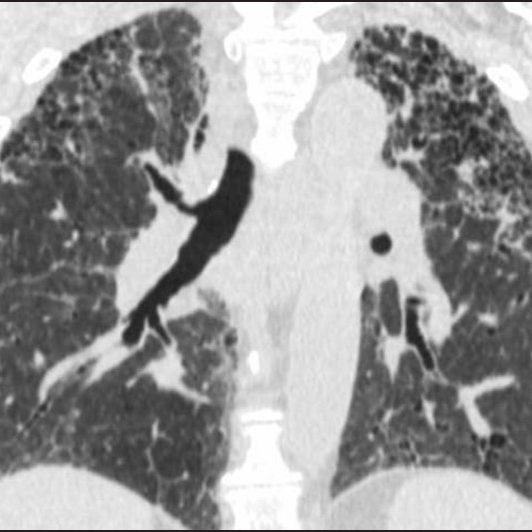
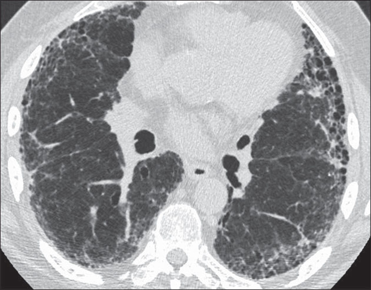
The goals of the radiologist in the evaluation of a patient with suspected pulmonary fibrosis are to determine whether a diffuse lung disease is present, determine the pattern of fibrosis, and provide an appropriate differential diagnosis. Usual interstitial pneumonia (UIP) pattern of pulmonary fibrosis is the most common ILD. UIP is most frequently idiopathic, but can also be secondary to connective tissue disease, medications, or exposure to asbestos [2]. Given the pervasiveness of this diagnosis, radiologists participating in the multidisciplinary diagnosis of patients with suspected ILD are frequently asked whether CT findings support a UIP diagnosis.Fortunately, guidelines can increase the confidence of radiologists in correctly identifying patients with UIP. The American Thoracic Society guidelines for the diagnosis of UIP pattern break down CT findings into four categories: UIP, probable UIP, indeterminate for UIP, and alternative diagnosis. The CT findings indicative of UIP pattern include subpleural and basal predominant fibrosis in addition to honeycombing, with or without traction bronchiectasis (Fig. 1).
Fig. 1—73-year-old man with idiopathic pulmonary fibrosis. HRCT scan shows usual interstitial pneumonia pattern of fibrosis characterized by subpleural and basal distribution of fibrosis with honeycombing.
This is to be distinguished from the probable UIP pattern, which is characterized by the same distribution of fibrosis including reticulation and traction bronchiectasis, but the absence of honeycombing [3].
The PPV of UIP pattern on CT for histologic UIP at surgical lung biopsy exceeds 90%, and as such, surgical lung biopsy is rarely performed when a confident diagnosis of UIP pattern can be made from imaging [3, 4]. For this reason, a diagnosis of UIP should only be made when the radiologist is confident that the imaging findings are consistent with this pattern, because often further diagnostic testing will not be pursued, potentially depriving the patient of the opportunity to receive the correct diagnosis. This distinction is not trivial; those diagnosed with UIP may be treated with antifibrotic medications and thus be subject to the side effects thereof. Not surprisingly, patients treated with antifibrotics for UIP will not be given immunosuppressive therapy, which could be a more appropriate treatment in the setting of another histologic diagnosis (e.g., nonspecific interstitial pneumonia) nor will an extensive search for exposures be pursued (e.g., as is done with patients with hypersensitivity pneumonitis).
Given the importance of correctly making a diagnosis of UIP and avoiding overdiagnosis of this entity, radiologists interpreting HRCT should be mindful of the potential pitfalls described in the following sections.
Correctly Distinguish Honeycombing From Mimics
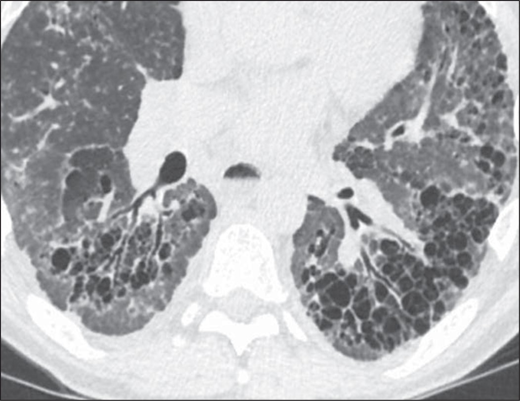
Honeycombing can be confidently diagnosed when there is a group of round clustered air-filled cysts in a row or cluster in the subpleural lung [5]. The subpleural involvement in honeycombing is critical in distinguishing it from other abnormalities. Multiple layers of cysts increase the reader’s confidence in honeycombing but are not required for diagnosis. Honeycomb cysts usually range in size from 3 to 10 mm and have relatively thick, well-defined walls [6]. In general, there is moderate agreement among radiologists for the presence of honeycombing, with kappa values ranging from 0.4 to 0.6 in one series comparing 43 different observers. There was disagreement on the presence of honeycombing in 29% of these cases [7]. Use of the above general rules for the features of honeycombing is helpful when distinguishing from common mimics. The most frequent findings mistaken for honeycombing include traction bronchiectasis, cystic lung disease, emphysema, and subpleural reticulation [8].To distinguish traction bronchiectasis from honeycombing, the shape of the air-filled structure should be noted. Airways in traction bronchiectasis are tubular in shape, which may be best seen on multiplanar reformatted images. Additionally, air-filled structures in the central or peribronchovascular lung are not consistent with honeycombing and are very likely a result of dilated airways (Fig. 2).
Fig. 2—Patient with scleroderma and fibrotic nonspecific interstitial pneumonia. Left, HRCT scan shows traction bronchiectasis mimicking honeycombing. Right, HRCT scan shows that air-filled structures spare subpleural lung.
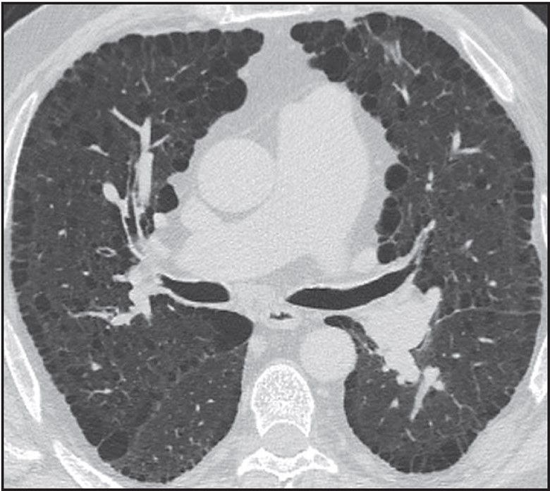
Destruction of airspaces in patients with emphysema can lead to the presence of air-filled structures in the subpleural lung; however, these structures can be distinguished from honeycombing by the overall size of emphysematous spaces that in general are larger than honeycombing cysts, the presence of paper-thin walls in emphysema in contrast to thicker walls of honeycombing, and the absence of other findings of fibrosis such as reticulation and traction bronchiectasis in patients with emphysema [9] (Fig. 3).
Fig. 3—HRCT scan shows patient with paraseptal emphysema with extensive involvement of subpleural lung, but without well-defined walls or other findings of fibrosis.
Cystic lung disease can be distinguished from honeycombing given that the cysts are often larger, scattered throughout the lung rather than clustered, and not subpleural in distribution. Shape can also be helpful in distinguishing cystic lung disease from honeycombing in that honeycomb cysts are round, whereas several cystic lung diseases are characterized by either oblong or elliptical cysts (Birt-Hogg-Dubé syndrome) or irregularly shaped cysts (Langerhans cell histiocytosis) [10].
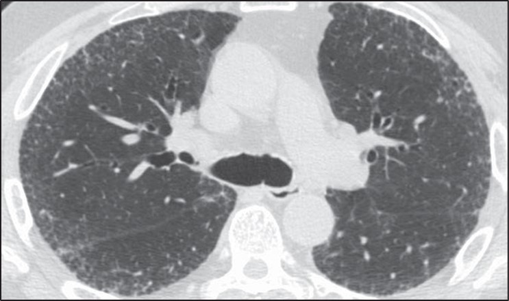
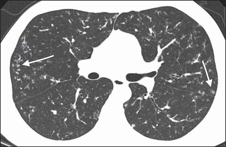
Reticulation or fine lines in the subpleural lung can also be mistakenly identified as honeycombing. To avoid this pitfall, radiologists should ensure that the subpleural abnormality is air density rather than lung density (Fig. 4).
Fig. 4—HRCT scan shows thin lines in subpleural lung in patient with pulmonary fibrosis characterized by diffuse reticulation. Abnormality in subpleural lung is lung density (same as more central lung parenchyma) rather than air density (for example in trachea), which is helpful in confirming that these findings do not represent honeycombing.
Identify Whether the Distribution of Fibrosis Is Subpleural and Basal
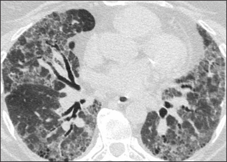
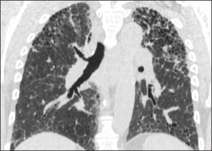
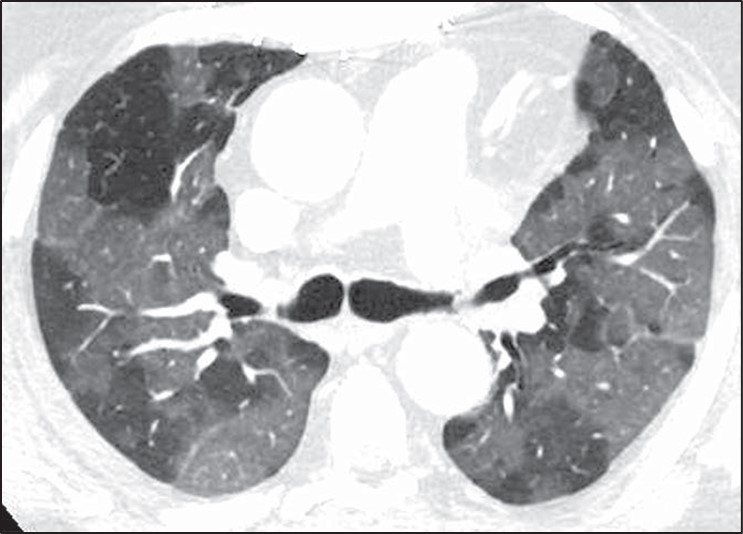
Fig. 5—Fibrosis with honeycombing in atypical distribution. Left, Axial HRCT scan shows diffuse fibrosis in association with ground-glass opacity. Diagnosis was hypersensitivity pneumonitis. Right, Coronal HRCT scan shows upper lobe–predominant fibrosis. Diagnosis was sarcoidosis.
Fibrosis that is diffuse in the axial plane or predominately in an upper lung, central, or peribronchovascular distribution may indeed be associated with honeycombing but nonetheless be caused by other entities such as nonspecific interstitial pneumonia, sarcoidosis, or hypersensitivity pneumonitis [11, 12]. Subpleural and basal distribution of fibrosis is essential to describing a pattern of fibrosis consistent with UIP at imaging. A percentage of cases with atypical distributions of fibrosis and honeycombing may be subsequently identified as UIP after biopsy; however, these cases are exactly those that benefit from surgical lung biopsy because there is a relatively high chance (70%) that another diagnosis will be found [12, 13].
Identify Inconsistent Findings
Numerous CT findings are of a diagnosis other than UIP pattern including the presence of significant ground-glass opacity, marked mosaic attenuation, nodules, and consolidation [13]. Each of these findings points the radiologist toward a diagnosis other than UIP. Patients with nonspecific interstitial pneumonia (i.e., ground-glass opacities), hypersensitivity pneumonitis (i.e., mosaic attenuation), sarcoidosis (i.e., nodules), and organizing pneumonia (i.e., consolidation) can all be identified by the presence of these features, and the presence of honeycombing should not detract from the CT findings that indicate these alternative diagnoses.
Overdiagnosis of Cystic Lung Disease
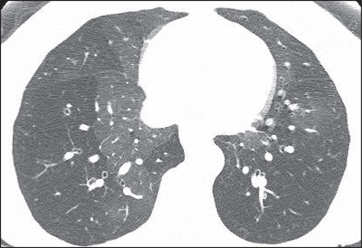
Many of the pitfalls in correctly identifying honeycombing and distinguishing honeycombing from mimics can also be applied to the correct diagnosis of cystic lung disease. When considering a potential diagnosis of cystic lung disease, it is important to again identify mimics: honeycombing, dilated airways and bronchiectasis, and emphysema. The extent of abnormality, from mild to severe, is also important to consider in this context. A few scattered pulmonary cysts may be considered in the spectrum of normal, particularly for older patients, and are most likely postinfectious rather than indicative of a cystic lung disease [14].Whereas the primary features of bronchiectasis (i.e., tubular shape) and honey- combing (i.e., thick walls, clustered, subpleural) make distinguishing these entities from cystic lung disease more straightforward, correctly distinguishing cystic lung disease from emphysema can be challenging. This challenge is in part because both entities can have very thin or imperceptible walls and can occur on a spectrum from mild to severe. The presence of the “central dot” sign in which the centrilobular artery is seen within an emphysematous space can be helpful in correctly distinguishing centrilobular emphysema from a cystic lung disease; however, this finding is not reliably seen in all regions of emphysema [15] (Fig. 6).
Fig. 6—Axial HRCT scan shows “central dot” sign in patient with centrilobular emphysema.
In general, pulmonary cysts are fewer in number, noncentrilobular in distribution, and have thicker or more perceptible walls compared with centrilobular emphysema [16]. Paraseptal emphysema and panlobular emphysema are less frequently mistaken for cystic lung disease because of their strongly subpleural distribution and overall extent respectively.
Distinguishing cystic lung diseases from one another can also be challenging; however, several key features including cyst shape, number, distribution, and classic demographic factors and associated findings can aid the radiologist in providing an appropriate differential diagnosis. Using these features allows the radiologist to narrow the differential diagnosis for a particular case to fit the specific CT features seen rather than including a long differential diagnosis consisting of all cystic lung diseases [17]:
Female sex, renal angiomyolipoma Pneumothorax, renal mass Cysts and nodules Smoker Ground-glass opacity, connective tissue disease
The presence of associated features may also be helpful in correctly identifying the presence and cause of a cystic lung disease when the abnormalities are mild and nonspecific.
Pitfalls in the Interpretation of Mosaic Attenuation and Small Airways Disease
Small airways disease may present a significant challenge in HRCT interpretation and typically manifests on HRCT as two main categories of findings: nodules or mosaic attenuation. Nodules may correspond to any of the following histologic findings: inflammation within the lumen of the airways, alveolar disease centered on the airway, or peribronchiolar interstitial inflammation. Diseases categorized by nodules are generally detected on HRCT with high sensitivity and are typically straightforward to classify.
Small airways obstruction causes hypoxia distal to the area of obstruction, resulting in regional areas of reflex vasoconstriction. Given that approximately 50% of lung attenuation is due to blood flow, regional reductions in perfusion result in a decrease in lung attenuation. These regional areas of decreased lung attenuation are described as “mosaic attenuation” or “mosaic perfusion.” More precisely, mosaic attenuation is a more general term and describes the presence of geographic areas of different lung attenuation but does not make a determination as to which lung is abnormal, whether the opaque or lucent lung. Mosaic perfusion, on the other hand, implies specifically that the lucent lung is abnormal and is the finding that most precisely corresponds to airways obstruction with reflex vasoconstriction [18].
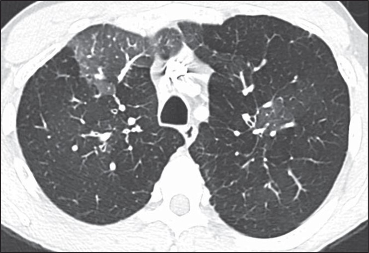
The differential diagnosis of mosaic perfusion is broad and encompasses a wide variety of both small airways diseases and pulmonary vascular diseases. It may be associated with other findings (e.g., nodules) or may be seen in isolation. The presence of mosaic perfusion is most helpful in formulating a differential diagnosis when seen in isolation, in which case it may be due to pulmonary vascular disease (mainly chronic thromboembolic disease), constrictive bronchiolitis, asthma, and hypersensitivity pneumonitis [19].Diseases characterized by isolated mosaic perfusion may present a significant challenge for several reasons. First, mosaic perfusion is a finding that is sometimes difficult to detect on HRCT. The subtle difference in attenuation frequently seen between the normal and more lucent lung is better observed when a narrow window is applied to the HRCT examination, accentuating the attenuation differences (Fig. 7).
Fig. 7—Mosaic perfusion and importance of windowing in high-resolution CT (HRCT). Left, Standard lung window in HRCT shows heterogeneous lung attenuation with subtle difference between opaque and lucent lung. Right, More narrow window accentuates difference between two lung attenuations and increases sensitivity for detection of mosaic perfusion.
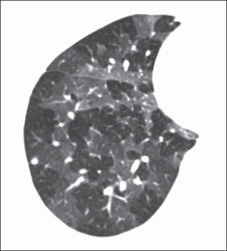
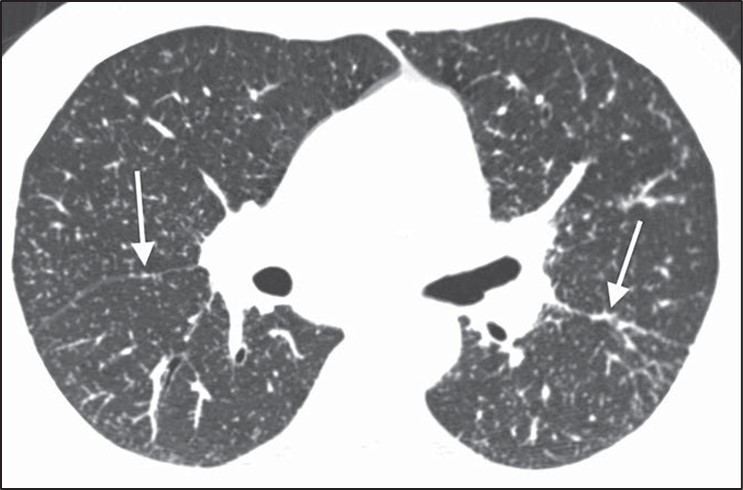
Second, when small airways or vascular diseases are diffuse in nature they result in a global and uniform decrease in lung perfusion. A diffuse HRCT abnormality is difficult to identify because there is no normal lung with which to compare the abnormality. This is most commonly seen in severe constrictive bronchiolitis [20]. Additionally, diffuse air trapping on expiratory CT is difficult to distinguish from poor timing or an inadequate respiratory effort. In these cases the HRCT scan may appear normal despite profound dyspnea and marked obstruction on pulmonary function tests. The diffuse but subtle decrease in lung attenuation is often not detected given its homogeneous nature.Mosaic perfusion (i.e., abnormal lucent lung) should be distinguished from ground-glass opacity (i.e., abnormal opaque lung), however, this distinction also has several pitfalls. Features that favor mosaic perfusion include sharp borders between the two regions of lung, smaller vessels in the lucent lung, and air trapping on expiration in the areas that were lucent on inspiration that only present in small airways disease (Fig. 8).
Fig. 8—Features of mosaic perfusion on high-resolution CT (HRCT). First two images, Axial HRCT scans show typical features of mosaic perfusion including sharp borders between opaque and lucent lung (first), larger vessels in normal more opaque lung (second), and air trapping on dynamic expiratory images. Third and fourth image, Paired inspiratory (third) and expiratory (fourth) HRCT images show heterogeneous lung attenuation on inspiration and air trapping on expiration.
None of these features are perfect in making this distinction, however. For instance, diseases characterized by ground-glass opacity may occasionally be geographic with sharp borders (Fig. 9).
Fig. 9—Axial high-resolution CT scan shows ground-glass opacity due to SARS-CoV-2 infection. Sharp borders between areas of opaque and lucent lung usually suggest that lucent lung is abnormal and pattern is mosaic perfusion. However, sharp borders may occasionally be seen in ground-glass opacity, such as in this case. Normal lung and areas of ground-glass opacity show marked difference in attenuation.
In these cases, the absolute difference in attenuation between the two regions of lung may be helpful. Mosaic perfusion typically results in a relatively subtle difference in attenuation between the diseased lucent lung and the normal opaque lung. Ground-glass opacity, on the other hand, typically shows a more marked difference in density between the two areas [21]. That being said, when mosaic perfusion results in significant shunting of blood away from the diseased areas, a greater difference in lung attenuation may be present. These cases are not infrequently misinterpreted as ground-glass opacity. Another challenge in the distinction between mosaic perfusion and ground-glass opacity is that many cases of mosaic perfusion will not show a significant difference in vessel size between the lucent and opaque lung. Last, pulmonary vascular diseases characterized by mosaic perfusion will not show air trapping on expiratory CT. Thus, expiratory CT is not helpful in the diagnosis of diseases such as chronic pulmonary embolism [22].
Pitfalls in the Interpretation of Diffuse Nodular Lung Disease
Formulating a differential diagnosis of diffuse nodular lung disease is done by identifying the distribution of nodules in relation to the pulmonary lobular anatomy. Three distributions have been described: perilymphatic, random, and centrilobular [23–25]. The perilymphatic distribution is characterized by patchy, clustered nodules that are concentrated most frequently in the peribronchovascular and subpleural interstitium. Random nodules will also be seen in the subpleural lung; however, they are not clustered but instead show diffuse homogeneous lung involvement. Centrilobular nodules are characterized by a distinct lack of nodules involving the subpleural interstitium.
The determination of the predominant pattern of diffuse nodular lung disease has several pitfalls. The perilymphatic pattern shows significant heterogeneity in the distribution of nodules. Although peribronchovascular and subpleural nodules are most typical, nodules in the interlobular septa, which also contain lymphatics, may predominate [26]. These cases may be confused for lymphangitic spread of tumor or pulmonary edema, although the thickening of the interlobular septa in pulmonary edema should be smooth, not nodular. The centrilobular interstitium is continuous with the peribronchovascular interstitium. Rarely, lymphatic diseases may have a predominance of centrilobular nodules overlapping with the centrilobular distribution (Fig. 10).
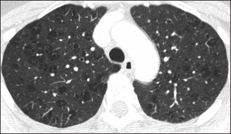
Fig. 10—Axial high-resolution CT scan shows centrilobular nodules in perilymphatic disease. Many centrilobular nodules (arrows) are present in this patient with sarcoidosis. Subpleural nodules reflect perilymphatic distribution of disease.
Although many centrilobular nodules may be present in lymphatic diseases, nodules should also be seen in the peribronchovascular or sub- pleural interstitium. This is in distinction to the centrilobular pattern in which only centrilobular nodules are present and no subpleural nodules should be seen. Lastly, diseases typically associated with a perilymphatic distribution of nodules (such as sarcoidosis) may occasionally show a fairly homogeneous involvement of the lung, mimicking a random distribution [27] (Fig. 11).
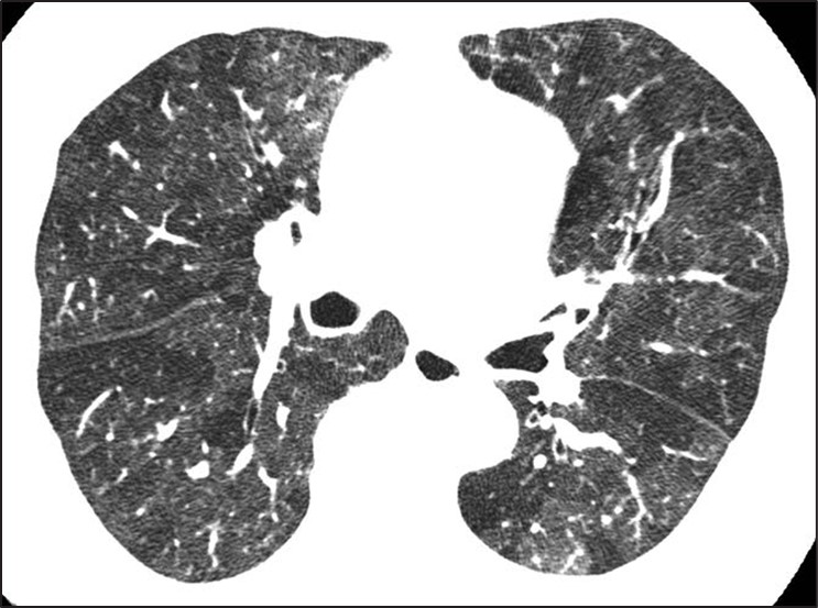
Fig. 11—Axial high-resolution CT scan shows perilymphatic distribution mimicking random nodules. Innumerable tiny nodules are present. Although pattern resembles random distribution, heterogeneous distribution in lung shows proportionally more nodules along fissures (arrows) than would be expected for random distribution.
A greater number of nodules in the subpleural or peribronchovascular interstitium may be the only clue that the distribution is perilymphatic.
Diseases for Which HRCT Has Limited Sensitivity
Certain categories of diseases may present with significant symptoms or pulmonary function test abnormalities but only manifest with mild HRCT abnormalities. Understanding the subtle imaging clues that may be present in these diseases is important in increasing the sensitivity of imaging for diagnosis. The two main categories of disease that show this discrepancy between symptoms and pulmonary function tests and HRCT manifestations of disease include small airways diseases and pulmonary vascular diseases. As discussed above, small airways diseases that manifest as isolated mosaic perfusion (e.g., constrictive bronchiolitis) may be difficult to detect on HRCT. The subtle increase in lung lucency associated with these diseases may be difficult to see, especially when the disease is diffuse in distribution [28]. Pulmonary vascular diseases such as pulmonary hypertension or chronic pulmonary embolism may also present with subtle findings. Centrilobular nodules or ground-glass attenuation or mosaic perfusion are often the only findings present and are typically much less severe than would be predicted by the patient’s advanced clinical symptoms. The lungs may appear completely normal in some patients with pulmonary vascular disease, in which case the only manifestation of pulmonary vascular disease may be extrapulmonary findings such as an enlarged pulmonary artery or right ventricular enlargement [29]. Lastly, pulmonary symptoms and pulmonary function test abnormalities might have one of several nonlung causes including pleural fibrosis, diaphragmatic dysfunction, and musculoskeletal abnormalities. All of these should be evaluated in patients with significant symptoms but no evidence of lung abnormalities on HRCT.
Awareness of common pitfalls in the diagnosis of ILD including the UIP pattern of fibrosis, cystic lung disease, airways disease, diffuse nodular disease, and lung diseases with subtle HRCT findings will better equip the radiologist to contribute to the multidisciplinary diagnosis of patients with ILD.
References
Hovinga M, Sprengers R, Kauczor HU, Schaefer-Prokop C. CT imaging of interstitial lung diseases. In: Schoepf UJ, Meinel FG, eds. Multidetector-row CT of the thorax. Springer, 2016:105–130
Wuyts WA, Cavazza A, Rossi G, Bonella F, Sverzellati N, Spagnolo P. Differential diagnosis of usual interstitial pneumonia: when is it truly idiopathic? Eur Respir Rev 2014; 23:308–319
Raghu G, Remy-Jardin M, Richeldi L, et al. Idiopathic pulmonary fibrosis (an update) and progressive pulmonary fibrosis in adults: an official ATS/ERS/ JRS/ALAT clinical practice guideline. Am J Respir Crit Care Med 2022; 205:e18–e47
Brownell R, Moua T, Henry TS, et al. The use of pre- test probability increases the value of high-resolution CT in diagnosing usual interstitial pneumonia. Thorax 2017; 72:424–429
Hobbs S, Chung JH, Leb J, Kaproth-Joslin K, Lynch DA. Practical imaging interpretation in patients suspected of having idiopathic pulmonary fibrosis: official recommendations from the Radiology Working Group of the Pulmonary Fibrosis Foundation. Radiol Cardiothorac Imaging 2021; 3:e200279
Hansell DM, Bankier AA, MacMahon H, McLoud TC, Müller NL, Remy J. Fleischner Society: glossary of terms for thoracic imaging. Radiology 2008; 246:697–722
Watadani T, Sakai F, Johkoh T, et al. Interobserver variability in the CT assessment of honeycombing in the lungs. Radiology 2013; 266:936–944
Arakawa H, Honma K. Honeycomb lung: history and current concepts. AJR 2011; 196:773–782
Devaraj A. Imaging: how to recognise idiopathic pulmonary fibrosis. Eur Respir Rev 2014; 23:215–219
Grant LA, Babar J, Griffin N. Cysts, cavities, and honeycombing in multisystem disorders: differential diagnosis and findings on thin-section CT. Clin Ra- diol 2009; 64:439–448
Abehsera M, Valeyre D, Grenier P, Jaillet H, Battesti JP, Braunerl MW. Sarcoidosis with pulmonary fibro- sis: CT patterns and correlation with pulmonary function. AJR 2000; 174:1751–1757
Silva CIS, Churg A, Müller NL. Hypersensitivity pneumonitis: spectrum of high-resolution CT and pathologic findings. AJR 2007; 188:334–344
Raghu G, Remy-Jardin M, Myers JL, et al. Diagnosis of idiopathic pulmonary fibrosis. an official ATS/ ERS/JRS/ALAT clinical practice guideline. Am J Respir Crit Care Med 2018; 198:e44–e68
Araki T, Nishino M, Gao W, et al. Pulmonary cysts identified on chest CT: are they part of aging change or of clinical significance? Thorax 2015; 70:1156–1162
Friedman PJ. Imaging studies in emphysema. Proc Am Thorac Soc 2008; 5:494–500
Lee KC, Kang EY, Yong HS, et al. A stepwise diagnostic approach to cystic lung diseases for radiologists. Korean J Radiol 2019; 20:1368–1380
Ferreira Francisco FA, Soares Souza A, Zanetti G, Marchiori E. Multiple cystic lung disease. Eur Respir Rev 2015; 24:552–564
Parambil JG, Yi ES, Ryu JH. Obstructive bronchiolar disease identified by CT in the non-transplant population: analysis of 29 consecutive cases. Respirology 2009; 14:443–448
Loverdos K, Fotiadis A, Kontogianni C, Iliopoulou M, Gaga M. Lung nodules: a comprehensive review on current approach and management. Ann Thorac Med 2019; 14:226–238
Gruden JF, Webb WR, Naidich DP, McGuinness G. Multinodular disease: anatomic localization at thin-section CT—multireader evaluation of a simple algorithm. Radiology 1999; 210:711–720
Shroff G, Konopka K, Chiles C. Perilymphatic pulmonary nodules: definition, differential diagnosis, and demonstration of the “pipe-cleaner” sign. Con- temporary Diagnostic Radiology 2013; 36:1–5
Rajagopala S, Sankari S, Kancherla R, Ramanathan RP, Balalakshmoji D. Miliary sarcoidosis: does it exist? A case series and systematic review of literature. Sarcoidosis Vasc Diffuse Lung Dis 2020; 37:53–65
Hansell DM. Small airways diseases: detection and insights with computed tomography. Eur Respir J 2001; 17:1294–1313
Kacprzak A, Burakowska B, Kurzyna M, et al. Predictive value of chest HRCT for survival in idiopathic pulmonary arterial hypertension. Respir Res 2021; 22:293
Center for Evidence-Based Imaging Brigham and Women’s Hospital
What would you do if your hospital was going to run out of iodinated contrast? Reduce the amount of IV contrast used for each CT scan? Administer multiple doses of IV contrast from a single-use vial? Defer non-urgent contrast-enhanced CT? Utilize alternative modalities, such as ultrasound, MRI, or PET/CT?
In March of last year, supply chain disruptions in China resulted in an unexpected 80% reduction in global supply of iohexol (Omnipaque, GE Healthcare). Hospitals needed to make immediate decisions about ways to conserve contrast. Otherwise, they may run out. The American College of Radiology [1], Radiological Society of North America [2], and American Hospital Association [3] released statements, and AJR continues to publish all of its research regarding the contrast media shortage as free and open access [4].
The situation was rapidly evolving, but getting more inventory wasn’t an option. As part of the response to the contrast shortage, our hospital system created and implemented an electronic health record (EHR) -based solution to help reduce iodinated contrast usage by targeting referring provider CT ordering patterns [5].
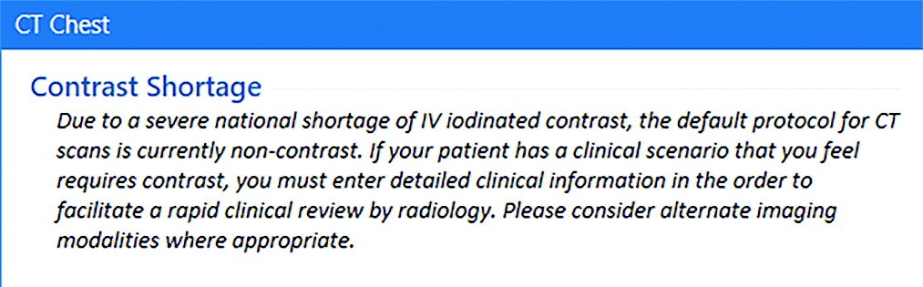
First, we added a sidebar to the ordering panel that presented an alert describing the shortage (Fig. 1), including the following strategies for imaging patients (Intervention 1; May 10, 2022):
Fig. 1—Screenshot from electronic health record shows sidebar text displayed to referring clinicians after placing orders for body CT (defined as CT of neck, chest, or abdomen and pelvis) that describes iohexol shortage and provides appropriate strategies for iodinated contrast media conservation.
Oncologic Imaging
Avoid contrast for chest CT done alone to assess metastatic disease, unless primary is thoracic malignancy
For chest/abdomen/pelvis restaging exams, consider combining non-contrast CT chest with abdominal MRI
Consider abdominal MRI for assessment of hepatic metastases
Non-Oncologic Imaging
CT for pulmonary embolism (PE)—utilize risk scoring methodology, such as Wells criteria or pulmonary embolism rule-out criteria (PERC), before pursuing CT
CT chest for lung parenchymal disease does not require IV contrast
In case of suspected musculoskeletal infection, use MRI
Emergency Imaging
Neuro
CTA head/neck—contrast needed to assess large vessel occlusion in patients within stroke treatment window. For subacute stroke outside window, please consider non-contrast head CT, followed by MRI, when appropriate
Reconsider CTA utilization for low-yield indications, including headache and dizziness
Thoracic
CT for PE—utilize risk scoring methodology (i.e., Wells or PERC)
CT chest for lung parenchymal disease doesn’t require IV contrast
Abdomen/pelvis
Pancreatitis and pyelonephritis—CT rarely indicated for these diagnoses
For primary hepatobiliary concerns, right upper quadrant ultrasound remains an excellent choice, unless high likelihood that CT also needed to explain symptoms
GI Bleeding—reserve CTA for patients with bright red blood per rectum or hemodynamic instability in whom acute intervention might be needed
Trauma
CT torso with IV contrast is needed to assess for parenchymal or vascular injury.
Consider non-contrast CT torso imaging (or radiography) in patients with low suspicion for parenchymal or vascular injury, such as elderly patients with ground-level fall and suspicion for rib fracture or thoracic/lumbar spine fracture
Next, we required referrers to enter additional clinical information into a free text field describing why iodinated contrast was needed for the CT (Intervention 2; May 16, 2022).
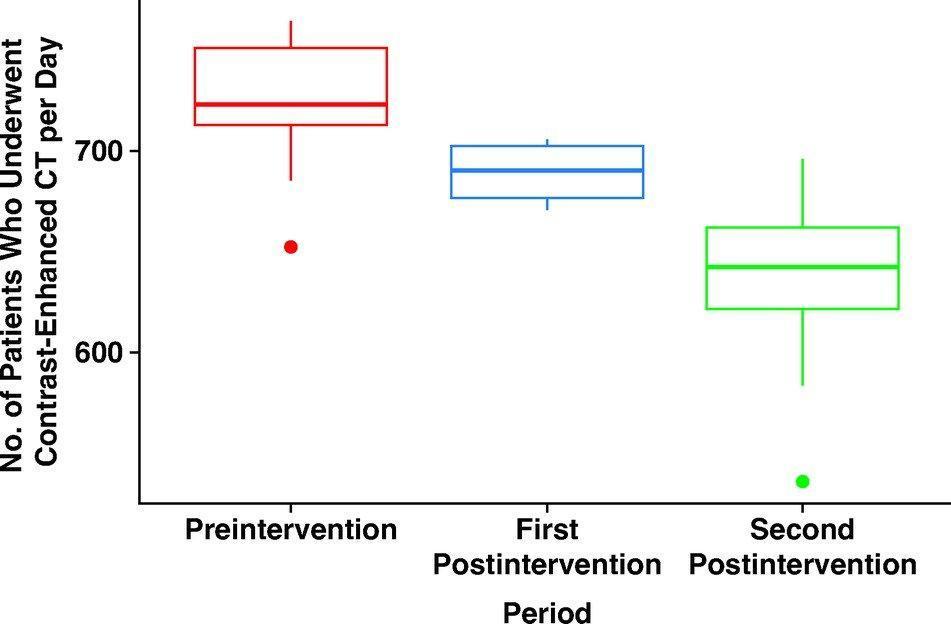
The number of patients undergoing contrast-enhanced CTs per day decreased from 726 prior to the interventions, to 689 after intervention 1, to 639 after intervention 2 (Fig. 2).
Fig. 2—Box-and-whisker plots show changes during preintervention and postintervention periods in number of patients who underwent contrast-enhanced CT examinations per day. Centerlines represent medians, ends of boxes represent interquartile ranges, ends of whiskers represent interdecile ranges, and dots beyond ends of whiskers represent outliers.
The overall number of patients undergoing CT per day decreased, as did the percentage of CT exams performed with IV contrast. These decreases were seen for all CT, as well as body CT alone (neck/chest/abdomen/pelvis). As expected, there was a decrease in requests for contrast-enhanced CT and a corresponding increase in requests for non-contrast CT.
In summary, an EHR intervention was able to reduce the number of contrast-enhanced CTs per day by 12%, the total number of CTs performed per day decreased 2.7%, and the percentage of CTs performed with IV contrast per day decreased from 53.8% to 48.6%. This simple intervention was implemented within weeks of the onset of the shortage and led to rapid practice change. Along with other conservation strategies, our health system was able to avoid rationing and continue near normal operations.
References
Wang CL, Asch D Cavallo J. Statement from the ACR Committee on Drugs and Contrast Media on the Intravenous Iodinated Contrast Media Shortage. J Am Coll Radiol 2022; 19:834-835
Grist TM, Canon CL, Fishman EK, Kohi MP, Mossa-Basha M. Short-, mid-, and long-term strategies to manage the shortage of iohexol. Radiol 2022; 304:2
Glazer DI, Lucier DJ, Sisodia RC. Electronic health record order entry–based interventions in response to a global iodinated contrast media shortage: impact on contrast-enhanced CT utilization. AJR 2022; 220:1
Over the last few years, we in radiology have faced incredible and unprecedented challenges in our day-to-day work, and this is true regardless of our specific work environments. Why? The pandemic, which has touched everyone, has had a profound impact on the workplace in general. It has changed how we work, approach work, and shaped our opinions of work. And it is not just the pandemic—it’s other phenomena: political polarization, social unrest, changes in home life and education, remote work. The pandemic and its effects led to a great resignation, and as a result, many of our sites are now understaffed. One in five doctors plan to leave their current practice in two years; two in five nurses plan to leave their practice in two years; one in three doctors expect to work less next year.
Health care workers have far greater demands now than in the pre-pandemic times. The delivery of health care has changed dramatically and quickly over the last few years. There is unprecedented “consumerism” in medicine now with a mandate to improve and rethink patient access, to provide more and better mental health services to our populations, and to have transparent pricing.
In radiology, whether you work in a large or small private practice, remotely by yourself, an academic department in a medical center, or part of a mega radiology practice, there has been a palpable shortage of radiologists. This shortage is fueled by a trend toward exclusive subspecialization with declining numbers of radiologists who can handle general work, ever-increasing expectations for service to our patients, referring doctors, hospitals, and health care systems. We have been stretched thinner. There is a desire by radiologists to have more flexible work hours or, simply stated, to work less hours overall compared to years past. There is a concern about what role artificial intelligence and machine learning will play; will we be displaced? Reimbursement has been decreasing relative to inflation and compared with other specialties. As a result of these realities and others, there is clear evidence of burnout among radiologists, similar to health care workers in other specialties. On top of that, sometimes, we find that the leaders in our organizations may be distant, or too corporate, or suffer from “toxic positivity,” which may be worse than “toxic negativity.”
There has been a steady headwind for years, but it now feels like a gale force wind. And a lot of this feels out of our control. So, goodness, how do we manage all of this?
Hold on, let’s take a breath. One strategy that we can embrace and control is to develop a culture of teams within our workplaces. In fact, I have titled this series “The Teamwork Imperative” because we must establish teamwork as a core value within the radiology workforce. I believe that if we foster a culture of teams, we can mitigate and shield ourselves from some of these headwinds.
Obviously, I’m not the first to suggest the importance of teams. It’s all over the blogs and press and our literature. In last year’s presidential address, Dr. Gary Whitman alluded to the importance of a culture of resiliency and teamwork [1]. I will also shout out to the 2021 president of our society, Dr. Jonathan Kruskal. Dr. Kruskal, along with colleagues and ARRS staff, launched RadTeams.org—an open-access website that helps radiologists establish, grow, and sustain high-functioning radiology teams, along with other aspects of imaging wellness and wellbeing [2]. Check it out. And you have to revisit the exciting ARRS Radiology Wellness Summit from the Annual Meeting that addressed, among many things, the importance of teams [3, 4].
Let me be clear. Here, when I say teams, I am specifically not referring to the “macro teams” that many of us find ourselves in. For example, at Duke Health, it is said that the 30,000-plus employees are my “teammates.” That very well may be true. But no, I am referring to your local and focal team. I am referring to the individuals that you rely on daily or weekly to deliver your work product. It’s the folks you huddle with. And the teams develop where you huddle. If you are in training, I am referring to your team of co-residents, your chief residents, maybe your program director or program coordinator who you lean on. If you are in a private practice, I am referring to those that you share physical space with, or perhaps switch call with, or the individuals you show difficult cases to, or the referring docs you have developed close relationships with, and who rely on you to deliver care. In an academic environment like mine, it might be the members of your subspecialty division. If done well, the division pulls together as a team to deliver care, service, teaching, and research.
Those divisions that have a culture of team are far more effective than those who are unable to act as a team. If you are lucky enough to have these local and focal teams (and these often form and evolve organically), many challenges at work open up and become more manageable and attainable. The clouds begin to lift. Specifically, your deliverables, whatever they may be, are far more easily and effectively achieved if you have your team and approach your work from the perspective of that team.
Work becomes more efficient and fulfilling and, frankly, more fun. The work becomes more manageable, with more aspects under your control. You become more engaged. And that then becomes an antidote to burnout. Teams, therefore, contribute to retention.
Coaches discuss this all the time. Just as Dr. Whitman was fond of quoting UCLA basketball coach John Wooden in his InPractice columns [5, 6], I’ll borrow here from Mike Krzyzewski, the legendary Duke basketball coach. “Coach K” famously talked about the five keys to an effective team and likened the keys to the fingers on a hand. Each finger is individual and can stand alone, but when the fingers come together into a fist, the fist proves to be much stronger than the sum of the individual fingers.
Communication, trust, responsibility, caring, productivity—my next installments of “The Teamwork Imperative” will discuss all five of these fingers and more, so please do stay tuned!
ARRS RadTeams website. About page. RadTeams.org/About. Accessed July 26, 2023
Kruskal J, Azour L, Goldin J. Introducing the ARRS Radiology Wellness Summit in Hawaii—Time to Get Serious! ARRS InPractice website. www.radfyi.org/radiology-wellness-summit-arrs-2023-hawaii. Published August 1, 2022. Accessed July 26, 2023
Kruskal J, Azour L, Goldin J. A Lighthouse for Radiology Wellness. ARRS InPractice website. www.radfyi.org/a-lighthouse-for-radiologists. Published November 14, 2022. Accessed July 26, 2023
Whitman GJ. Be a Primary Radiologist. ARRS InPractice website. www.radfyi.org/primary-radiologist. Published August 5, 2022. Accessed July 26, 2023
Whitman GJ. Repairing the World. ARRS InPractice website. www.radfyi.org/repairing-the-world. Published November 4, 2022. Accessed July 26, 2023
Sadia R. Qamar, Ramin Hamidizadeh, Samad Shah, Patrick McLaughlin, Anto Sedlic, Savvas Nicolaou
Department of Radiology University of British Columbia
Memoona Mian
Department of Radiology University of Arkansas for Medical Sciences
Prompt and early diagnosis is vital for timely treatment of traumatic cardiac emergencies. Myocardial rupture is a rare cause of immediate death after blunt cardiac trauma, with only 0.3–1.1% of patients with trauma reaching the emergency department (ED) [1]. Pericardial tears caused by deceleration forces or rib cage fractures are uncommon after blunt chest trauma, with a frequency of 0.3–0.5%. Rarely, valvular dysfunction can be seen due to an abrupt raised intracardiac pressure against a closed valve resulting from sudden rise in intraabdominal pressure translating into the heart causing valve cusp avulsion or tear [2]. Penetrating trauma can result in pericardial injuries further complicated by life-threatening conditions including partial or complete transdefect cardiac herniation or luxation with a mortality rate as high as 67%.
Plain radiographs will show pneumopericardium, hydrothorax or hemothorax, and mediastinal hematoma. Echocardiography will show abnormal valve function; wall motion abnormalities with decreased left ventricular ejection fraction; and pericardial effusion, signs of cardiac tamponade, or both. CT will show pneumopericardium, pericardial effusion, pericardial or myocardial laceration or rupture, cardiac herniation or luxation with associated SVC obstruction or right heart strain, valvular cusp avulsion or tears, coronary artery dissection or rupture, and associated rib cage fractures, retained foreign bodies, bullet fragments, and wound tracks [3] (Fig. 1).
Fig. 1—29-year-old man with gunshot wound. Left, Axial four-chamber cardiac CT image shows bullet fragment (arrow) abutting left ventricular side wall at mid cardiac level without myocardial penetration. Right, Mid ventricle short-axis color-coded functional cardiac CT image depicts reduced perfusion (white arrow) consistent with myocardial contusion. Black arrow indicates bullet fragment.
Imaging Pearls and Pitfalls in Cardiac Trauma
Any pericardial effusion detected in the acute trauma setting is presumed to be hemopericardium until proven otherwise. CT provides valuable information about the possible nature of pericardial effusions on the basis of the attenuation measurements of the collection. Coronary artery injuries are rare (in less than 2% of chest trauma cases), with left anterior descending artery being most commonly injured [4]. Penetrating cardiac trauma can result in pericardial injuries, which can result in partial or complete transdefect cardiac herniation or luxation with mortality up to 70% [5]. Portable supine studies in ED are suboptimal with overlying artifacts, which limits evaluation. TEE is invasive and difficult to perform in patients with acute craniocervical injuries. Cardiac MRI in trauma is primarily useful as a problem-solving tool after patients are admitted, especially to delineate the extent of myocardial contusion, regional infarction, wall motion abnormality, and valvular dysfunction.
Limitations of MDCT
First, CT involves use of ionizing radiation which increase the radiation exposure in the population. Second, the quality of MDCT images suffers with fast heart rate and high calcium burden. Finally, the patients with arrhythmias, ectopy or misregistration ECG artifacts degrade the image quality and limit evaluation. Optimizing techniques should be incorporated to counter these limiting factors.
Reduction of Radiation Dose
Radiation dose can be reduced with a prospective ECG-gated technique with narrow window acquisition, ECG tube current modulation, and limited pulse windows; tube voltage reduction based on body mass index; automated tube voltage reduction based on topogram attenuation profile; adaptive collimation limiting helical over spiral scanning; and iterative reconstructive techniques to reduce noise and ultimately reduce dose.
Optimizing Quality of Cardiac CT
Several steps can be taken to optimize the quality of cardiac CT studies. A heart rate of less than 65 beats/min can be achieved by administering 5–20 mg of β-blocker (metoprolol) IV or 50–100 mg by mouth 1 hour before the CT. Lowering the heart rate widens diastole and decreases beat-to-beat variability. Coronary arterial dilatation for optimal visualization can be achieved by administering 0.4–0.8 mg of nitroglycerin sublingually 5 minutes before contrast injection. Reconstruction algorithms can be used to reduce beam-hardening artifacts from iodine that mimic ischemia (Fig. 2).
Fig. 2—CT images show beam-hardening correction (left) and utility of B23 kernel (right) as it reduces beam-hardening artifact from iodine in left ventricle and thoracic aorta, affecting posterior inferior aspect of left ventricle wall mimicking infarct.
Edge-enhancing reconstruction algorithms can be used to reduce noise caused by extensive coronary calcifications or coronary stents.
Emerging Applications and Outlook
Coronary Atherosclerotic Plaque Characterization
The rationale behind growing efforts to accurately characterize a vulnerable, predominantly lipid-rich, plaque is its grave association with ACS and SCD. Novel attenuation-based application of dual-energy CT (DECT) has shown promising results when correlated with histologic findings. Spectral attenuation curves for material characterization are generated using attenuation values of a specific material for each and every monochromatic energy ranging from 40 to 140 keV [6]. Lipid-rich atherosclerotic plaques share the known attenuation curve of fat, in which attenuation decreases with lower monochromatic energy, thus differentiating lipid-rich plaques from fibrous plaques [7].
CT-Derived Fractional Flow Reserve
Coronary blood-flow volume effectively provides an estimation of lesion-specific ischemia. Recent vigorous advancements in digital analysis of fluid dynamics allow noninvasive assessment of coronary flow on the basis of mathematic models. CT-derived fractional flow reserve (FFR) calculates lesion-specific FFR using static coronary CT data without additional radiation or modification in image acquisition protocols. Studies have found that CT FFR shows 90% sensitivity and nearly 83% specificity for lesions with moderate stenosis causing ischemia [38]. A multicenter prospective trial showed 73% specificity and 90% sensitivity for CT FFR in diagnosing obstructive CAD compared with conventional angiographic FFR [8].
CT Myocardial Perfusion and Viability
Myocardial perfusion is one of the most important prognostic indicators for patient outcome and management of CAD. CT myocardial blood pool analysis using myocardial iodine content is a promising dynamic technology. DECT color-coded iodine maps permit sensitive detection of myocardial perfusion by depicting myocardial blood pooling [9]. The perfused myocardium takes up iodine, but no iodine uptake is seen in the infarcted myocardium (Fig. 3). Assessment of myocardial viability predicts successful revascularization therapy.
Fig. 3—Myocardial ischemia in 51-year-old man. Left and right, Mid ventricle short-axis cardiac CT image (left) and iodine perfusion map (right) show decreased subendocardial iodine uptake (arrows) in inferior basal ventricle, suggesting perfusion defect consistent with myocardial ischemia.
MDCT is a viable, reliable, and potentially effective imaging modality in evaluation of coronary and noncoronary cardiac emergencies. Cardiac CT efficiently rules out CAD in patients with low to intermediate risk who present with acute chest pain in the ED and accurately predicts midterm adverse outcome. With integration of innovative applications like morphologic plaque characterization, coronary FFR and myocardial perfusion, cardiac CT will be able to offer unprecedented benefits, ranging from triage to treatment decisions in the ED.
References
Mirvis SE. Imaging of acute thoracic injury: the advent of MDCT screening. Semin Ultrasound CT MR 2005; 26:305–331
Farhataziz N, Landay MJ. Pericardial rupture after blunt chest trauma. J Thorac Imaging 2005; 20:50–52
Sohn JH, Song JW, Seo JB, et al. Pericardial rupture and cardiac herniation after blunt trauma: a case diagnosed using cardiac MRI. Br J Radiol 2005; 78:447–449
Bruschi G, Agati S, Iorio F, Vitali E. Papillary muscle rupture and pericardial injuries after blunt chest trauma. Eur J Cardiothorac Surg 2001; 20:200–202
Prêtre R, Chilcott M. Blunt trauma to the heart and great vessels. N Engl J Med 1997; 336:626–632
Beckman JA, Ganz J, Creager MA, Ganz P, Kinlay S. Relationship of clinical presentation and calcification of culprit coronary artery stenoses. Arterioscler Thromb Vasc Biol 2001; 21:1618–1622
Min JK, Leipsic J, Pencina MJ, et al. Diagnostic accuracy of fractional flow reserve from anatomic CT angiography. JAMA 2012; 308:1237–1245
Yoon YE, Choi JH, Kim JH, et al. Noninvasive diagnosis of ischemia-causing coronary stenosis using CT angiography: diagnostic value of transluminal attenuation gradient and fractional flow reserve computed from coronary CT angiography compared to invasively measured fractional flow reserve. JACC Cardiovasc Imaging 2012; 5:1088–1096
Han R, Sun K, Lu B, Zhao R, Li K, Yang X. Diagnostic accuracy of coronary CT angiography combined with dual-energy myocardial perfusion imaging for detection of myocardial infarction. Exp Ther Med 2017; 14:207–213