
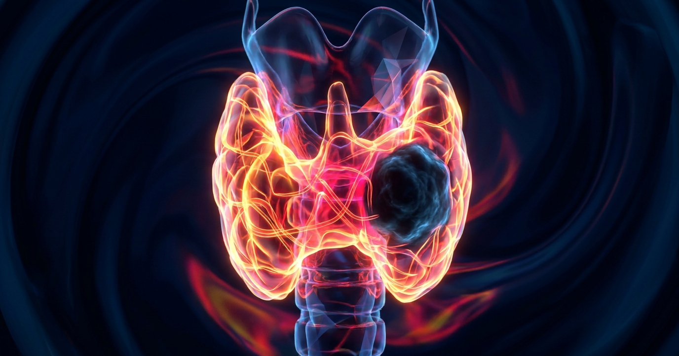
Cold Case: Radionuclide imaging for Graves’ disease can obscure an underlying malignancy when hyperactive tissue surrounds a non-functioning thyroid nodule. Precipitated by smoking, diet, stress, or illness, clinically, Graves’ oftens presents alongside extra-thyroidal manifestations: orbitopathy, dermopathy, acropachy, etc. So, as Dr. Katherine Zukotynski explained at ARRS26, workup combines both sonographic and radiotracer techniques:
- Ultrasound: Usually shows an enlarged, diffusely hypoechoic, and diffusely hypervascular thyroid gland.
- Scintigraphy: Protocol utilizing I-131 or pertechnetate shows sky-high radionuclide uptake with diffuse, homogeneous activity throughout the entire gland.
Archetypal Graves’ scintigraphy does not preclude structural lesions. Patients can present with Graves’ while harboring nodules, too.
- Sky-high diffuse uptake across the gland can draw attention away from parenchymal defects.
- A focal region exhibiting decreased radiotracer uptake—a cold nodule—embedded within a diffusely active gland.
RadFYI: If scintigraphy exhibits classic Graves’, yet reveals a focal area of decreased activity, follow up with US for coexisting cancer.
Leave a Reply