
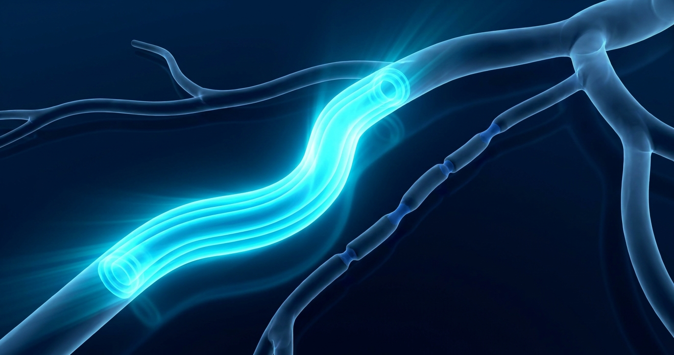
Summary: Whereas lumen-only looks show overlapped narrowing in both conditions, high-res, contrast-enhanced VWI reveals fundamental differences in wall thickening, enhancement, as well as temporal persistence.
Against the Wall: Having to differentiate CNS vasculitis from reversible cerebral vasoconstriction syndrome (RCVS) is as common as it is critical. “That one comes up a lot,” Dr. Hediyeh Baradaran noted in her ARRS Quick Bytes video. Thankfully, intracranial vessel wall imaging (VWI) exists to aid in your DDX.
- Vasculitis
- Enhancement & Thickening: Characterized by concentric vessel wall enhancement and marked wall thickening.
- Longitudinal Persistence: Both arterial narrowing and contrast enhancement persist across multiple follow-up exams.
- Remodeling Pattern: Typically demonstrates a higher negative remodeling index.
- RCVS
- Enhancement & Thickening: Shows multifocal arterial irregularities with no enhancement (or only rare, subtle wall thickening without significant contrast uptake) on pre- and post-contrast VWI.
- Longitudinal Resolution: Arterial narrowing does not persist over time, resolving on repeat imaging—“as the name implies,” said Dr. Baradaran.
Leave a Reply