Recognizing this hallmark pattern can spare unnecessary confusion, guiding the right surgical call.
The Big Picture

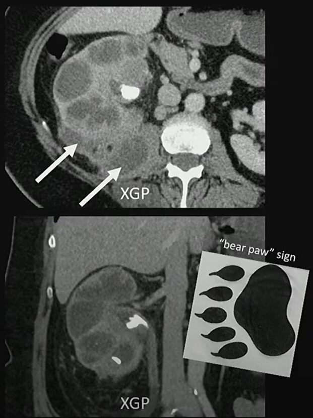
Xanthogranulomatous pyelonephritis (XGP) is a rare, destructive renal infection, most often in women with staghorn calculi. It replaces functioning parenchyma with lipid-laden macrophages and inflammatory tissue, often extending beyond the kidney.
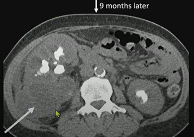
Nine months later, we see much more exuberant inflammation—soft-tissue thickening extending out from the kidney into the retroperitoneum.
Patient underwent renal scintigraphy prior to getting a nephrectomy, and we can see that that right kidney is not contributing to the renal function.
Key Takeaways
- Classic appearance: Enlarged kidney, staghorn calculi, and a contracted pelvis—the “bear paw.”
- Functional loss: Renal scintigraphy often shows little or no contribution from the affected kidney . . .
| Parameter | Left Kidney | Right Kidney |
|---|---|---|
| Peak Time | 6.7 minutes | 2.3 minutes |
| Relative Function (Integral, 1–3 min) | 85% | 15% |
- Extrarenal extension: Look for inflammation tracking into the retroperitoneum, psoas, or paraspinal muscles.
- Definitive treatment: Because the kidney is typically nonfunctional, nephrectomy is the standard.
Challenges Ahead
- Distinguishing XGP from renal cell carcinoma or pyonephrosis can be difficult without correlating imaging and functional data.
- Awareness of extrarenal spread is crucial to surgical planning.
- Early recognition can prevent unnecessary biopsy or delay in definitive management.
Presented by Anup Shetty, MD, during ‘Radiology Case Review: Genitourinary Imaging,’ part of the ARRS 2025 Annual Meeting. Watch the full session now: arrs.org/am25
Bottom Line
When you see that “bear paw” (i.e., staghorn calculus, perinephric inflammation, and an enlarged, poorly functioning kidney), think XGP!