
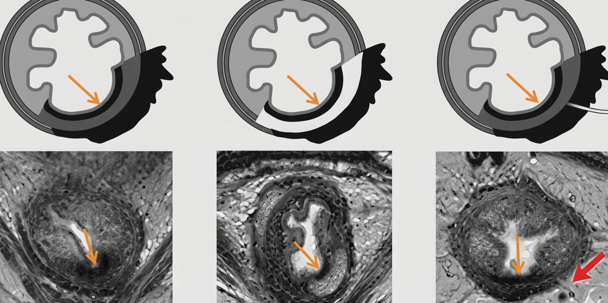
The split scar sign, first described in 2020, is a powerful predictor for sustained complete clinical response and pathological complete response in rectal cancer patients following treatment.
Bio Marker: The split scar sign helps rads identify patients who have responded exceptionally well to therapy—often preventing the overcalling of residual tumor. And as Professor Kirsten Gormly of the Royal Australian and New Zealand College of Radiologists (RANZCR) illustrated during the ARRS 2026 Global Partner Society Course, to determine if a split scar sign is positive, simply use the continuous line rule:
- Positive—If you can draw a continuous thin inner black line over the scar with a marker (or “texter” as it’s called down under) without lifting it off the image, the sign is present.
- Negative—If you have to lift that
markertexter, or if the scar is so thick it requires a bigger texter, the sign is in absentia.
Additional features from ARRS and RANZCR’s Rectal MRI: Best Practices from Ocean to Ocean to further set straight the split scar sign:
- Inner Winner: The most critical component is the continuous thin inner scar.
- Optional Outer: Whereas this sign is so named for its split look, you don’t always need an outer scar, especially if the original tumor was not T3.
- Virtue Signals: Look for intermediate signal in the central muscle and the potential for mucinous degeneration (i.e., higher signal) or perforating vessels to be present within the layers.
- Black Mass: The black layers on MRI represent submucosal collagen and fibrosis merging with muscle cells.
RadFYI: Misinterpreting these thin scars as residual tumor is a common pitfall. Mastering this T2 assessment allows for more accurate posttreatment restaging and better informed clinical management.
Leave a Reply