
Summary: MRU is indeed invaluable when you want to avoid ionizing radiation, à la pregnant patients. But it should be leveraged, too, as an essential second-line problem solver for atypical renal and urothelial masses.
GU Pro: Sure, CTU remains the daily, fast, first-line workhorse for genitourinary imagers. Still, standard scans can struggle to characterize atypical tumors or complex tissue changes. Enter, then, multiparametric MR urography, offering multi-layered tissue characterization to narrow your DDX.
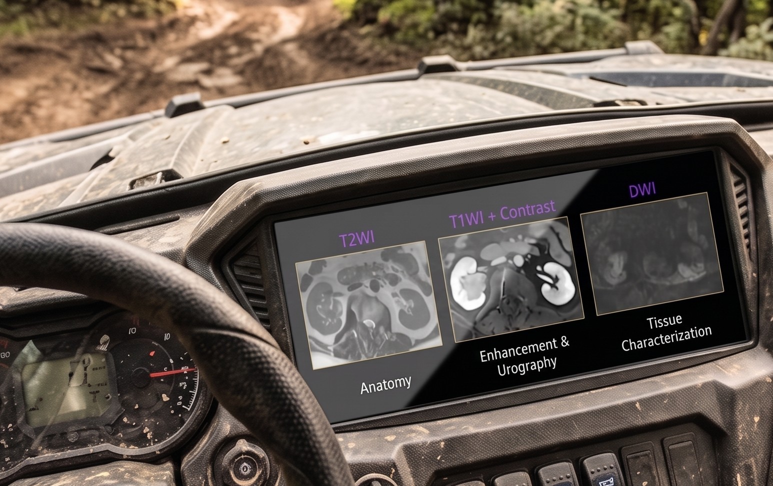
By combining multiple specialized sequences, Dr. Myles Taffel noted during our longitudinal course Mastering Genitourinary Imaging & Interventions, MRU allows rads to analyze pathology from several angles across distinct terrain:
- T2-Weighted Anatomy: Delivers detailed structural imaging comparable to what is obtained with CT.
- Multi-Phase Contrast: Captures multiple enhancement phases, including early dynamic post-contrast and late urographic phases, which may not be feasible on CT.
- DWI: Highlights cellular diffusion restriction to pinpoint exact areas of pathology.
- Intrinsic T1: Provides native tissue contrast to further assist in detailed characterization.
As Taffel recalled, the real utility of MRU’s multiparametric sequences shines in those really tough cases, including off-roaders:
- ID Lymphoma Mimics: A renal hilum mass can mimic standard urothelial cell carcinoma. However, when MRU shows the mass extending along the ureter without significant hydronephrosis—coupled with restricted diffusion on DWI—the diagnosis of lymphoma becomes clear.
- Bladder Subtleties: In a patient presenting with hematuria, a standard T2 sequence might only show vague bladder wall enhancement. Adding DWI and early dynamic post-contrast sequences provides the high-contrast resolution needed to confidently pinpoint exactly where the malignancy lies.
Leave a Reply