
Where’s Waldo? Subcutaneous lipomas are notoriously easy to miss on cross-sectional imaging because they match the exact density and signal intensity of surrounding tissue. DuringARRS26, Dr. Mark Murphey detailed the diagnostic principles he uses to confidently spotsubtleties.
- If you see nothing within the first 5 milliseconds of perusal, you are almost certainly dealing with a subcutaneous lipoma.
- It’s not unlike searching for Waldo—you’re trying to ID fat against a background of fat.
The Monotony of It All! Up to 71% of lipomas consist of simple, monotonous fat with no significant architectural complexity across CT, MRI, or US.
- Compare directly to adjacent normal subcutaneous fat on T1-weighted scans.
- On US, lipomas exhibit echotexture similar to fat and, crucially, show no evidence of significant blood flow on Doppler.
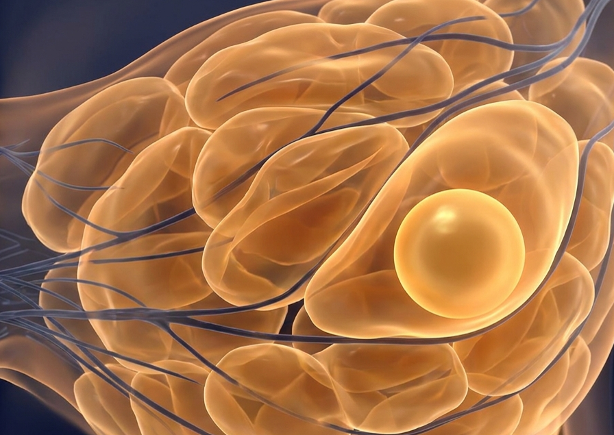
Ask Yourself: Is this lesion more, or less, complex than its counterpart? Normal fat consists of lobules divided by connective tissue septa (grossly, these appear as greasy, monotonous yellow tissue composed of adipocytes uniform in size and shape). And should your mass look less complex than its surroundings, “it can’t be very bad,” Dr. Murphey said.
- Ca. 85% of subcutaneous lesions can be diagnosed by physical exam alone, but rads are increasingly relied upon as the primary examinator.
- Markers placed by techs over the palpable point of concern are vital for localization.
RadFYI: Evaluate relative complexity against adjacent normal tissue. If it looks identical to—or even simpler than—normal fat and lacks vascularity, your DDX is done.
Leave a Reply