
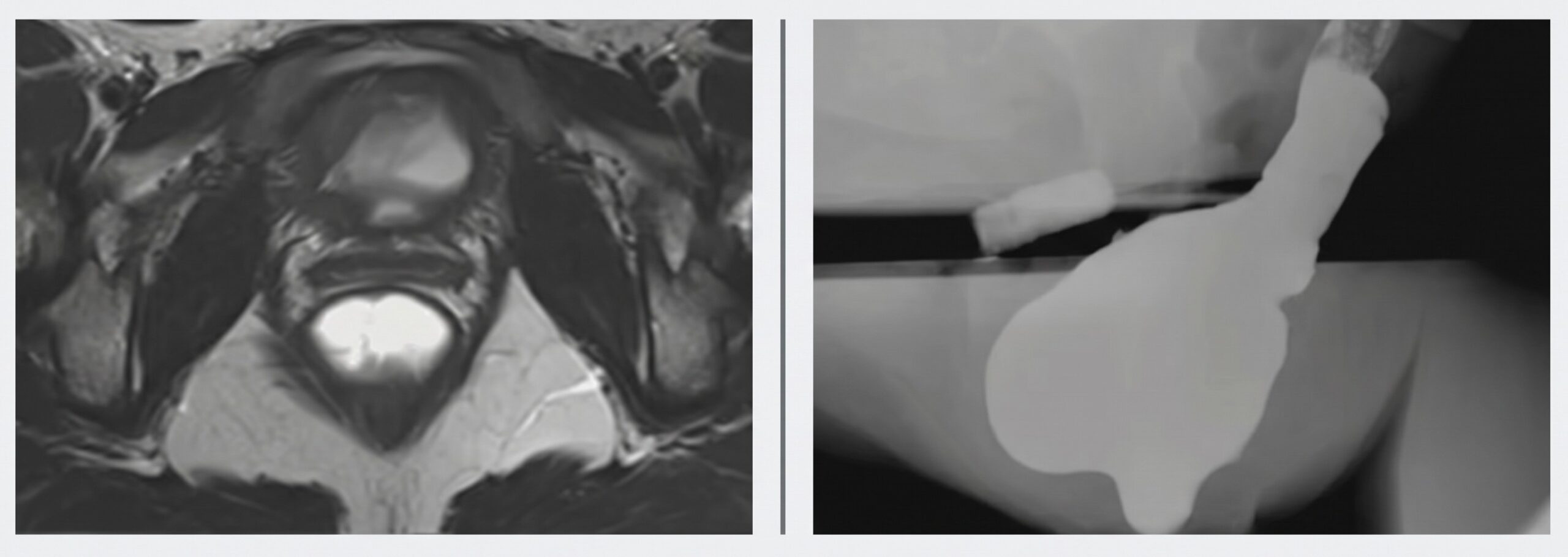
Summary: Whereas modality selection depends on patient tolerance, provider preference, and scanner availability, MR defecography provides unmatched soft-tissue resolution and functional detail to guide targeted clinical and surgical management.
Dare to Compare: A physical exam alone doesn’t always capture a pelvic floor disorder’s underlying dysfunction. Dynamic MR defecography does, though, delivering high-yield multiplanar imaging that directly informs surgical decision-making.
Pelvic prolapse, relaxation, dyssynergic defecation—all stem from chronic elevations in intra-abdominal pressure or structural disruptions, Dr. Christine Boatright noted during the Annual Meeting.
Percent Points: Whoppingly, 94% of MR defecography exams indicate posterior compartment complaints, à la chronic constipation and obstructive defecation.
- 92%—Correlation between MR defecography findings and suspect pelvic floor pathologies.
- 86%—Proportion of surgical cases where MR defecography provides added operational value.
MR Defecography: Performed with the patient supine using rectal gel to simulate defecation, it requires no oral small-bowel, vaginal, or bladder contrast.
- MRI offers superior soft-tissue contrast, allowing direct visualization of the levator ani muscle complex, pelvic ligaments, pubic symphysis, bladder, uterus, and rectum.
Fluoroscopic Defecography: Performed upright on a specialized chair (providing a more physiological posture), it requires oral barium swallowed an hour prior for small-bowel opacification, rectal contrast, and an optional contrast-soaked vaginal tampon, but provides limited soft-tissue detail.
Evacuation Dynamics: Patient movement during evacuation helps categorize the underlying etiology. Prominent pelvic movement suggests pelvic floor relaxation, whereas minimal pelvic movement or an inability to evacuate rectum content points toward dyssynergic defecation.
- MRI can uniquely reveal levator ani muscle atrophy and structural disruptions that remain invisible on fluoroscopy.
Leave a Reply