Author: Logan Young

  • Neurological Manifestations of COVID-19: Insights for the Diagnostic Radiologist

    Neurological Manifestations of COVID-19: Insights for the Diagnostic Radiologist

    Published January 4, 2021

    avatar

    Ian Y. M. Chan

    Department of Medical Imaging
    Western University

    avatar

    Tyler Cossetto

    Department of Medical Imaging
    Western University

    avatar

    Bradley Davis

    Department of Medical Imaging
    Western University

    avatar

    Victor Lam

    Department of Medical Imaging
    Western University

    avatar

    Timo Krings

    Division of Neuroradiology
    University of Toronto

    The coronavirus disease (COVID-19) pandemic is now the most significant pandemic in over a century. Severe acute respiratory syndrome coronavirus 2 (SARS-CoV-2), the virus that causes COVID-19, affects multiple body systems by binding to the angiotensin-converting enzyme 2 (ACE2) receptors on cells. Although pulmonary manifestations of COVID-19 have garnered much attention, its neurological impact can be as devastating. Neurological manifestations include ischemic stroke, intracranial hemorrhage, encephalopathy, and peripheral neuropathy. Neurological symptoms are common and include headache, dizziness, myalgia, seizures, weakness, strokes, and alterations of consciousness. Although rarely isolated, disorders of smell and taste have also been seen in COVID-19. For some patients, cognitive symptoms have been found to last for months following infection. Radiologists must be attuned the neurological manifestations of COVID-19, as early identification of these findings may help to guide management and reduce disease transmission.

    Ischemic Stroke

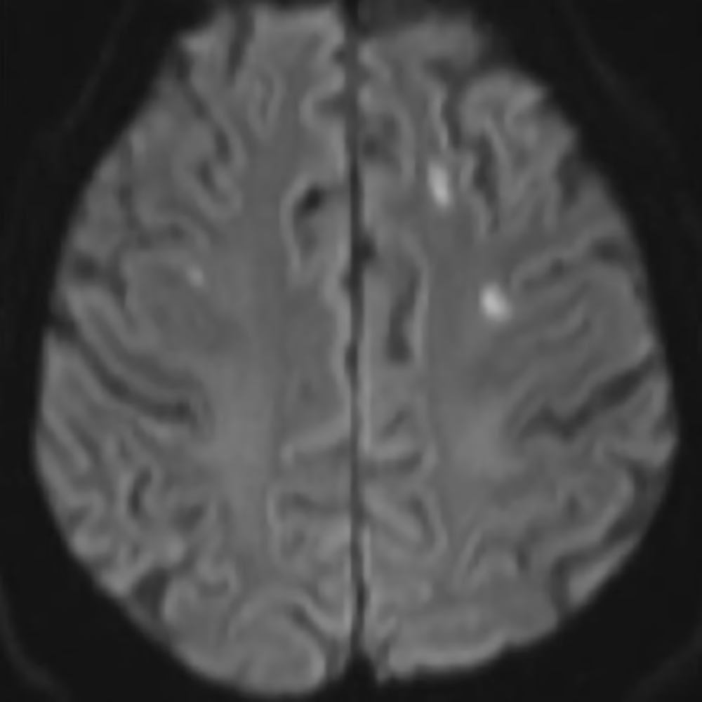
    The incidence of ischemic stroke associated with COVID-19 in hospitalized patients is between 0.4% and 2.7% but may vary with infection severity. The association between ischemic stroke and COVID-19 has been proposed since the outset of the outbreak, fueled by the rationale that infectious processes like COVID-19 are prothrombotic through an endotheliitis-mediated mechanism [Fig. 1].
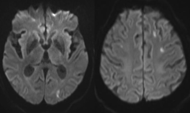
    Initial concern was raised following an increased incidence in large vessel occlusion (LVO) strokes. In April 2020, an early case series published in the New England Journal of Medicine highlighted five cases with LVO stroke in New York City and suggested that LVOs may be a presenting feature of COVID-19 in young adults. A case-control study from New York City published in AJR showed that LVOs were present in 32% of code stroke patients with COVID-19, compared to 15% without COVID-19. Recently, a systematic review by Canadian researchers confirmed that 1.5% of patients admitted to hospitals with COVID-19 suffered an ischemic stroke with a resultant mortality of 35% for all stroke types. More concerning, in patients younger than 50, half had no other symptoms of infection at the time of ischemic stroke onset, and 69% presented with LVO.

    By contrast, 17 stroke centers in Europe and North America reported a 32% reduction in endovascular thrombectomy (EVT) procedures during the pandemic. The number of patients undergoing neuroimaging for stroke in the United States also decreased by 39%. Proposed reasons for this discrepancy include reluctance of patients to seek care due to fear of contracting COVID-19 and an overwhelmed health care system. With this divergent data, strong epidemiological studies are needed to determine the underlying reasons for these statistics.

    Intracranial Hemorrhage

    A growing number of studies have investigated the relationship between COVID-19 and intracranial hemorrhage (ICH). The incidence of ICH in COVID-19 ranges from 0.2% to 0.9%. ICH has a much higher likelihood of occurring in hospitalized patients with COVID-19 who receive anticoagulation or extracorporeal membrane oxygenation.

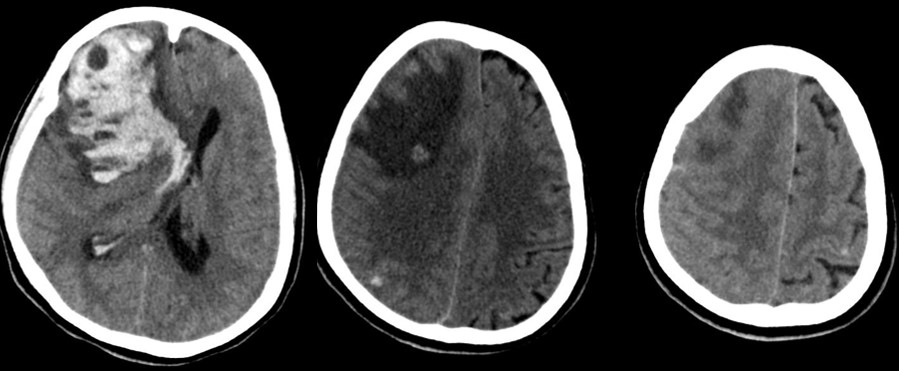
    Intracerebral and subarachnoid hemorrhage, as well as hemorrhage from cerebral venous thrombosis, have been reported in COVID-19. The majority of intracerebral hemorrhages are lobar with deep structure and infratentorial hemorrhages being less common. Microhemorrhages in the juxtacortical white matter and corpus callosum (particularly the splenium), multilobar microhemorrhages, and single large lobar hemorrhages have been observed.

    At this time, it is unclear if ICH seen with COVID-19 is causative or coincidental. Two prominent hypotheses suggesting causation point to the pathophysiology of sepsis and the activation of the renin-angiotensin system leading to endovascular disruption. A retrospective case series, which suggested these findings are coincidental, showed that most patients with ICH and COVID-19 could be attributed to prior trauma and known suspected hemorrhage before contracting the virus. This highlights the challenge in accurately attributing ICH events to COVID-19 [Fig. 2].

    Encephalopathy

    Encephalopathies have gained increasing attention as unusual manifestations of COVID-19 and harbingers of more severe disease. Similar coronavirus disease, such as severe acute respiratory syndrome (SARS) and Middle East respiratory syndrome (MERS), have shown a similar propensity to cause acute encephalopathy. The prevalence of this condition ranges from 0.04% to 0.2% for COVID-19 patients. Acute encephalopathy was associated with more severe infection and worse clinical outcomes in hospitalized patients, which makes identification important.

    The etiology of acute encephalopathy associated with COVID-19 is not entirely clear. Cases have been reported in the literature. Meningoencephalitis was shown in a young adult patient without significant respiratory disease. Leukoencephalopathy and acute hemorrhagic necrotizing encephalopathy were noted in case reports. Encephalitis was also seen in an adolescent patient with severe infection

    The primary imaging finding of encephalopathy was nonspecific cortical T2/FLAIR hyperintensities with associated restricted diffusion. Bilateral FLAIR hyperintense thalamic lesions have been characteristically described in acute necrotizing encephalopathy. Imaging features of posterior reversible encephalopathy syndrome and hypoxic-ischemic encephalopathy, as well as exacerbation of demyelinating disease, were also seen. Kihira et al. described multiple cases of white matter disease related to hypoxic leukoencephalopathy, acute disseminated encephalomyelitis, and direct viral encephalitis attributed to COVID-19. A serious condition linked with COVID-19 known as multisystem inflammatory syndrome in children (MIS-C) can cause neurocognitive symptoms, and in four cases, MRI revealed signal abnormalities in the splenium. Consequently, radiologists must have a high index of suspicion for a variety of encephalopathies in patients with COVID-19.

    Peripheral Neuropathy

    Various studies have identified peripheral neuropathy related to COVID-19. Indeed, since ACE2 receptors are found in the olfactory epithelium, the neuroinvasive potential of SARS-CoV-2 has been a topic of investigation.

    Anosmia may be the most common neurological symptom. Inflammatory obstruction of the olfactory clefts is known to impair olfaction. CT and MR images in patients with COVID-19 have shown obstruction of the olfactory clefts, which has been thought to lead to anosmia. Imaging of infected patients has also shown olfactory bulb atrophy and MRI signal changes that can resolve on follow-up imaging. 

    Although rare, Guillain-Barré syndrome (GBS) has also been reported in COVID-19. Comprising a heterogeneous group of immune-mediated polyneuropathies, GBS classically presents as rapid symmetrical ascending muscle paresis or paralysis. To date, at least 73 cases of GBS have been reported in patients with COVID-19. GBS associated with COVID-19 has been shown to exhibit asymmetrical thickening and hyperintensity of the post ganglionic roots supplying the brachial and lumbar plexuses on STIR images. Facial nerve, nerve root, and leptomeningeal enhancement have been reported. Of note, MRIs of the brain and spine in these patients are most often normal. Other neuromuscular conditions, such as generalized myoclonus and rhabdomyolysis, have also been rarely reported.

    We have highlighted a number of important neurological manifestations of COVID-19. Our understanding of this novel coronavirus and its neurological manifestations continues to improve, and the epidemiology between these conditions and COVID-19 are frequently being refined. We remain hopeful that the management and prevention of COVID-19 will continue to advance with the development of new therapeutics and vaccines in the coming months and beyond. As radiologists, we should be cognizant of the fact that imaging has crucial role to play in diagnosing many of these neurological manifestations. Moreover, long-term sequelae of these conditions will often necessitate further follow-up imaging, ensuring our continued involvement in patient care for the foreseeable future.

  • A Practical Primer for Dual-Energy CT

    A Practical Primer for Dual-Energy CT

    Published November 23, 2020

    avatar

    Mayur Virarkar

    Department of Neuroradiology
    University of Texas Health Science Center at Houston

    avatar

    Priya Bhosale

    Department of Diagnostic Radiology
    University of Texas MD Anderson Cancer Center

    Dual-energy CT (DECT) is a state-of-the-art technology that simultaneously processes data from multiple photon energies in a single CT acquisition. The principle of dual energy dates back to the 1970s, whereas the first clinical DECT scanner was available in 2006. However, the utilization of DECT in routine clinical practice has grown over the past decade owing to increased scanner availability from vendors and multiple new applications of DECT techniques. Furthermore, the postprocessing DECT data using commercially available software have resulted in the generation of virtual monoenergetic or monochromatic images ranging from 40 to 200 keV. These images can also be used to produce spectral attenuation curves, scatterplots, histograms, and effective atomic number (Zeff). Various technical approaches of DECT imaging are currently available through different vendors, such as single source rapid-kVpswitching DECT (GE Healthcare, WI), single source helical DECT (Siemens Healthineers, Germany), single source twin-beam DECT (Siemens Healthineers), dual source DECT (Siemens Healthcare, Germany), single source sequential DECT (Toshiba, Japan), and dual layer DECT (Philips Healthcare, Netherlands).

    In a typical CT, two different materials (e.g., calcium and iodine) may demonstrate similar CT attenuation values when subjected to a single radiation beam; however, these materials behave differently when exposed to different energy levels, as in dual energy CT. Atomic number (i.e., Z) is an important parameter determining the CT attenuation values. For example, higher Z materials are more susceptible to the photoelectric effect than lower ones. The commonly used contrast agent iodine is discernible at low kiloelectron volt values, and these properties can be useful in distinguishing iodine from other body materials, such as calcium and water. Soft tissues, such as muscles and organs, have weak photoelectric effects and less variation in their attenuation values at different energies.

    DECT postprocessing techniques produce different types of reconstructed images with multiple clinical functions. These images include mixed material-specific images, such as water, iodine, or fat images, virtual monochromatic and virtual monoenergetic images (VMIs) generated for a single energy level. Spectral attenuation curves display particular ROI energy values on the x-axis (range, 40–140 keV) and mean attenuation values on the y-axis. Scatterplots are generated by comparing the ROI attenuation values with water concentrations. Histogram displays the frequency of values for a single ROI variable. The materials can be differentiated from one another on the basis of their calculated Zeff values (i.e., virtual atomic numbers). The calculated Zeff takes into account the unique nature of the materials over the range of energies in DECT (40–140 keV).

    Researchers have explored the utilization of DECT in identification and characterization of various tumors in the body. VMIs are more advantageous in identification and characterization of liver lesions than conventional CT. Material-specific images, such as iodine images, aid in assessing hypervascular metastases from uterine sarcomas. In addition, these images may be able to detect remote small subdiaphragmatic perihepatic implants. The retroperitoneal lymph nodes are better visualized on iodine-enhanced images than conventional CT images. Studies have shown lower iodine uptake on DECT in metastatic than in normal and inflammatory lymph nodes, guiding the diagnosis of lymph node metastases. Osseous metastases with soft-tissue components are also better visualized on iodine-enhanced images. VMI has also been beneficial in identifying incidental pulmonary embolism (PE) in oncologic patients. 40-keV VMI images have been shown to improve objective image quality of the pulmonary vessels, along with increased diagnostic confidence in the diagnosis of incidental PE. Virtual unenhanced DECT images offer valuable tools for improving the diagnosis of pediatric abdominal neoplasms—helping to identify or validate the presence of tumoral calcifications and hemorrhage, appropriate lesion delineation, and differentiate an abdominal mass from adjacent contrast- filled bowel or abdominal organs. DECT has also been documented to identify vascular and perfusion abnormalities due to hypoxemia related to coronavirus disease 2019 (COVID-19).

    There are few clinical studies in the current literature assessing the diagnostic ability of DECT in gynecological malignancies. These studies support using low energy for assessing endometrial cancer invasion, characterization of ovarian masses with internal septation and mural nodularity, and identification of calcified peritoneal implants and remote serosal perihepatic implants. Iodine maps are useful for assessing response after chemoradiation in cervical cancer patients, peritoneal implants, and nodal and osseous metastases, as well as distinguishing benign and malignant ovarian tumors. Water maps obtained from DECT are useful in distinguishing high- and low-grade ovarian tumors. Currently, there are numerous and amazing new applications of DECT being investigated in clinical studies. Spectral photon-counting CT with enhanced image quality is being translated successfully into clinical studies. There have been recent developments in new nanoparticle contrast agents with specific disease biomarkers for dual-energy and multi-spectral CT. All of that being said, there is a need for more prospective trials to explore the true potential of this innovative and promising technology, so as to make DECT a true multiparametric imaging modality in the future.

  • Precision Prospectus: Limiting Variability in PET Interpretation

    Precision Prospectus: Limiting Variability in PET Interpretation

    avatar

    Daniel C. Sullivan

    Professor Emeritus, Department of Radiology
    Duke University Medical Center

    Variability in the interpretations of clinical imaging studies is a problem that has been recognized for decades and has been extensively documented in the radiology literature. It’s a problem that pervades all imaging modalities. Patients should get the same result if they go to the radiology department any day of the week. Sadly, that is too often not the case. The reasons for this day-to-day variability are complex and reflect the use of different scanners, software, technologists, local operating procedures, and different radiologists. However, to be in synch with the continuing emergence and maturity of precision medicine, and to meet the expectations of referring physicians and our patients, the field of radiology must more strongly strive to improve the reproducibility of clinical imaging results for each individual patient.

    One of the generally accepted benefits of precision medicine is to monitor a patient’s response to therapy and adjust, tailor, or change the patient’s health care plan according to the degree of or lack of response. Unfortunately, the traditional subjective, qualitative interpretation of clinical imaging examinations, based on visual inspection of the images, results in marked interreader and intrareader variability. This frequently makes it difficult to be confident across serial scans when determining whether a given patient’s condition has improved, worsened, or stayed the same.

    One strategy to reduce variability is to extract objective, reproducible, quantitative results from clinical imaging scans. Since all clinical imaging studies today are digital, this is feasible. One clinical setting where referring physicians particularly want objective measurements delineating change in the burden of disease is oncology. Oncologists want objective measurements of both tumor size (whether from CT or MRI) and metabolic activity (from FDG-PET scans).

    Reproducible, quantitative standardized uptake value (SUV) results from FDG-PET scans are increasingly viewed as important in clinical oncology—both in routine clinical practice, as well as in clinical trials. In 2010, we surveyed several hundred oncologists at National Cancer Institute-funded cancer centers about tumor measurements. Ninety-four percent expected tumor size measurements to be provided routinely, and more than half also expected SUV to be provided from FDG-PET scans.

    Recommendations to use FDG-PET scans as part of the staging workup for solid tumors have been included in several chapters of the eighth edition of the American Joint Committee on Cancer’s Cancer Staging Manual. These panels of expert oncologists recognize that SUV from FDG-PET scans likely conveys important diagnostic and/or prognostic information, but lack of standardization makes it impossible at present to determine appropriate thresholds or cut-points to guide clinical decision-making. They therefore recommend that all FDG-PET scan reports in breast cancer patients should contain SUV values for the primary tumor and SUV for the primary tumor, as well as hilar and mediastinal nodes in patients with lung cancer [6]. They further recommend that these SUV values be extracted from the medical record by cancer registrars, so that large databases can be developed to determine relevant thresholds. It is sobering to consider that another medical specialty is promulgating recommendations to collate imaging data to inform their clinical decision-making. One could argue that the radiology profession should be initiating such data collection and analysis.  

    In 2018, the American College of Radiology (ACR) Quality Measures Technical Expert Panel, recognizing the increasing clinical importance of objective SUV measurement, approved a quality performance measure entitled “Use of Quantitative Criteria for Oncologic FDG PET Imaging”, which says, in part: “Final reports for FDG PET scans should include at a minimum…at least one lesional SUV measurement OR diagnosis of ‘no disease-specific abnormal uptake.’” In other words, providing an accurate SUV result for every patient with cancer is now an expected performance measure by the ACR. Obtaining accurate and reproducible SUV measures requires attention to a range of specifications that target hardware, software, personnel, and procedures. In 2007, the Radiological Society of North America (RSNA) formed the Quantitative Imaging Biomarkers Alliance (QIBA). QIBA now has more than 20 committees developing standards, called Profiles, for a variety of quantitative imaging biomarkers. One QIBA Profile deals with SUV from FDG-PET scans. Rigorous attention must be paid to all potential sources of variance to obtain reproducible, clinically meaningful SUV results.

    Adherence to these specifications is entirely possible in nuclear medicine departments that prioritize the quality of their results. Conformance to these specifications would lead to a significant improvement in the reproducibility of SUV measurements, thus greatly improving their clinical usefulness. This will translate into a major benefit to patients in this era of precision medicine.

    The comments in this article are focused on the need for accurate and reproducible quantitative results in oncologic FDG-PET scans; however, the medical literature clearly supports the need for similar reproducible quantitative imaging in several other clinical areas Wallis RS, Maeurer M, Mwaba P, et al. Tuberculosis—advances in development of new drugs, treatment regimens, host-directed therapies, and biomarkers. Lancet Infect Dis 2016; 16:e34–46
    Loomba R. Role of imaging-based biomarkers in NAFLD: recent advances in clinical application and future research directions. J Hepatol 2018; 68:296–304
    Schrantee A, Ruhé HG, Reneman L. Psychoradiological biomarkers for psychopharmaceutical effects. Neuroimaging Clin N Am 2020; 30:53–63
    Mobasheri A, Saarakkala S, Finnilä M, Karsdal MA, Bay-Jensen AC, van Spil WE. Recent advances in understanding the phenotypes of osteoarthritis. F1000Res 2019; 12:2091
    Thomas MR, Lip GY. Novel risk markers and risk assessments for cardiovascular disease. Circ Res 2017; 6:133–149
    . All current clinical images contain quantitative information. We must make use of the digital techniques available to us to extract that quantitative data and make radiology more precise.

  • Extravasation Reporting: The Scientific Case for Regulatory Change

    Extravasation Reporting: The Scientific Case for Regulatory Change

    avatar

    David W. Townsend

    Professor of Radiology (Retired), National University of Singapore
    Director (2010–2018), Singapore Clinical Imaging Research Center

    Dr. Townsend receives royalties from Siemens for the co-invention of the PET/CT and receives no compensation from Lucerno Dynamics as a voluntary scientific adviser.

    When performing an imaging study involving the injection of a radioactive compound into a patient, it is implicitly assumed that the compound is injected directly into the circulation, without any infiltration or extravasation into the tissue surrounding the injection site. Failure to achieve this goal will have a number of unintended consequences, depending upon the extent of the extravasation. Unfortunately, until recently, such extravasations have not been monitored, and in the cases where they occurred, the magnitude and extent were unknown.

    Recently, I authored an article highlighting that such radiopharmaceutical misadministration resulting in extravasation is exempt from medical event reporting, owing to an outdated 1980 internal policy of the Nuclear Regulatory Commission (NRC). This exemption applies even when the radiation dose to tissue locally exceeds the NRC threshold for event reporting. The policy was based on the assumptions that extravasations are a “frequent occurrence” and are “virtually impossible to avoid”;  assumptions that are no longer valid today.

    Since much of my career has been spent developing instrumentation and reconstruction algorithms for PET imaging, let us consider the imaging consequences resulting from the extravasation of an injection of a PET radiopharmaceutical. Obviously, its significance will depend upon the extent of the extravasation: the fraction of the radioactivity that remains locally at the injection site compared with that entering the circulation. Since the interpretation of the PET image assumes that all the radiopharmaceutical entered the circulation at the moment of injection, extravasation may affect the image both qualitatively and quantitatively. The reduced volume of radiotracer in the circulation may increase image noise that then obscures small, low uptake pathologies resulting in misinterpretation. Further, the uptake in a volume of tissue, such as a tumor, is estimated relative to the injected dose per unit weight of the patient, so that if the true dose injected is incorrect due to extravasation, the estimate of uptake in the tumor is also incorrect. This, again, potentially leads to misinterpretation of the study, particularly when it is used to assess response to a specific therapy.   

    In addition to these well-known effects on the image quality, there can also be a significant change in the dose distribution of the injected radiopharmaceutical. The majority of imaging studies meet the criteria of low, or very low, radiation dose to the patient. This assumes that the injection is directly into the vein and the radioactivity distributes uniformly throughout the body. Obviously, depending on the radiotracer, uptake in certain organs may be higher than others, but overall, a typical equivalent dose to tissue at the injection site is less than 1 mSv. Extravasation of the injection will change this distribution, resulting in potentially much higher doses at the site of the injection. In a study of 36 significant extravasations of injections for diagnostic imaging, all of them exceeded the NRC medical event reporting threshold of 0.5 Sv, and 80% of them also exceeded the 1-Sv limit that the nuclear medicine community takes as the threshold for an adverse tissue reaction. The dosimetry estimates in this study are based upon patient-specific biological clearance that assesses the dose in a 5-mL sample of tissue. However, as mentioned previously, the outdated 1980 NRC policy does not require such extravasations to be reported as a medical event, even when there is the possibility of tissue damage to the patient.

    Within the context of radiation protection, such a contradictory approach makes little sense: a 1-Sv radiation dose externally resulting from radioactivity spilled on the patient is reportable, whereas a 1-Sv dose internally resulting from extravasation of an injection is not reportable. Obviously, a critical consideration is how often do such extravasations arise during the normal practice of nuclear medicine, and is it a significant problem? Almost 20 million nuclear medicine studies are performed in the US each year, and therefore, even a small percentage of extravasated injections represents a large number of patients. However, most centers do not monitor the quality of the injection, and therefore, their extravasation rate is unknown. Twelve centers have published eight studies of 3,254 patients acquired between 2003 and 2017 and identified an average rate of 15.5% Hall N, Zhang J, Reid R, Hurley D, Knopp M. Impact of FDG extravasation on SUV measurements in clinical PET/CT. Should we routinely scan the injection site? J Nucl Med 2006; 47:115P
    Bains A, Botkin C, Oliver D, Nguyen N, Osman M. Contamination in 18F-FDG PET/CT: an initial experience. J Nucl Med 2009; 50:2222
    Krumrey S, Frye R, Tran I, Yost P, Nguyen N, Osman M. FDG manual injection verses infusion system: a comparison of dose precision and extravasation. J Nucl Med 2009; 50:2031
    Osman MM, Muzaffar R, Altinyay ME, Teymouri C FDG dose extravasations in PET/CT: frequency and impact on SUV measurements. Front Oncol 2011; 1:41
    Silva-Rodriguez J, Aguiar P, Sánchez M, et al. Correction for FDG PET dose extravasations: Monte Carlo validation and quantitative evaluation of patient studies. Med Phys 2014; 41:052502
    Muzaffar R, Frye SA, McMunn A, Ryan K, Lattanze R, Osman MM. Novel method to detect and characterize (18)F-FDG infiltration at the injection site: a single-institution experience. J Nucl Med Technol 2017; 45:267–271
    . This work could only include imaging studies where the injection site is within the imaging field-of-view, and therefore, the actual extravasation rates could be higher. This finding was supported by a multi-center study published in 2019, of which I was a co-author, incorporating 2,531 patients from seven centers. 

    Such statistics invalidate the assumptions upon which the 1980 NRC policy was based: that extravasations are frequent and impossible to avoid. An average rate of 15% implies many centers have extremely low rates, and some centers have higher rates. The fact that there is a wide disparity of rates suggests that they are not impossible to avoid—some centers avoid them almost entirely. The studies in which I have been involved identified factors such as the tools used for the injection, the technique, and the experience of the technologist that influence the probability of extravasation, rather than any patient-specific factor. Consequently, with better tools, improved technique, and more experience, extravasation rates can be reduced to very low levels. We also showed that such improvements are sustained, and the rates do not increase again over time. If all centers monitored their extravasation rates and implemented an appropriate training program, such rates could be kept extremely low.

    Based on these considerations, it is time for the NRC to update their 1980 policy to be consistent with all patient radiation exposure exceeding 0.5 Sv as reportable, whether external or internal. Such a change would be in the interests of patient safety and would encourage centers to monitor their extravasation rates and maintain them at very low levels. In addition, the quality of the imaging study would be improved, both from a qualitative and quantitative perspective. By reducing rates to very low levels, any increased administrative demand on the center to report extravasations would be kept to a minimum.

    The NRC is considering a petition to update their 1980 policy, for which they are currently accepting public comment. As stated above, such a change would protect patient safety and improve the quality of imaging studies. Although the petition would require reporting of significant extravasations where the estimated tissue dose exceeds the 0.5-Sv threshold, it also suggests a 12-month regulatory reporting grace period to allow all centers to monitor and improve their injection techniques. It has been shown that even levels of 15% or above can be reduced (and maintained) at less than 1% with appropriate effort. Consequently, supporting this petition for change will introduce consistency into the NRC policy for radiation exposure, and it will further encourage centers to resolve an issue that compromises both patient safety and the diagnostic quality of the imaging study.

    As a final point, it should be noted that all the above considerations apply even more so to therapeutics involving injected radioactive compounds, where an extravasated dose may have profound consequences for the patient.

  • Prostate Imaging During COVID-19

    Prostate Imaging During COVID-19

    Here are the highlights from #ARRSProstateChat, a tweetchat discussion on how COVID-19 has affected breast imaging.

    How did the initial wave (Spring 2020) of COVID-19 impact prostate MRI, targeted biopsy and prostate cancer diagnosis at your institution?
    Which strategies, initially, were used to maintain clinical care while reducing risk of exposure to patients and staff?
    What was the short term impact on prostate MRI, targeted biopsy and prostate cancer diagnosis at your institution after the initial wave of COVID-19?
    With a resurgence of COVID-19 cases (Fall 2020), which strategies employed during the initial wave are still being used and which have been modified at your institution?
    What will be the long term impact on prostate cancer diagnosis, treatment and outcomes for men with prostate cancer due to the COVID-19 pandemic?
  • Breast Imaging During COVID-19

    Breast Imaging During COVID-19

    Here are the highlights from #ARRSBreastChat, a tweetchat discussion on how COVID-19 has affected breast imaging.

    Breast Cancer Awareness Month highlights the fact that breast cancer does not go away during a pandemic. How is your institution encouraging continued breast imaging despite the pandemic?
    What measures have your team put in place to ensure the safety of women who attend screening mammography during the pandemic?
    What changes have your team put in place that you see continuing after the pandemic is over?
    What advice do you have for women who are eligible for screening mammography?
  • Legacy Interrupted—Patient Shielding in Diagnostic Imaging

    Legacy Interrupted—Patient Shielding in Diagnostic Imaging

    Published October 9, 2020

    avatar

    Rebecca M. Marsh

    Associate Professor & Medical Physicist
    University of Colorado School of Medicine

    avatar

    Michael Silosky

    Associate Professor, Department of Radiology
    University of Colorado School of Medicine

    In mid-2018, we wrote and submitted a manuscript, “Patient Shielding in Diagnostic Imaging: Discontinuing a Legacy Practice,” to AJR. In it, we discussed the practice of shielding patients during medical imaging exams within the context of current technology and scientific knowledge. We proposed that the benefits to patients are negligible, while the associated risks can be substantial, and that the “legacy” practice should be abandoned. Our article was accepted by AJR and set aside for the April issue that would have a special focus on medical physics.

    We were certainly not the first to question the continued use of patient gonadal shielding, but its print publication ended up coinciding with two other pertinent announcements. On April 1, 2019, the Food and Drug Administration announced a proposal to remove the portions of the Code of Federal Regulations that recommended the use of gonadal shielding. The next day, following the spring meeting of their board of directors, the American Association of Physicists in Medicine (AAPM) released a position statement that outlined the scientific justification for removing patient gonadal and fetal shielding from routine clinical use. This confluence of events firmly established April 2019 as the unofficial “Month of Patient Shielding” and revealed that the time had come for the imaging community to fully address this topic.

    What followed has been an animated debate about the role of patient gonadal and fetal shielding. We had written our article to advocate for science-based clinical practice but were unprepared for what the surrounding discussion would reveal—a broad spectrum in perceived effectiveness of patient shielding and awareness and interpretation of available scientific evidence. We will highlight some of this by looking at the response to the “Month of Patient Shielding” and provide some thoughts about a path forward.

    Response

    Several professional organizations have joined efforts to reconsider patient shielding practices through their endorsement of the AAPM position statement. These include American organizations such as the American College of Radiology (ACR), Health Physics Society, Image Gently Alliance and international organizations both near and far. In March 2020, the British Institute of Radiology (BIR) released an extensive guidance document that, like the AAPM position statement, states that current radiography technology and scientific evidence supports the removal of patient gonadal and fetal shielding from all radiograph-based imaging, including fluoroscopy, computed tomography (CT), and mammography. Notably, the BIR document expands this stance to include dental radiography.

    Responses from state regulatory bodies have been mixed—some have removed the patient shielding requirements from their regulations (Oregon), while others have doubled down on their gonadal shielding requirements (Ohio). In some states, regulatory changes are in progress. For example, in California, the Radiologic Technology Certification Committee has voted to remove the section of state regulations that had previously required gonadal shielding. Other states have decided to delay consideration of changing state regulations until the release of a pending statement from the National Council on Radiation Protection and Measurements.

    Conversations have also revealed misconceptions about regulatory and accreditation bodies’ positions on patient shielding. Neither the Joint Commission nor the ACR accreditation programs even mention patient shielding. And although gonadal shielding is often seen as a federal requirement or law (21 CFR), it is not. The section about gonadal shielding that appears in 21 CFR, added in 1976, is a recommendation. While it is not, nor has it ever been, a requirement, the addition in 21 CFR led almost every state to introduce gonadal shielding into their local regulations. Interestingly, the most recent version of the Suggested State Regulations prepared by the Conference of Radiation Control Program Directors in 2015 makes no statement about patient shielding. Further, every state that has (or had) a regulation about gonadal shielding contained language that allowed not shielding a patient, if it could compromise the diagnostic quality of the exam. We posit that this condition is met for every exam where the gonads are within or near the imaging field of view—the presence of patient gonadal shielding greatly increases the risk of degrading image quality.  

    While many professional organizations and individuals united in efforts to redefine the role of patient shielding, this support was not universal. Some comments brought our professional and personal competence into question. On social media, our article was described as “a fluff article” and “very irresponsible and ill-informed.” We were deemed suitable recipients of the “Idiot of the Year Award,” accused of lacking a basic understanding of science. Importantly, these comments often revealed misconceptions about patient shielding and x-ray physics in general, including the idea that shielding increases patient dose by reflecting x-rays back towards the patient (it doesn’t) or that off-axis radiation contributes substantially to patient dose (also not true). This discussion highlighted differences in definitions of “common knowledge” within and between medical imaging subspecialists, including physicists, radiologic technologists, radiologists, and regulators.

    Perhaps most importantly, the responses to our article revealed where we had failed to communicate effectively. Some interpreted the article as an accusation that radiologic technologists are incompetent, despite our intention to point out that even under ideal circumstances, it is nearly impossible for shields to fully cover the gonads (or fetus) without also obscuring adjacent anatomy, increasing x-ray tube output, or negatively affecting image quality. We were highlighting a deficiency in patient shielding policies, not in radiologic technologists’ skill.

    Overall, there was substantial concern that if gonadal and fetal shielding were removed from the clinic, patients and parents would be up in arms, fating facilities that made this change to become outcasts of the medical imaging world, disgraced and destitute. Some argued that the perceived safety provided by shielding outweighs any potential negative effects on image quality or dose. While these are legitimate concerns, and they ought to be considered and addressed, we maintain that it is insufficient reason to abandon the overarching goal of advocating for science-driven clinical care. (As an aside, patients’ response to the discontinuation of patient shielding in the facility where we work—first implemented in the fall of 2018—has been remarkably mild.)

    Further Research Developments

    As mentioned, recognition of the problems associated with patient shielding was not novel. Our paper was based on the work of others, accompanied by a logical discussion of the associated science. However, one criticism that was levied in response to our paper was, “Where’s the science to support this?” We encourage anyone seeking more information to review the reference list included in the initial article. Further, we direct them to more recent articles that address various relevant topics, including measurement of the increase in female gonadal dose when shielding is used in conjunction with automatic exposure control; a review of gonadal doses in radiography and the effect on patient shielding guidelines; and quantification of fetal dose with and without patient shielding. Notably, all of this research reinforces the existing evidence that the benefits of patient gonadal and fetal shielding are negligible, especially when compared with the associated risks.

    Future Work

    It has become apparent that while physics can (and in our opinion should) lay the foundation for clinical practice, any real change must address the less tangible aspects of patient care and professionalism. How will patients and parents respond to such a change? How will the imaging community communicate this change with other medical professionals, including referring physicians? What are the regulatory and legal ramifications? What effect will there be on certification exams and educational programs? Does not providing patient shielding conflict with certifying bodies’ codes of ethics?

    Luckily, when the position statement was formally released, AAPM had the foresight to form a multi-society committee that would focus on communication and educational efforts across subspecialties, including radiologic technologists, educators, regulators, physicians, and physicists. One of us (RM) was asked to chair this committee, which also has representation from more than a dozen professional organizations. The Communicating Advances in Radiation Education for Shielding (CARES) committee represents the creation of a virtual table where these discussions can take place. To date, this has resulted in a strong social medial presence, inter-society dialogue, and an FAQs document. Further educational materials are under development.

    We were not the first, nor will this article be the last, to question whether patient gonadal and fetal shielding improves patient safety. However, developments over the past 18 months leave us confident that there are many people who care deeply about patient safety and are committed to finding a reasonable path forward. We are also hopeful that this multi-society cooperation in addressing a clinical problem is representative of how the medical imaging community can approach similar topics in the future. While we each view the practice of medical imaging through the lens our own subspecialty, patients are best served when our clinical practice is formed from our collective knowledge, skills, and experience.

  • Girl with a Plague Earring?

    Girl with a Plague Earring?

    Published October 22, 2020

    Lead white appears white on x-ray due to high atomic weight. Since lead white is a ground component, x-ray shows marks where ground was spread over the canvas using a knife. Other paints containing large amounts of lead white are also opaque. (Courtesy of Mauritshuis)

    We know not who she was, nor any verified details about her relationship with the Dutch Golden Age master. But thanks to radiographic fluorescence scanning and reflectance imaging spectroscopy, we know now that the lead ore in that white orb hanging hookless from the left lobe of Vermeer’s 355-year-old Girl with a Pearl Earring came from England. Aided by digital microscopy and a macro x-ray fluorescence map for iron, conservators at The Hague’s Mauritshuis Museum also revealed how Vermeer’s lustrous black background was originally a set of folded emerald curtains—scant proof to confirm the plot of Tracy Chevalier’s novel-cum-film (Scarlett Johansson is Colin Firth’s maid there in Delft), but sufficient to suggest the artist was faithfully rendering a real girl circa 1665. Historically, that year is far more telling because back in England, where basic lead carbonate had been mined for white pigment since antiquity, some 100,000 of Charles II’s subjects were dead. Lasting a little more than a year, the Great Plague of 1665–1666 was the last widespread outbreak of bubonic plague in England during the 400-year Second Pandemic.—Logan Young

  • The History of Radiology Journals, in the Wake of COVID-19

    The History of Radiology Journals, in the Wake of COVID-19

    Published October 22, 2020

    avatar

    Michael S. Huckman

    Professor Emeritus of Radiology
    Rush University Medical Center

    A year ago, when I was asked to prepare a talk on the history of radiology journals, little did I think that, because of a pandemic, a new chapter would be added to the history of medical journals in general, that of how public anxiety would be assuaged by the dissemination of medical knowledge via journals and mass media, in print and online. Academic radiology journals have set high standards of peer review and avoidance of conflicts of interest for over 100 years, but these are time-consuming processes at a moment when there is instant demand for new medical knowledge in so many fields.

    Just this June, while I was writing, the rush to publish resulted in the retraction of studies from respected publications such as New England Journal of Medicine and Lancet because the authors could not verify a new database on which the results depended and, in other instances, the results of published articles were considered statistically invalid. 

    To radiology’s credit, the history of its academic journals has always been tied to how they assure scientific honesty, how fraud might potentially creep in, and how peer review attempts to prevent its appearance. Whether in print or online, these journals try to bring us legitimate validation of the newest developments in our field, simultaneously providing an archive of the history, organization, and expanding scope of our profession.

    Medical journals are no stranger to pandemic. During a smallpox outbreak in Paris in 1820, there was uncertainty about the effectiveness of the new vaccine. Journalists were invited to debates at the Academie Royale de Medecine and quickly relayed their versions of them, setting a panicked public atwitter. The Academie sought to control the message of its debates, publishing its own accounts, which led to the establishment of the academic journals we have today. It took until the mid-1900s for the concept of peer review, with its panels of outside experts and deliberate processes of revision and resubmission, to be widely adopted.

    Even in their earliest incarnations, the speed of disseminating information via journals was considerably slower than that of less reliable mass media—and those speeds, and often gaps, have increased over generations of medical practice. A recent New York Times article observed that during a pandemic, journals are pressured to be not only relevant, but also responsive to the urgency of the circumstances and maintain their standards, which require caution. In January 2020, 50 papers on the novel coronavirus, COVID-19, had been published. By the start of June 2020, 17,000 published papers about COVID-19 were listed in the National Library of Medicine’s database, referred to by some as an “infodemic.” Respected journals have quickly disseminated COVID-19-related articles free of charge online, and new websites with names like bioRxiv  (pronounced “bioarchive”) and medRxiv have included many of-the-moment studies that did not complete peer review; last I checked, these two sites were listing over 4,000 papers on COVID-19.

    No matter how trying the times, radiology has kept peer review at the heart of its academic journals. The system is not perfect, often taking a year from submission of an article to publication. Our major journals now shorten those times using electronic submission and review, but formal peer review will, and should, always remain a deliberative, time-intensive process. A recent article in Journal of the American Medical Association highlights the trade-off between learning and doing during a pandemic—that is do something (treat the patient) or learn something (appropriately test a new technique or therapy), a dilemma often referred to as the “exploitation-exploration trade-off.” Radiology journals have successfully navigated our advancing profession through the landmarks, and hazards, of this evolving debate between quick change and cautious optimism, or skepticism, for more than a century.


    Archives of Clinical Skiagraphy was the first radiology scientific journal and appeared in May 1896. Its founder and editor was Sydney D. Rowland (1872-1917), a medical student at St. Bartholomew’s Hospital in London. The first issue comprised 16 pages with an article by Rowland on the method of taking a radiograph, or skiagram as he called it, and six photographic plates. Seven months later the journal became Archives of Skiagraphy. Skiagraphy was a term proposed by Rowland that literally means “shadow writing,” derived from the Greek terms skia (σκια) and grapho (γραφω). His first editorial stated, “The object of this publication is to put on record…the most striking applications of the new photography to the needs of medicine and surgery.” In 1897, the journal was renamed, again: British Journal of Radiology.

    The earliest radiology journals that appeared in the United States were the precursors of the present American Journal of Roentgenology (AJR) and Radiology. Dr. Heber Robarts (1852-1922), a surgeon for the Great Northern Railway, established a laboratory in St. Louis for the further study of the x-ray phenomenon. His ambition was to establish an American periodical devoted to the interests of this new medical specialty. His American X-ray Journal was initially a monthly magazine, first published in May 1897, devoted to practical x-ray work and allied arts and sciences. Robarts noted that the application of the “new rays” allowed for “the ease and certainty of aiding diagnosis which has advanced more in the past 12 months than any previous hundred years.” He went on to say “that no advertisements shall appear in this journal that savor of quackery, deception, or fraud,” an early manifestation of the intended integrity of a medical journal. In 1900, Robarts organized “the Roentgen Society of the United States” and was chosen as the first president of the society. The ranks soon became infiltrated by commercial interests, and Robarts disposed of the journal’s control.

    In 1906, the American X-ray Journal’s editorship passed to Dr. Preston Manasseh Hickey (1865-1930) of Detroit, a pathologist, otolaryngologist, and skilled amateur photographer who, with a colleague, purchased one of the first x-ray machines in Michigan. In 1922, Hickey was appointed chair of radiology at the University of Michigan; he is counted among the founding members of the American Roentgen Ray Society (ARRS). ARRS started publishing the scientific presentations at its meetings as Transactions, and after 1906, in the American Journal of Roentgenology and Radium Therapy, AJR’s predecessor. Hickey was editor from 1906 until 1916, and he was responsible for the terms “roentgenology,” “roentgenogram,” and “radiograph.” He was also instrumental in standardizing radiographic nomenclature and report structure.

    Digitized by Google from the library of Harvard University and uploaded to the Internet Archive.

    At this point, a new radiology society gave rise to a second credible journal. ARRS was considered an “Eastern” society with most of its meetings held in that part of the country.  Attendance for Hickey’s colleagues west of the Alleghenies was difficult, and many “Western” radiologists were ineligible for membership according to ARRS’ professionally elitist criteria. Some sensed that there should be a place in organized radiology where young physicians were encouraged to develop, so Dr. Edwin C. Ernst (1885-1969) of St. Louis convened a meeting in St. Louis in 1915. His organization was originally known as the Western Roentgen Society, and in 1922, its name was changed to the Radiological Society of North America (RSNA). RSNA’s 1923 articles of incorporation included a plan “to maintain a journal in order that legally the society should own and control, RADIOLOGY,” which was then beginning publication. Dr. Maximilian John Hubeny (1880-1942) was editor from the journal’s inception until his departure in 1931.  Under Hubeny’s editorship, Radiology was immediately established as a quality medical journal, and he supervised its first 16 volumes. AJR and Radiology remain the flagship publications of our profession.

    In more recent decades, as sub-specialization and significant technologies like CT reshaped the profession, different types of radiology journals appeared, the predecessors of today’s several respected publications targeted to particular imaging techniques and specific anatomic areas or diseases of an organ system. In 1977, once CT had advanced past its initial novelty and rarity to become widely accepted, Journal of Computer Assisted Tomography was first published by Raven Press as a technique-oriented journal dedicated to emission and transmission tomography, under the editorship of Dr. Giovanni Di Chiro (1926-1977) of the National Institutes of Health. Soon after in 1980, Dr. Melvin Figley (1920-2010) at the University of Washington, editor of AJR, helped establish the sub-specialized American Journal of Neuroradiology (AJNR) as a joint project of ARRS and American Society of Neuroradiology (ASNR). Dr. Juan Taveras (1919-2002), a founder of ASNR in 1962 before moving to Massachusetts General Hospital, was AJNR’s first editor. Selected articles were reprinted in AJR a month after they appeared in AJNR, elevating neuroradiology’s academic credibility among the various neurosciences and setting a precedent for successful organ system-oriented radiology journals in other sub-specialties.


    The long history of responsible medical journalism is the pride of radiology. Ethical conduct by editors and reviewers is mandatory in academic journals, and authors are always responsible for honest conduct of research. Conclusions are evaluated based on credible data.

    There is, admittedly, a recent proliferation of for-profit journals, many of which are internet-based, which charge authors a fee for publication, have questionable peer review, and draw advertising away from established academic journals. It is increasingly our responsibility as radiologists, particularly in the rapidly changing times COVID-19 makes so immediately familiar, to respect and protect the legitimate journals our specialty has so long esteemed—the traditional publications highlighted in this article and the many recent efforts space limits my mentioning—as the primary sources of information for our practices in an era when expansive, even sensational, mass-media coverage of new techniques, cures, and devices rushes all of us ahead.


    This article was first published in the Summer 2020 edition of ARRS’ SRS Notes. The opinions expressed in InPractice magazine are those of the author(s); they do not necessarily reflect the viewpoint or position of the editors, reviewers, or publisher.

  • Of Scrubs and Screens: My Life as a Rad Tech and Actor

    Of Scrubs and Screens: My Life as a Rad Tech and Actor

    Published April 8, 2021

    avatar

    Michael John Benzaia

    R.T. (R)(CT)


    Who knew that 15 years ago when I was walking into my first clinical at a hospital in New York that my knowledge and medical skills would later land me on television? My emergency department experience as a radiologic and CT technologist has ensured that my portrayal of characters as an actor on The Mindy Project, Shameless, How to Get Away With Murder, and General Hospital have remained truthful. This experience has also led to medical consulting and—in the midst of a pandemic, ultimately—a pivotal role as a coronavirus disease (COVID-19) compliance officer..

    Growing up in the Hudson Valley of New York, I was exposed to a variety of different activities and pastimes. I focused a lot of my energy outside of school on the soccer field and my local theater. When the time came to decide on a major, my mother’s oncologist gave me the opportunity to shadow his work. Seeing the compassion for his patients, something sparked within me. At that moment, I knew radiology would be the perfect fit, that it would allow me to utilize both sides of my brain. Today, I get to show my empathy for others while working in a fast-paced environment, where each day presents new challenges to tackle.

    Throughout all of this, I notably remember one other spark that never quite left me. I was 10 years old when my aunt Rosalie took me to see a stage performance of Hammerstein’s 1994 revival of Show Boat. I can still see myself sitting in that large seat, mesmerized by what was playing out on the wooden stage above me. I didn’t necessarily know I wanted to be an actor at that moment; I just knew that whatever this magic was I was experiencing, well, I must gain more of it.  

    Life continued on. I saw more shows, and I started to develop a strong connection to the arts. When it was time to get serious about my career path, I had some decisions to make. My family has always been understanding and open to allowing me to make my own life choices. Although I decided to put my love for theater on the shelf to pursue my interests in the medical field, I knew performing would come knocking again. I just didn’t know the knock would be so loud.

    Anyone who personally knows me knows that I’m a bit of an overachiever. I think it would be safe to say a high percentage of imaging professionals tend to be. Within the first two years of working as a rad tech, I gained my CT license, my bachelor’s degree in radiological sciences, as well as an MBA. I earned the title of lead technologist, in charge of everything from patient complaints to personnel scheduling to resource allocation for the department. I was thriving and truly enjoying my career choice. In life, I’ve discovered that when something is meant for you, it will eventually find its way to your doorstep. Our job is to just pay attention because, sometimes, it doesn’t come delivered in the package we had expected.

    A friend of mine knew that I had this secret passion for performing and asked me to submit an audition tape. I specifically remember being so green that I recorded my tape from the hospital bathroom, dressed in my scrubs.

    Somehow, they saw something in me and called me into the casting office for a follow-up. (I have since learned that this additional meeting is called a callback, a very good thing.) This second audition did not lead to that specific role but instead another role that was casting. That’s how I’ve seen things materialize in my life. I think it’s coming directly for right field and then, last-minute, a line drive down the middle.

    Later, I gained a scholarship to the Stella Adler Academy of Acting in Los Angeles, California. Alumni include Mark Ruffalo, Salma Hayek, and Benicio Del Toro. This experience changed everything for me. The way I looked at myself, others, and the world around us all would never be the same. I was able to deepen my empathy and my understanding of the human experience. During this period, I practiced hard at fully living in the moment. Experiencing a given moment was something that I was able to use in both of my careers. Too often, we are in 20 different places in our minds. Now, when I’m with my patients, I make sure to be there fully for them. And when I’m on set, I block out everything happening around me, from sound crew movement to camera equipment lights. This ensures that I can live as my character as truthfully as humanly possible.

    In several ways, my medical experience has been my secret weapon. I often find myself on set suggesting medical corrections to the director. I’m sure many radiologists and rad techs alike can relate. We’re watching a television show, and the doctor says a brain MRI has shown him to a specific diagnosis, but he holds up a CT scan of the abdomen. We find ourselves wanting to stand up and scream at the screen! Every time I walk onto a set as an actor, the director and medical consultant are happy because they know I understand the medical world. Casting offices also feel comfortable hiring me for roles thanks to my extensive understanding of our terminology. This background has helped me book jobs not just as an actor, but also as medical consultant, ensuring that what’s being portrayed on screen is medically sound. Most recently, I have gained a newer job title: COVID-19 compliance officer. 


    The Screen Actors Guild-American Federation of Television and Radio Artists, our labor union, quickly came up with on-set safety protocols to allow productions to continue creating content without risking exposure to COVID-19. This fast action kept so many individuals working and able to survive financially through this past year. A COVID-19 compliance officer is now mandatory for every production crew—a staple I believe will not be gone overnight, unfortunately. COVID-19 compliance officers’ tasks include everything from screening for the virus to enforcing masking and other protocols set by the Centers for Disease Control and Prevention.   

    Whether seated behind a CT scanner or navigating the fast-paced world of television, there is no cap to what modern-day radiologic technologists can accomplish. During this unprecedented time, it is reassuring to know that when I sent in my application to x-ray school all those years ago, I made the right decision, indeed.


  • The Journey to AI for COVID-19 Radiography

    The Journey to AI for COVID-19 Radiography

    Published September 29, 2020

    It all began with The Roentgen Fund® grant for deep learning cardiac MRI

    avatar

    Albert Hsiao

    Associate Professor of Radiology, University of California-San Diego
    2019 ARRS Scholar

    A specific form of artificial intelligence (AI), called a convolutional neural network (CNN), is rapidly becoming a standard tool for analysis of biomedical images, including radiography, CT, and MRI. Within only a few years, many laboratories have grown to adopt this technology to answer specific medical questions, make technical advances, or enhance clinical workflows. For example, CNNs have shown a remarkable ability to detect lung cancer on CT scans or diabetic retinopathy on fundoscopic photographs. One important reason for this is simplicity and ease of use, as CNNs are capable of learning characteristics or “features” of disease without being explicitly programmed. Another reason for rapid adoption of CNNs is remarkable generalizability, as they can be taught to be insensitive to artifacts, body habitus, technique, or even modality (CT or MRI). Just like humans.

    The remarkable power of CNNs has led some physicians to conjure up mythical battles between man and machine, leading radiologists to reflect on how CNNs contribute and provide diagnostic value in medical imaging. With time, many of us now see this technology as a way to enhance our practices, reducing the clutter and labor that limit our ability to engage our higher cognitive skills to diagnose and manage our patients. There are few clinical arenas in diagnostic radiology that are more interesting but beset by more clutter and labor than cardiac imaging. That is, in fact, one of the reasons I was reluctant to pursue this field, initially choosing intervention instead. Fortunately, I was guided to take notice that where there are challenges, there are often extraordinary opportunities.

    Several years ago, we began exploring the potential of CNNs to simplify the process of performing and interpreting cardiac MRI. The idea was simple. There were too few technologists proficient in cardiac MRI, and the ability of radiologists to carefully supervise these examinations has been compromised by diminishing reimbursements and increasing clinical volumes. However, if we could use AI to automate the manual tasks on the scanner, we might be able to both improve image quality and reduce the amount of training needed for our technologists. AI could serve as our conduit of knowledge to improve the quality of the imaging we provide. Our efforts quickly showed promise, and we began integrating our approach into clinical software and the MRI scanner with research grants and partnership with GE Healthcare.

    The ARRS Scholarship provided me an opportunity to take this project in a new direction, considering it not just as a clinical end-product, but also exploring fundamental questions about how we bring AI to our practices. Specifically, in our proposal entitled “Adaptive Artificial Intelligence for the Acquisition and Analysis of Multiplanar MRI,” we asked: What tools do we have at our disposal to ensure that our algorithms would work well across multiple field strengths and technological improvements in MRI? Imaging equipment is constantly changing and improving, and AI algorithms need to grow and adapt, just as we do. We had some hints that CNNs could have the flexibility to continuously learn. With my brilliant students, we are looking at this from several angles, asking several key questions. Can we predict which images the AI algorithm will fail to process correctly? If so, can we selectively collect these images to teach our AI algorithm to learn from these cases and improve its performance? The results, so far, are promising, and we are preparing a manuscript on this topic for submission.

    We also looked at this from another perspective. There are far more x-rays in my practice than cardiac MRIs. Data are king when it comes to developing AI. Further, early and atypical pneumonias can be readily missed by trainees and even by experienced faculty. Could we use the same approach that we use to identify the mitral valve on cardiac MRI to find pneumonia on radiography? Could we enhance our x-rays with color, the same way we use 4D Flow to enhance cardiac MRI? Yes and yes.

    We did not anticipate that coronavirus disease (COVID-19) would become so prevalent and change our lives so dramatically, but as it emerged, we had the opportunity to test our AI algorithm on x-rays from some of the first patients diagnosed with COVID-19 pneumonia. Surprisingly, it tracked well with the severity of pneumonia. We have since received funding and support from the University of California Office of the President, National Science Foundation, NVIDIA/Groupware, Amazon, and Microsoft AI for Health to further explore this promising approach to AI.

    We did not seek initially to develop an AI algorithm to better diagnose and manage COVID-19 when exploring our strategy for cardiac MRI. However, it certainly goes to show that benefits from investments in research and education are not always linear and predictable. They create opportunities for young clinicians and researchers to tackle important questions that can ultimately shape the future of our field.


    Help The Roentgen Fund® provide support to talented young radiologists with your tax-deductible gift. 100% of your donation will go to funding scholarship and fellowship programs. Receive a special thank-you gift from ARRS when you contribute $200 or more, in addition to many other donor benefits.

  • Fear and Hope in Indiana: One Rad Tech’s Look at COVID-19

    Fear and Hope in Indiana: One Rad Tech’s Look at COVID-19

    Published August 25, 2020

    avatar

    Lizzy Rainey, R.T. (R)

    Franciscan Health Lafayette

    A radiologic technologist and respected painter in Lafayette, Indiana, Lizzy Rainey transforms complex medical images into what she calls “landscape anatomy.” Since 2008, her paintings have appeared on the covers of American Society of Radiologic Technologists scientific publications more than 21 times. Today, her work hangs in the Pentagon and in medical centers across the country.


    My story is about fear. It’s also about hope.

    I’ve been a radiologic technologist for nearly 40 years. At first, I felt confident we had a handle on this virus. I knew we could simply use our protective equipment, and it would keep us safe.

    I’ve survived working with devastating diseases before: TB, HIV, H1N1, meningitis, CDIFF, strep, influenza. In the past, we always had PPE, the proper training, and the knowledge of how to keep safe.

    This time, it didn’t take long to realize the world had changed overnight. Now, our protective tools are rare and guarded. For the first time in 40 years, I feel there is a chance some of us may not survive this one. The fear is very real.

    My coworkers and I continue to work with first-contact patients in the ER and patients on the floors, while using the best protective gear possible. But we often have to search for it, demand it, beg for it, and even attempt to create our own.

    Someone told me, “It’s like when you’re on vacation and run out of clean clothes, you wear your best dirty shirt.” But that statement offers little comfort when our lives depend on what we wear. As the days go by, more of the appropriate gear has started to become available. But not knowing when or if this situation will come to an end, or if our PPE will always be there, creates unrelenting stress for all of us.

    I’ve already seen my coworkers frightened, panicked, in tears. Yet everyone has somehow found their self-control and overcome the worst of their anxiety. So far, none of my colleagues have refused to do their job, even with their very real concerns.

    As a grandmother and technologist in my late fifties, working weekends in a busy hospital, I wonder when I will safely hug my kids and grandkids again. Even if they lift the social distancing order, I’m committed to keeping away from people for their safety.

    I find myself counting the days of the virus incubation period every week, wondering if I will have symptoms before the week ends and I return to the ER, just to begin counting the days again.

    There are no definite answers right now. My fear is real, but so is my hope. My hope comes from my faith in God, and I also paint. Yes, I paint my feelings and that brings me great hope. I’ve been painting portraits of my hospital colleagues and other technologists in their protective gear. Not necessarily portraits praising the individual but demonstrating the spirit of the job.

    The paintings show them in the midst of the crisis, working in Indianapolis, Indiana’s Methodist Hospital, draped in protective gear. One painting captures a moment after the successful completion of a dangerous procedure at my hospital, Franciscan Health in Lafayette, Indiana. My colleagues strike a pose, smile, and flash a peace sign. All this while enjoying the job. I hope, through my paintings, you can feel their compassion, purpose, and, yes, hope—even through the fear we feel every day.

    Lizzy Rainey’s essay and paintings are shared in collaboration with the American Society of Radiologic Technologists (ASRT), the premier professional association for the medical imaging and radiation therapy community. With more than 157,000 members, ASRT has a profound commitment to the ongoing support and advancement of radiologic technologists.n