Category: Latest Posts

  • DECT to the Edges: Photon-Counting CT in the ED

    DECT to the Edges: Photon-Counting CT in the ED

    Dual-energy CT (DECT) has become a workhorse in emergency imaging, particularly for iodine mapping and virtual noncontrast applications. But as Aaron Sodickson, MD, reveals in a presentation now available in the ARRS Quick Bytes library, conventional DECT systems come with a quiet limitation: dual-energy information is not uniformly available across the entire field of view. Photon-counting CT (PCCT) changes that.

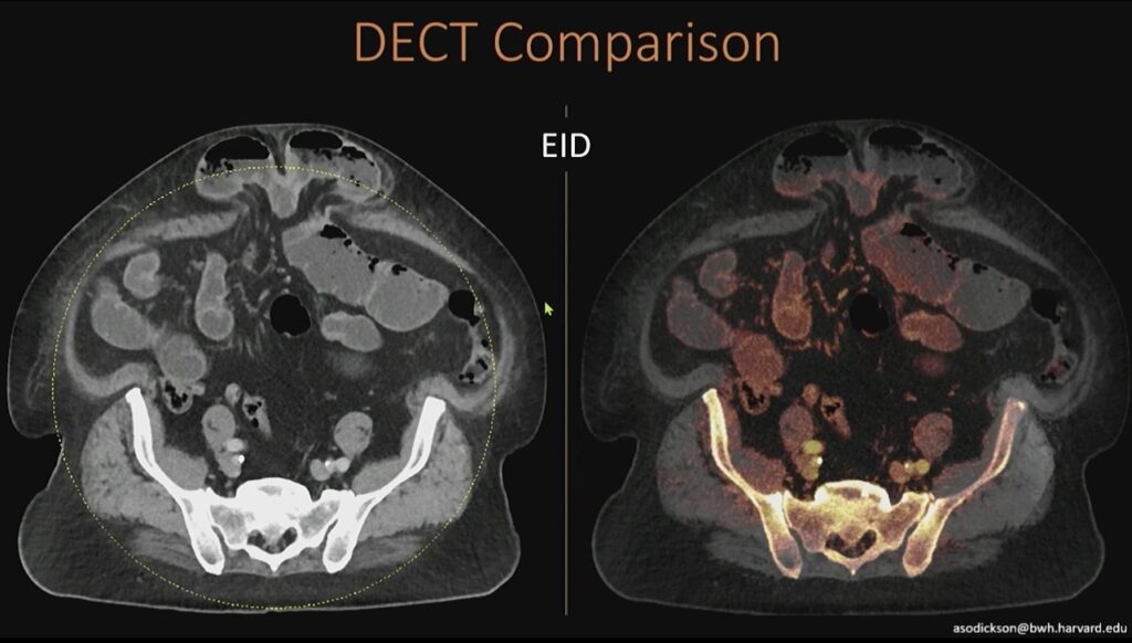
    Left: What you see here is that on our existing high-end dual-energy scanners, we have a slight limitation, which is that we don’t get dual-energy information outside this yellow-dotted circle. Right: While we have iodine content centrally, we don’t have anything out in the periphery.

    There Are Limits: On many high-end dual-energy scanners, iodine maps are only reliable within a central circular region. Outside that area, dual-energy information is lost. In large patients or peripheral anatomy, this can mean incomplete iodine characterization and diagnostic uncertainty.

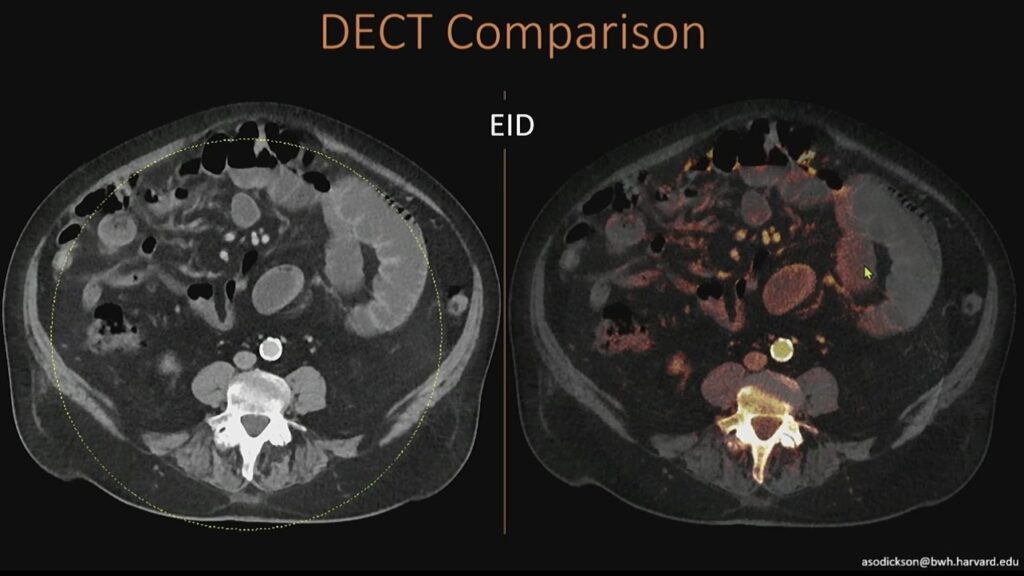
    Here’s another example of the same thing, where you can see that we’ve lost our information out at the edges.

    In practice, you may see clean iodine signal centrally, but nothing at the edges—simply because the system cannot acquire dual-energy data beyond that geometric constraint.

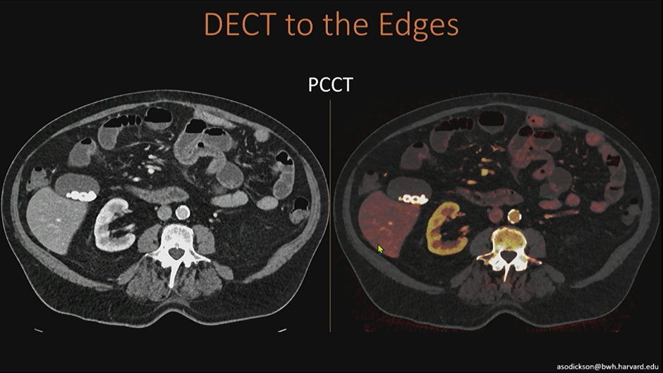
    PCCT Is Built Different: PCCT acquires spectral information directly at the detector. Because it does not rely on paired detector geometries or source-based separation, dual-energy information is available across the entire field of view, meaning:

    • Iodine maps extend fully to the periphery
    • Large patients no longer fall outside the dual-energy zone
    • Edge anatomy benefits from the same spectral data as the center

    In the ED, where patient size and positioning are unpredictable, this matters.

    On the photon-counting scanner, all of this dual-energy information content is acquired at the detector, so we don’t geometrically need the two CT detectors that are being used in our conventional scanner. Thus, we can get dual-energy information all the way out to the edges, even in large patients outside that field of view.

    Better-Behaving Noise: Another practical advantage of PCCT is cleaner iodine post-processing. DECT maps may show speckled signal in non-enhancing structures—algorithmic noise that can mimic low-level enhancement.

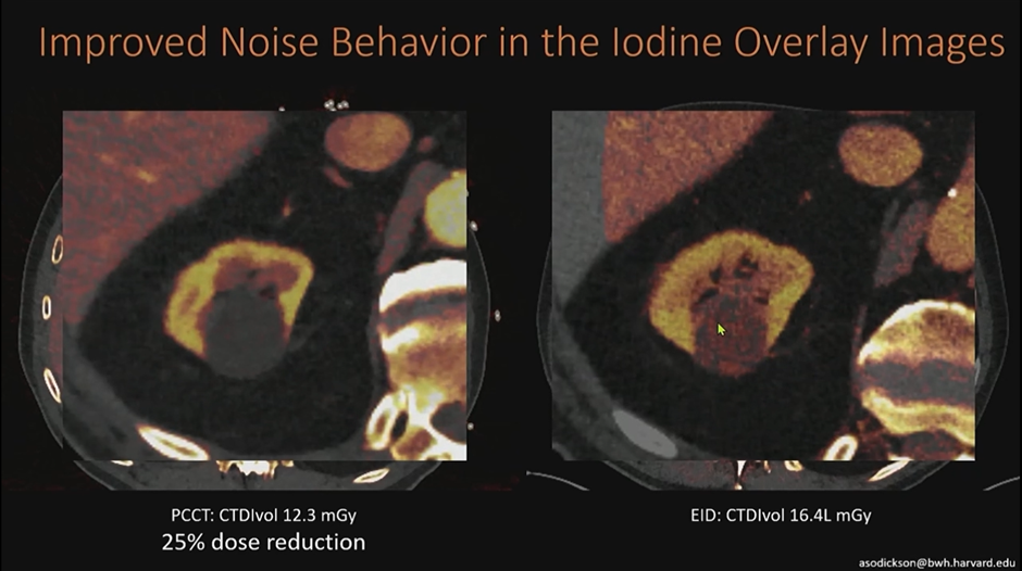
    In this example below of a renal cyst, conventional DECT shows scattered orange signal that is not true enhancement. On PCCT, that noise is substantially reduced, allowing confident classification of a benign, non-enhancing complex cyst.

    Looking at this renal cyst on a conventional scan, you see some noise in the iodine post-processing content (e.g., green arrow in orange speckle). There isn’t actually enhancement within this cyst; this is noise in the algorithm. On the photon-counting scanner, we’re able to knock down that noise to see much more cleanly that this is a non-enhancing, benign, complex cyst.

    Why This Matters in the ED: Emergency radiology rewards speed and certainty; PCCT improves both by:

    • Preserving iodine data across entire image
    • Reducing false-positive enhancement
    • Increasing confidence in lesion characterization
    • Decreasing need for follow-up imaging

    Bottom Line: Photon-counting CT removes the “edge problem” of conventional DECT. By delivering full-field spectral data with improved noise characteristics, PCCT strengthens iodine-based interpretation—exactly where ED imaging needs it most.

  • Volunteering: All for the Organization or Do Volunteers Derive Benefits?

    Volunteering: All for the Organization or Do Volunteers Derive Benefits?

    This president’s column will be a departure from my last two discussing serendipity in radiology and medicine. Instead, I would like to focus on a topic that I have thought about for a long time but never dug deeply into the surrounding research; that is, the benefits of volunteering to the volunteer. An organization like the American Roentgen Ray Society (ARRS), simply put, cannot survive without volunteers. ARRS has amazing staff, but like all medical societies, Roentgen Ray relies heavily on its physician and scientist volunteers to organize programs, moderate sessions and speak, govern and peer review abstracts and articles, etc. So, are there benefits of volunteering besides the tangible work that the society derives? Meaning, do volunteers derive benefits from volunteering? The answer is, yes!

    And I will divide these benefits into several categories, including academic currency, individual wellbeing and mental health, impact on physical health and mortality, and impact on workplaces and employers.

    Academic Currency

    For members of a medical society who are also members of an academic department, the opportunities afforded by a society to lead or be a member of committees, to organize or teach learning sessions, to review journal articles, etc. are all ways to prove your academic worth. When a physician or scientist is considered for promotion, they are asked to put together their CV and service, teaching, and research portfolios for review. Depending on the track for promotion (this varies from institution to institution) some form of service on a local (the institution at which you work), regional (state medical societies), national (ACR, ARRS, RSNA, or specialty society) or international level is important and, in some cases, may be required for promotion. Generally, the higher the level of promotion desired (full professor over associate professor over assistant professor over instructor), the more service is required at a regional and national level. Further, engagement with academic colleagues at meetings ensures an individual will be able to suggest names of people to support their quest for promotion with a letter of recommendation. In other words, it is difficult at most institutions to attain the rank of full professor without this volunteer work.

    Individual Wellbeing and Mental Health

    Controversy exists as to whether volunteering really improves individual subjective wellbeing. There is the question of whether happier people are more likely to volunteer, potentially skewing any survey data that cannot find a way to control for baseline wellbeing. Lawton et al. published a paper in 2021 specifically addressing this issue in a large population (household survey datasets totaling over 245,000 people) in Great Britain [1]. Their study found that controlling for baseline levels of reported wellbeing and happiness, there is still a statistically significant improvement in wellbeing equivalent to a salary increase of £911 per person per year; that is, a person who volunteers would have to experience a salary loss of £911 to bring them to the same average happiness level of a person who does not volunteer. They further found that frequency of volunteering matters; volunteering weekly or more is almost twice as beneficial as volunteering several times a year and that volunteering once a year or less does not improve wellbeing.

    Other studies have focused on various aspects of wellbeing. For example, a study of 3,351 adults conducted by UnitedHealth Group found that volunteering lowered stress levels in 78%; volunteers in this study were more likely than non-volunteers to report feelings of calm and peace, good energy levels, and increased sense of purpose [2]. Studies have also shown a positive association of volunteering with increased sense of purpose, optimism, and positive affect [3]. Volunteers also had higher average CASP scores (control, autonomy, self-realization, and pleasure) regardless of the gross domestic product of their country of residence [4]. Further, one study found that the effects of volunteering stop when individuals stop volunteering, supporting a causal relationship [5].

    Impact on Physical Health and Mortality

    The impact of volunteering on mental health is well-documented. What about physical health and by extension, mortality? Kim et al. published a study in 2020 examining data from 12,998 people over 50 living in the US; the dataset is considered diverse and nationally representative [3]. When adjusting for variables such as socioeconomic status, physical health, health behaviors, personality and other psychosocial factors, their study found that individuals who volunteered at least 100 hours per year (which is equivalent to just under 2 hours a week) compared to those who did not volunteer at all were 44% less likely to die, 17% less likely to have new physical limitations and higher self-rated health. Further, volunteering at this level was also associated with a 12% increased likelihood of frequent physical activity. The effects of volunteering on health were also documented in an analysis of 6 longitudinal panel surveys of Europeans including 267,212 people [6]. In this “mega-analysis,” volunteers had a 13% health advantage (health score 68 in volunteers versus 60 in non-volunteers) and the advantage positively correlated with age (no advantage in individuals less than 40, 26% in individuals in their 70s and 35% in individuals in their 80s). This study also found that the benefits of volunteering were greater in those who experienced poorer health at baseline. Finally, an unusual study of salivary cortisol levels [7] found that in a cohort of 340 middle aged and older adults, cortisol output decreased on days the individuals did volunteer work compared to days they did not meaning their levels of stress hormones were reduced. The authors hypothesize that this positive effect on cortisol may be one mechanism by which volunteering improved health.

    Impact on Workplaces and Employers

    Beyond the work that is done for an organization by volunteers, are there benefits to corporations and other employers? If people who volunteer outside the workplace are both mentally and physically healthier, an employer can expect lower healthcare costs; further, if workers are less stressed, they are “more present and engaged” elevating job performance [2]. Volunteering can also bring new skills to the workplace, including time management and people skills as well as job specific skills depending on the type of volunteering that is pursued. And, if volunteering with colleagues is encouraged, stronger workplace relationships can result in increased teamwork and goodwill in the workplace.

    Not only do our ARRS volunteers have a huge impact on the ARRS, but also the act of volunteering has a huge impact on our volunteers. Thus far, the ARRS has done little to recognize in a meaningful and purposeful manner all the ways in which our volunteers contribute to and enrich our society; we have many volunteers who have given their time and expertise for 20 years or more. In my next column I will introduce and discuss a new volunteer recognition program that will roll out at the 2026 ARRS annual meeting. I hope to see you all in Pittsburgh!


    References

    1. Lawton RN, Gramatki I, Watt W, Fujiwara D. Does volunteering make us happier, or are happier people more likely to volunteer? Addressing the problem of reverse causality when estimating the wellbeing effects of volunteering. Journal of Happiness Studies 2021;599-624
    2. UnitedHealth Group. Doing Good is Good for You: 2013 Health and Volunteering Study. 2013
    3. Kim ES, Whillans AV, Lee MT, Chen Y, VanderWeele TJ. Volunteering and subsequent health and well-being in older adults: an outcome-wide longitudinal approach. American Journal of Preventive Medicine 2020;59:176-186
    4. Morwski L, Okulicz-Kozaryn A, Strelecka M. Elderly volunteering in Europe: the relationship between volunteering and quality of life depends on volunteering rates. Voluntas 2022:33;256-268
    5. Matthews K Nazroo J. The impact of volunteering and its characteristics on well-being after state pension age: longitudinal evidence from the English longitudinal study of aging. Journal of Gerontology: Social Services 2021:76;632-641

  • Liver Fat Quantification: Billing, Workflow, and Real-World Use

    Liver Fat Quantification: Billing, Workflow, and Real-World Use

    Fatty liver has become so common that trainees often see steatosis more frequently than a normal liver. During the latest AJR Forum on Quantitative Ultrasound, David Fetzer, MD, asked Roentgen Fund recipient Theodore Pierce, MD, a practical question regarding liver fat assessment:

    Are practices billing for liver fat quantification, and is it performed alone or bundled with other exams?

    The Big Picture

    Liver fat quantification is increasingly embedded in routine abdominal imaging. But billing practices, reimbursement, and workflow integration vary widely across institutions. Dr. Pierce noted that while they do code and submit billing for fat quantification, reimbursement remains inconsistent at this stage.

    Key Takeaways

    Billing is performed, even though reimbursement is not yet reliable. Fat quantification is coded whenever performed. Reimbursement is infrequent but expected to improve as adoption grows and payers recognize the clinical value.

    Standalone liver fat quantification exists, but it is rarely used. Although offered as its own CPT-coded exam, most clinicians prefer to order it in combination with either a limited right upper quadrant ultrasound or with both RUQ ultrasound and elastography. The combination is determined by the referring clinician and the specific clinical question.

    Interpretation requires clinical and imaging context. As Dr. Fetzer emphasized, fat quantification values are interpreted alongside B-mode appearance and elastography stiffness to evaluate steatosis, fibrosis, and inflammation. The combined data provide a more accurate assessment than any single technique alone.

    Bottom Line

    Liver fat quantification is billable, clinically valuable, and most informative when paired with RUQ ultrasound and elastography. Utilization is increasing, reimbursement frameworks are evolving, and the technique is moving toward becoming a routine component of hepatic imaging.

  • Midgut Malrotation and Volvulus in Children: US or Upper GI?

    Midgut Malrotation and Volvulus in Children: US or Upper GI?

    In smaller hospitals, choosing between ultrasound or upper GI for suspected malrotation can feel high-stakes. During the latest AJR Live Webinar, Jonathan Dillman, MD, and HayThuy Nguyen, MD, tackled a key question:

    Should community hospitals perform both ultrasound and upper GI—or is one enough before transferring a child?

    The Big Picture

    Even though ultrasound for midgut volvulus performs incredibly well in published studies, your institutional comfort and consistency matter. Smaller centers may see fewer neonates, and that affects how confident technologists and radiologists feel with real-time sonographic anatomy.

    Dr. Nguyen’s advice: Start with both. Build confidence. Then taper.

    Key Takeaways

    Ultrasound + Upper GI Can Be Complementary, At First

    • Even at large centers, both exams are often paired early on.
    • Not because ultrasound underperforms, but because every institution needs their own “local data.”
    • Once your team demonstrates consistently accurate ultrasound performance, you can safely drop routine upper GI.

    See a Whirlpool Sign? Call Surgery!

    A positive whirlpool sign on ultrasound is highly specific. No need to wait for additional imaging; direct referral to pediatric surgery is appropriate.

    No Whirlpool, but Symptoms Persist → Consider Upper GI or Transfer

    Ultrasound may still be equivocal in some infants. Upper GI remains a helpful confirmatory test when the diagnosis is uncertain but suspicion stays high.

    Don’t Forget: CT or MRI Can Make the Diagnosis for Older Kids

    Dr. Dillman emphasized that adolescents or older children being scanned for unrelated reasons may still show:

    • SMV/SMA reversal
    • Swirling mesentery
    • Engorged mesenteric vessels
    • Collaterals

    Even a noncontrast CT for renal stone can incidentally reveal the vascular swirl. MRI—especially rapid MR protocols—can also depict abnormal vascular orientation.


    Bottom Line

    Start with both ultrasound and upper GI, if your institution needs to build confidence. Once your team demonstrates reliable ultrasound performance, ultrasound alone is often sufficient…and a positive whirlpool sign should trigger immediate surgical evaluation.

  • Scimitar Syndrome with Horseshoe Lung: Key Imaging Clues

    Scimitar Syndrome with Horseshoe Lung: Key Imaging Clues

    Scimitar syndrome represents a distinctive form of right lung partial anomalous pulmonary venous return. It is classically associated with right lung hypoplasia, abnormalities of the right pulmonary artery, and a characteristic anomalous vein draining into the systemic venous system. As Abbey J. Winant, MD, MFA, illustrates in “Pediatric Thoracic Vascular Disorders: Congenital to Acquired Pathology,” CTA plays a central role in defining venous anatomy and identifying associated anomalies.

    What Defines Scimitar Syndrome?

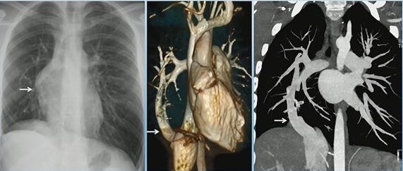
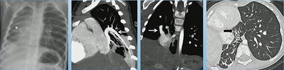
    Scimitar syndrome (RLL PAPVR) with R Lung hypoplasia

    The hallmark is a right lower lobe pulmonary vein draining anomalously—most often into the inferior vena cava, but occasionally into the inferior right atrium. This vein produces the classic “scimitar” appearance on chest radiography and cross-sectional imaging. Children often have concurrent right lung hypoplasia, which alters airway and vascular proportions.

    Scimitar Syndrome (“Scimitar Vein”)

    Recognizing Associated Findings

    In addition to partial anomalous pulmonary venous return, scimitar syndrome often presents with:

    • Hypoplastic right lung
    • Hypoplastic right pulmonary artery
    • Systemic arterial supply to portions of the right lung
    • Bronchial abnormalities, including bronchiectasis

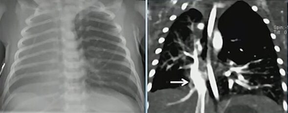
    Scimitar Syndrome with Horseshoe Lung: Hypoplastic right lung, PAPVR to RA, horseshoe lung, R lung bronchiectasis

    One of the most notable associations is horseshoe lung, seen in approximately 80% of cases. Horseshoe lung consists of a parenchymal isthmus connecting both lungs across the midline, usually posterior to the heart. When present, it further reinforces the diagnosis and alerts the radiologist to search for additional congenital anomalies.

    Additional Congenital Abnormalities to Consider

    Although not seen in every case, associated developmental abnormalities may include:

    • Extralobar sequestration
    • Vertebral anomalies
    • Diaphragmatic defects
    • Cardiac malformations

    Their presence can significantly influence management, operative planning, and follow-up.

    Why Does CTA Matter?

    CTA provides the most comprehensive view of the venous drainage pattern, systemic arterial contributions, and bronchial architecture. It allows precise localization of anomalous veins and helps differentiate scimitar syndrome from other types of partial anomalous pulmonary venous return.

    Bottom Line

    Scimitar syndrome is more than an anomalous pulmonary vein. Its constellation of findings—right lung hypoplasia, anomalous venous return, and frequent association with horseshoe lung—requires careful, structured evaluation. CTA remains the best tool to clarify anatomy and guide clinical management.

  • Open-Source AI for Radiology Reporting: Barriers and Practical Workarounds

    Open-Source AI for Radiology Reporting: Barriers and Practical Workarounds

    Large language models (LLMs) are reshaping radiology, but their integration into the reading room is far from straightforward. 2026 ARRS Annual Meeting Categorical Course Director Yee Seng Ng, MD, outlines the most significant barriers to adoption and why the solutions are more complicated than they appear.

    Security Slows Adoption

    Most widely available LLMs live on proprietary cloud platforms. Sending protected health information (PHI) outside a hospital network creates immediate compliance issues, and current guidelines from major organizations explicitly prohibit using public LLMs for protected patient information. Even if models claim not to store or reuse data, rads cannot verify how patient information is handled, refined, or monetized.

    Private Solutions, But Not for All

    Institutions can build private LLM instances behind their firewall, but this requires substantial infrastructure, IT support, and vendor partnerships—resources generally limited to large academic centers. Local installations using open-source models (via interfaces such as webUI) avoid cloud exposure but introduce new challenges: maintenance, computing requirements, and accessibility across the department.

    Where Is NLP Already Helping?

    Even though LLMs aren’t widely used at the point of care, rads are already benefitting from improved natural language processing (NLP) embedded in commercial reporting tools, including

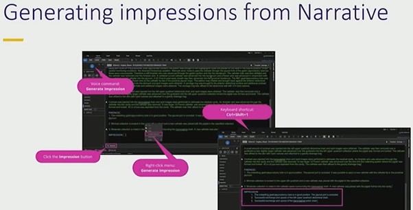
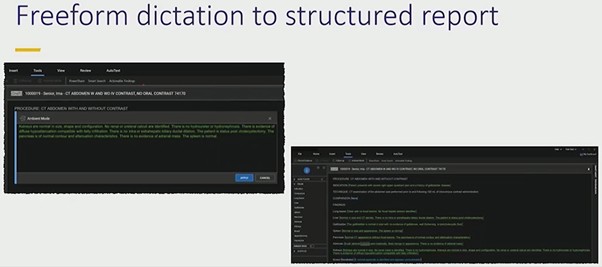
    • Automated impression generation from narrative text
    • Converting freeform dictation into structured reports
    • Organizing sentences under correct headings

    These features accelerate reporting and reduce cognitive load without exposing PHI externally.

    Error Prevention Still Matters

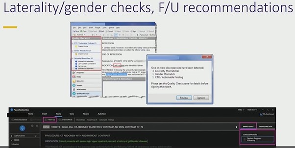
    Simple NLP tools remain some of the most valuable. PowerScribe’s laterality and gender checks prevent avoidable mistakes that can undermine confidence in a report. Tools that flag mismatched anatomy—such as referencing a prostate in a female patient—provide immediate, low-friction safety nets that rads consistently appreciate.


    Bottom Line

    Security and workflow realities remain the biggest obstacles to adopting LLMs for radiology reporting. Until private, institution-controlled LLMs become practical and widely available, rads will continue to rely on integrated NLP tools that improve.

  • Orbital Trauma Reporting: Why Markowitz and Manson Matter

    Orbital Trauma Reporting: Why Markowitz and Manson Matter

    When evaluating orbital trauma, one detail rads should address is the Markowitz and Manson (M&M) classification. As Blair A. Winegar, MD, explains, this system focuses on the degree of comminution in the region of the lacrimal fossa and helps predict whether the medial canthal tendon is likely to be injured.

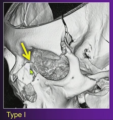
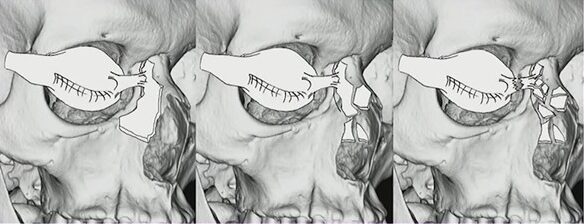
    Left: Type I, Intact medial canthal tendon connected to single large fracture fragment; Center: Type II, Intact medial canthal tendon connected to single comminuted fracture fragment; Right: Type III, Disrupted medial canthal tendon with severe comminution about the lacrimal fossa

    What M&M Describes

    The key question is whether the lacrimal fossa remains intact or is significantly fragmented.

    • Intact lacrimal fossa: Low likelihood of medial canthal tendon injury.
    • Heavy comminution in the lacrimal fossa: Higher suspicion for medial canthal tendon disruption, which may require surgical repair.

    Why Does It Matter?

    The medial canthal tendon anchors at the anterior lacrimal crest. When that region is fractured extensively, the surgical team needs to prepare for possible tendon repair. Including this observation in your rad report sets appropriate expectations and guides planning for reconstruction.

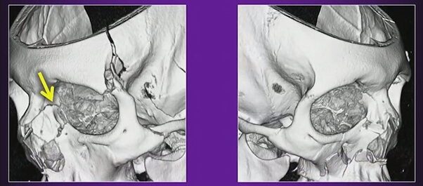
    Left: Type I; Right: Type II

    Practical Approach

    On CT, evaluate the anterior lacrimal crest and adjacent lacrimal sac fossa.

    Describe whether this region is intact, minimally displaced, or extensively comminuted. Explicitly link substantial comminution to the potential for medial canthal tendon involvement.


    Bottom Line

    In orbital trauma, reporting the Markowitz and Manson classification provides actionable information. Identifying comminution in the lacrimal fossa helps surgeons anticipate medial canthal tendon repair and improves communication between rads and the operative team.

  • Keeping Remote Radiologists Connected

    Keeping Remote Radiologists Connected

    The promise of remote radiology is reading from anywhere. The reality? It can feel like you’re reading from an island. In a talk at the ARRS 2025 Wellness Symposium, William Moore, MD detailed NYU’s seven-year mission to end isolation for its 60-person remote radiology team.

    Why it matters: Disconnected radiologists are far more likely to leave their jobs, which can lead to staffing issues and a loss of institutional knowledge.

    Cracking the Engagement Code

    Every journey has moments of discovery, NYU’s came when they figured out what truly connected people.

    • They found the magic formula for meetings. Useful, can’t miss education sessions, like interesting case conferences or non-punitive peer learning sessions provided opportunities for meaningful connection.
    • Go big or stay home. Remote staff would skip out on virtual mixers, but made time to attend department parties and CME conferences that built camaraderie.
    • Fast, flawless technology. To make remote work successful, you must invest in hardware and software that makes the remote experience as seamless as being on-site.

    Battles Still Being Waged

    Dr. Moore highlighted several ongoing issues:

    • Solutions can create new problems. A move to split teams into remote and in-person divisions solved one conflict but created organizational silos.
    • Old tensions linger. Conflicts over goals and roles are still a challenge, showing that policies alone don’t solve turf wars.

    The takeaway: The answer to remote isolation isn’t one static fix; it’s a dynamic work culture. Teams thrive by making sure no one is left behind.

  • The Koala in the Reading Room

    The Koala in the Reading Room

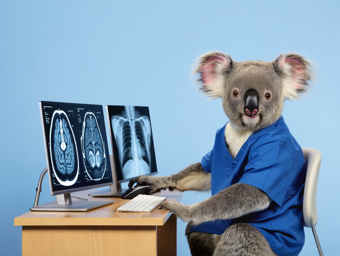
    Why do we need a complex algorithm to tell us an image’s texture? In a recent R3 Author Interview, Hyun Ko, MD, lead author of an R3 article on radiomics, explained that our eyes can be misleading.

    Her perfect example: a koala. Most people imagine a koala as a cute, fluffy animal. But its actual texture is rugged like a doormat.

    Why it matters: This is precisely what radiomics is intended to address. It’s designed to identify when the look of a lesion doesn’t match its underlying texture.

    • We see the forest, but radiomics can analyze the individual trees.
    • Human eyes miss the detailed interpixel relationships that are beyond the limits of perception.
    • This hidden texture could be the key to “characterizing lesions, predicting behavior, and detecting mutations” in ways simple size or attenuation metrics can’t.

    The bottom line: The promise of radiomics is seeing what the human eye can’t. To make this promise a reality, strategic shifts are needed to produce meaningful evidence to show it’s ready for clinical adoption.

  • Riding the Wave: How Data-Driven Scheduling Can Transform Your Radiology Practice

    Riding the Wave: How Data-Driven Scheduling Can Transform Your Radiology Practice

    At the ARRS 2025 Wellness Symposium, William Moore, MD, delivered a compelling presentation on a strategy that is reshaping radiology workflows for the better: wave scheduling. While the traditional definition involves scheduling multiple patients at the top of the hour, Dr. Moore explained how he has adapted this concept to create a more balanced, efficient, and predictable environment for radiologists. The core idea? To move away from a reactive “feast or famine” workday and towards a steady, manageable flow of cases.

    What is Wave Scheduling in Radiology?

    In the context of a radiology department, wave scheduling isn’t about grouping patients, but about building systems that create a “wave” of studies ready for interpretation. This ensures that when radiologists begin their day, a backlog of cases is already waiting for them.

    One of the most significant pain points this system addresses is the morning lull. “When [physicians] show up in the morning and there’s nothing on the list, you want to hear some angry people,” Dr. Moore noted. To combat this, his outpatient centers continue scanning late into the evening, well after the radiologists have finished reading for the day. This ensures a robust worklist is available first thing in the morning, allowing physicians to be productive from the moment they arrive.

    A similar logic is applied to the inpatient and Emergency Department (ED) settings. Overnight readers are instructed to leave non-critical ICU cases, such as follow-ups for line placement, for the morning team. This provides an immediate queue of work, while urgent reads are, of course, handled immediately overnight.

    Letting Data Drive the Schedule

    The successful implementation of wave scheduling is impossible without one critical component: data. Dr. Moore stressed the need for analytics to understand workflow patterns and make informed decisions. By analyzing historical data, his department uncovered predictable peaks and troughs in case volume, both throughout the week and across the year.

    Weekly Trends: The data revealed a clear pattern: Mondays are consistently the busiest day of the week, with case volume tapering off towards Friday.

    • The Problem: An evenly staffed week would leave radiologists overwhelmed on Monday and underutilized on Friday.
    • The Solution: “We have anywhere between one to two extra people on a Monday,” Dr. Moore explained. As the week progresses, more radiologists are allocated academic or administrative time. This matches staffing levels to the actual workload, balancing the week for the entire team.

    Seasonal Trends: Analysis of yearly data showed predictable lulls and surges. For instance, August is consistently a slow month, while volume explodes in September and October before dipping again in December.

    • The Problem: Strict vacation policies can lead to burnout and retention issues, especially when staff want time off during busy periods.
    • The Solution: By identifying August as a reliably slow period, the department can confidently approve vacation requests during this time. “We have the opportunity using our data to schedule our physicians in a meaningful way so that they can get time off with their family and try to keep them,” said Dr. Moore. This data-backed flexibility is a powerful tool for improving physician wellness.

    The Four-Part Plan for Success

    Dr. Moore concluded by boiling the process down to a clear, iterative cycle:

    1. Make a Plan: Use data to design a workflow and staffing model that anticipates patient volume.
    2. Execute the Plan: Implement the schedule and the technology to support it.
    3. Tweak It (Endlessly): This is not a “set it and forget it” solution. Continuously adjust distribution rules, staffing levels, and schedules based on performance and new data.
    4. Get Feedback: The most crucial step. “You must get feedback from your radiologists,” Dr. Moore urged. “If you don’t, you can’t possibly understand what you’re going to do.”

    By embracing wave scheduling, departments can create a system that smooths out the chaotic peaks and valleys of the workday. It’s a data-driven, flexible approach that not only enhances efficiency but also directly contributes to radiologist wellness by creating a more predictable and manageable work environment.

  • Strategies To Retain Your Workforce

    Strategies To Retain Your Workforce

    With more than 1,700 radiology job openings nationwide and many practices actively hiring, retaining rads has become a growing concern. During the ARRS Wellness Summit, Dr. Jay Parikh of University of Texas MD Anderson Cancer Center emphasized that this challenge predates the COVID-19 pandemic. Turnover rates were already rising due to increasing imaging volumes, workflow changes, and mounting operational pressures.

    Burnout Drives Turnover: Burnout, recognized by the World Health Organization as a consequence of chronic, poorly managed workplace stress, remains a central factor. Studies estimate burnout prevalence in radiology between 37% and 80%, depending on subspecialty. Dr. Parikh highlighted research showing that rads experiencing burnout are twice as likely to consider leaving their jobs compared with those who are not. This direct link makes burnout a retention issue, not just a wellness concern.

    Fulfillment as Counterbalance: Dr. Parikh dubbed professional fulfillment as the most effective antidote to burnout. Fulfillment is shaped by cultural wellness, efficient practice environments, and personal resilience. He challenged the notion that burnout stems from insufficient physician resilience, noting evidence that physicians often demonstrate higher resilience than the general population. Instead, system-level issues—workload intensity, organizational culture, and operational inefficiencies—play a larger role.

    Operation and Leadership Roles: Operational decisions strongly influence rad wellbeing. While managing workload and improving efficiency are essential, Dr. Parikh cautioned against “over-efficiency,” which removes the slack time necessary for reflection, creativity, and thoughtful interpretation. Flexible scheduling, adequate staffing, and financial stability are critical, but so is recognizing radiologists as human capital rather than interchangeable labor.

    Leadership quality also has a measurable impact. Leaders who model self-care, promote psychological safety, and support professional growth can reduce burnout and improve job satisfaction across teams.

    Training Works, Really: Dr. Parikh cited data showing that structured leadership training—focused on emotional intelligence, resilience, and burnout awareness—improves teamwork and reduces work–life conflict. These programs benefit not only individual leaders but also the departments and cultures they shape.

    Bottom Line: Rad retention depends less on individual toughness and more on culture, leadership, and system design. Addressing burnout requires intentional investment in professional fulfillment, operational balance, and leadership development. Practices that prioritize these areas are more likely to build stable, engaged radiology teams. And keep them.

  • Fall: A Time for Renewal

    Fall: A Time for Renewal

    Fall is my favorite season, a time of change that invites us to slow down, reconnect, and nurture our wellbeing. As the air turns crisp and the leaves shift to rich hues, the season offers a unique opportunity to embrace balance and self-care.

    The cooler temperatures make outdoor activities more inviting. Whether it’s a brisk morning walk or a weekend hike through the changing foliage, spending time outdoors in fall can improve mood and reduce stress. Nature’s beauty in this season also inspires mindfulness—being present in the moment, whether it’s during a walk or while enjoying a hot cup of tea.

    Fall is also a season of nourishment. With harvests of pumpkins, apples, and squash, it’s a perfect time to incorporate warm, hearty meals that fuel both body and soul. Seasonal produce supports immunity and helps prepare us for the cooler months ahead. (See here for my favorite butternut squash soup recipe!)

    As the days grow shorter, it’s natural to embrace rest. Fall is ideal for creating or refining evening routines that promote relaxation, such as reading, meditation, or enjoying a calming tea before bed. Prioritizing sleep and rest during this season help to restore energy and prepares us for winter.

    Finally, fall encourages us to let go, just as the trees shed their leaves. It’s a time for reflection, to release stress or habits that no longer serve us, and to set new intentions as we approach the year’s end.

    By aligning with the rhythm of the season, we can nurture our wellbeing and find peace in the transition that fall brings.

    Lily M. Belfi, MD, FACR

    Professor of Clinical Radiology

    Director of Medical Student Education

    Division of Emergency/ Musculoskeletal Radiology

    Weill Cornell Medicine

    In “Words of Wellness” on www.radfyi.org/, members of the ARRS Wellness Subcommittee share what “wellness” and “wellbeing” mean in their own clinical practices, research focuses, and everyday lives.

    Dr. Belfi’s ARRS “Sound of Wellness” Playlist Selection:

    Carolina In My Mind

    You may also be interested in
    https://www.radfyi.org/2023/09/20/words-sounds-of-wellness-dr-sherry-wang/