Category: Latest Posts

  • Pancreatic Neuroendocrine Tumor Imaging

    Pancreatic Neuroendocrine Tumor Imaging

    Summary: Effective evaluation requires reliance on structured imaging that’s razor-focused on cyst size, count, location, and calcification.

    Nothing But PNET: Diversity is on full display with cystic pancreatic neuroendocrine tumors (PNETs), requiring a structured approach to calcification and enhancement, as well as clinical presentations. Detailing these distinctions during our online course Radiology Case Review–Abdominal Imaging, Mark Sugi, MD, noted that, typically, functioning tumors present early vis à vis clinical symptoms. Meanwhile, non-functioning tumors show up later and larger and undergo necrotic degeneration.

    • Intratumoral: Punctate, mural, or non-mural calcifications should serve as signs for PNETs.
    • Obstructionist: Even if a lesion obstructs the pancreatic duct, the presence of intratumoral calcification should arouse suspicion for PNET.
    • Pattern Profile: PNETs characteristically demonstrate post-contrast enhancement, frequently exhibiting eccentric or rim-enhancing patterns.

    Beware of Imitations: A circumscribed rim-enhancing lesion with subtle punctate calcification can closely mimic duodenal diverticulitis. Alternatively, ductal adenocarcinoma developing in the setting of pancreatitis with calcifications presents a similar kind of overlap.

    • Insulinomas? In patients with multiple endocrine neoplasia type 1, insulinomas display as rim-enhancing, low-attenuation masses in the pancreatic body or tail with high uptake on ⁶⁸Ga-DOTATATE PET.

  • Esophageal Motility and Gastric Pathology

    Esophageal Motility and Gastric Pathology

    Summary: Take that extra second to inspect the entire stomach and gastric pouch on CT and MRI, leverage DWI sequences, and correlate manometry with fluoroscopy to detangle complex dysmotility.

    Hard to Digest: Upper GI motility disorders and subtle gastric lesions don’t fit into neat tracts. Misidentifying esophageal patterns, or overlooking the excluded stomach after bariatric surgery, too often cause diagnostic delays, persistent symptoms, as well as missed malignancies.

    As Dr. Kristina Flicek directed during ARRS26, look beyond the narrows to differentiate primary disorders from secondary postoperative changes. And keep an eye out for the nuances of gastric pathology on cross-sectional CT and MRI:

    • Achalasia: Characterized by aperistalsis of the distal two-thirds of the esophagus and failure of the lower esophageal sphincter (LES) to relax, you get that classic bird beak sign.
      • Giving adequate contrast during a timed esophagram uses hydrostatic pressure to force open the LES, helping rule out a fixed obstruction or pseudoachalasia from GE junction adenocarcinoma.
    • EGJOO: Impaired transit across the EGJ with elevated LES relaxation pressure on manometry, albeit normal esophageal body peristalsis on fluoro? That’s esophagogastric junction outflow obstruction.
      • Stems from structural—strictures, hiatal hernias, epiphrenic diverticula, fundoplication wraps—or non-structural causes (e.g., chronic opioid use, which disrupts nitric oxide releasing neurons, leading to unopposed excitatory cholinergic input and spastic LES contraction).
    • POSED: A secondary achalasia developing after Roux-en-Y gastric bypass or sleeve gastrectomy, post-obesity surgery esophageal dysfunction is driven by elevated pressure or decreased compliance of the proximal pouch.
      • Fluoro demonstrates disruption of the primary stripping wave, proximal escape, and distal motor incoordination (manometry frequently confirms type II achalasia pattern).

    More Roux-en-Y Complications:

    • GJ Fistula: An abnormal tract communicating between the gastric pouch and the excluded remnant stomach, usually, patients present with weight regain or reduced satiety.
    • Marginal Ulcers: Outpouchings or small air foci right at the gastrojejunal anastomosis caused by gastric acid irritating vulnerable jejunal mucosa that’s readily identifiable on fluoro and targeted cross-sectional CT.

    Exclusions Apply! Pathology in the bypassed remnant stomach (including large bleeding masses or mucosal lesions) can be completely missed on standard upper endoscopy, if cross-sectional imaging is not performed.

    • Persistent antral thickening should not be dismissed as benign simply because it appears static over consecutive months; signet cell adenocarcinoma can masquerade as stable wall thickening.
      • DWI is crucial for highlighting wall restriction, effectively distinguishing true gastric adenocarcinoma from poor stomach distension.
  • Wrist Arthrography Contrast Distribution

    Wrist Arthrography Contrast Distribution

    Summary: Focusing on contrast compartmentalization helps pinpoint full-thickness ligamentous and TFCC tears, pivoting away from small-joint pitfalls.

    Small Joint, Big Problem: It’s a two-part question in single-joint wrist arthrography: where is contrast supposed to be, versus where is it not? As Dr. Joseph A. Delic reminded us during ARRS26, fluoroscopy doesn’t merely confirm needle placement; it directly aids decision-making for subsequent MRI sequences.

    Strict Boundaries: In a normal single-joint radiocarpal injection, contrast remains entirely confined within the radiocarpal joint.

    • Normality in sequence shows zero contrast extension through the scapholunate or lunotriquetral intervals, and no contrast leaking into the distal radioulnar joint (DRUJ).

    Interval Training: Linear contrast extending through the scapholunate interval indicates a full-thickness ligament tear. When tears involve the dorsal band, volar band, and membranous segment, contrast insinuates throughout the proximal and distal carpal rows.

    • Contrast extending into the DRUJ during injection would suggest a full-thickness TFCC central disc tear.
  • CLEAR Framework in Breast Imaging

    CLEAR Framework in Breast Imaging

    Summary: Rushing to definitives on imperfect studies leads to cognitive errors, overcalling, and inappropriate assignment (e.g., BI-RADS 3 should never be assigned at screening; baseline findings with incomplete evaluation require BI-RADS 0 for additional workup).

    Mastering Mystery: An unknown diagnosis does not equal an unknown process; you needn’t know the precise pathology to determine your safest next step. Introduced by ARRS Distinguished Educator Dr. Tanya Moseley during our Mystery to Mastery longitudinal course, here’s how her C-L-E-A-R acrostic’s stepwise methodology transforms clinical uncertainty into structured reasoning:

    • C—Context: Anchor in Facts
      • Gather patient age, symptoms, indication, modality, technique, priors, etc.
    • L—Localize: Boundaries, First
      • Pinpoint an abnormality’s region, compartment, and epicenter, before attempting to characterize it.
    • E—Examine: Objective Descriptions
      • Systematically evaluate morphology, density, echogenicity, or enhancement, then document key negatives.
    • A—Alternatives: Defensible DDX
      • Avoiding the too obscure, rank 2–3 reasonable possibilities (summation artifact vs. focal asymmetry vs. obscured mass).
    • R—Resolve: Execute Safely
      • Determine whether the patient needs FFDM, DBT, targeted US, prior comparison, or even reassuring observation.

    Case in Point: Single-View Asymmetry

    • Scenario: In Moseley’s case, a baseline screening mammogram in an asymptomatic patient revealed a focal density in the middle posterior left breast, visible on only one view.
    • Reasoning: Persistence, morphology, and second-view correlate couldn’t be confirmed.
    • Action: Incomplete exam assigned BI-RADS 0 and recommended mammo, DBT, and US—rather than guessing or misapplying BI-RADS 3.
  • Azygos Valve Contrast Pooling

    Azygos Valve Contrast Pooling

    Summary: Before diagnosing a calcified right paratracheal lymph node or metastasis, verify whether that heavy brightness is simply transient contrast pooling inside an azygos arch venous valve.

    Trapper John, MD: Got high attenuation in the right paratracheal space on contrast-enhanced chest CT? Careful. As Dr. Brent P. Little explained during ARRS26, contrast material often becomes trapped behind a normal venous valve in the azygos arch—creating a bright focal density that mimics pathology:

    • Anatomy of Venous: A normal valve with two leaflets sits in the region of the azygos arch.
    • Delayed Clearance: Whereas contrast washes out from the rest of the thoracic vasculature, stagnant contrast trapped behind the valve leaflets can remain for several minutes.
    • It’s Not Unusual: Trapped contrast presents in the right paratracheal space with a distinct gestalt (sometimes demonstrating a subtle fluid/contrast level).
    • The Pit: In patients with calcifying malignancies, this high-density contrast retention can be wrongly suspected as a calcified metastatic node.

  • Periprosthetic Soft-Tissue Masses

    Periprosthetic Soft-Tissue Masses

    Summary: Pairing artifact-reduced imaging with consistent, rad-forward language helps hone reportage that can effectively guide surgical decision-making.

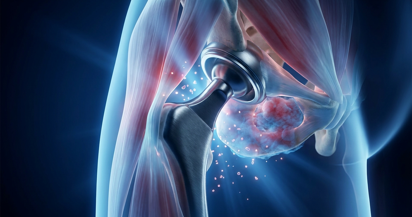
    Particle Problem: All functional joint arthroplasties create wear particles that can trigger a host immune response, leading to inflammatory tissue damage around hardware. Because terminology describing these soft-tissue abnormalities is used inconsistently across literature and practice alike, as Dr. Jeremiah R. Long reminded us during ARRS26, clarity is critical:

    • Quality Control: Implement metal artifact reduction techniques across both CT and MRI to better detect and characterize lesions.
    • A Priori Advantage: Track lesion size and progression over time to assist surgeons in determining whether/when to revise.
    • Clinical Context: Query the EMR for history, labs, operative details, etc.
    • Imaging-Centric Language: Utilize “umbrella terms” appropriate for the specialty—pseudotumor and adverse local tissue reaction (ALTR), specifically.

    Jettison the Jargon: Dr. Long suggested that we refrain from using gross, intraoperative, or pathological terminology, à la metallosis or trunnionosis.

  • Neonatal Pneumatosis: Why Specificity Beats Sensitivity

    Neonatal Pneumatosis: Why Specificity Beats Sensitivity

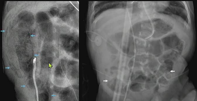
    RadFYI: Portal venous gas is one of the most critical catastrophes that can develop as a result of pneumatosis, so do call on the classics here: branching lucencies within the liver.

    Not Nothing: Reading neonatal abdominal radiographs straddles that line between the overly sensitive “call me, maybe” and the specific and definitive. As Dr. Andrew T. Trout reminded us during the ARRS Online Course Radiology Case Review – Pediatric Imaging, calling pneumatosis is not a benign diagnostic event.

    • Usually, a positive read prompts clinicians to rest the baby’s gut, make them nothing by mouth, and initiate total parenteral nutrition.
    • Since such interventions carry their own risks, prioritize specificity over hypersensitivity.

    Clear-cut cases present with obvious intramural collections of linear or curvilinear gas across multiple loops…or the presence of portal venous gas!

    • Trout’s Tip: Do not call pneumatosis unless you see clear, indisputable intramural lucencies (either linear or curvilinear) in the context of other concerning clinical findings.

    Grey Zone: When faced with subtle, bubbly lucencies in the hemi-abdomen, obtaining a delayed, follow-up radiograph is a highly reasonable and helpful strategy. Over time, true pneumatosis will progress to show more convincing, clearly intramural curvilinear lucencies on a delayed radiograph.

  • Nodeworthy: It’s N2a—and b—Now for Lung Cancer Staging

    Nodeworthy: It’s N2a—and b—Now for Lung Cancer Staging

    Summary: Any contralateral mediastinal nodal metastasis or any supraclavicular/scalene adenopathy remains classified as N3, regardless of the primary tumor location.

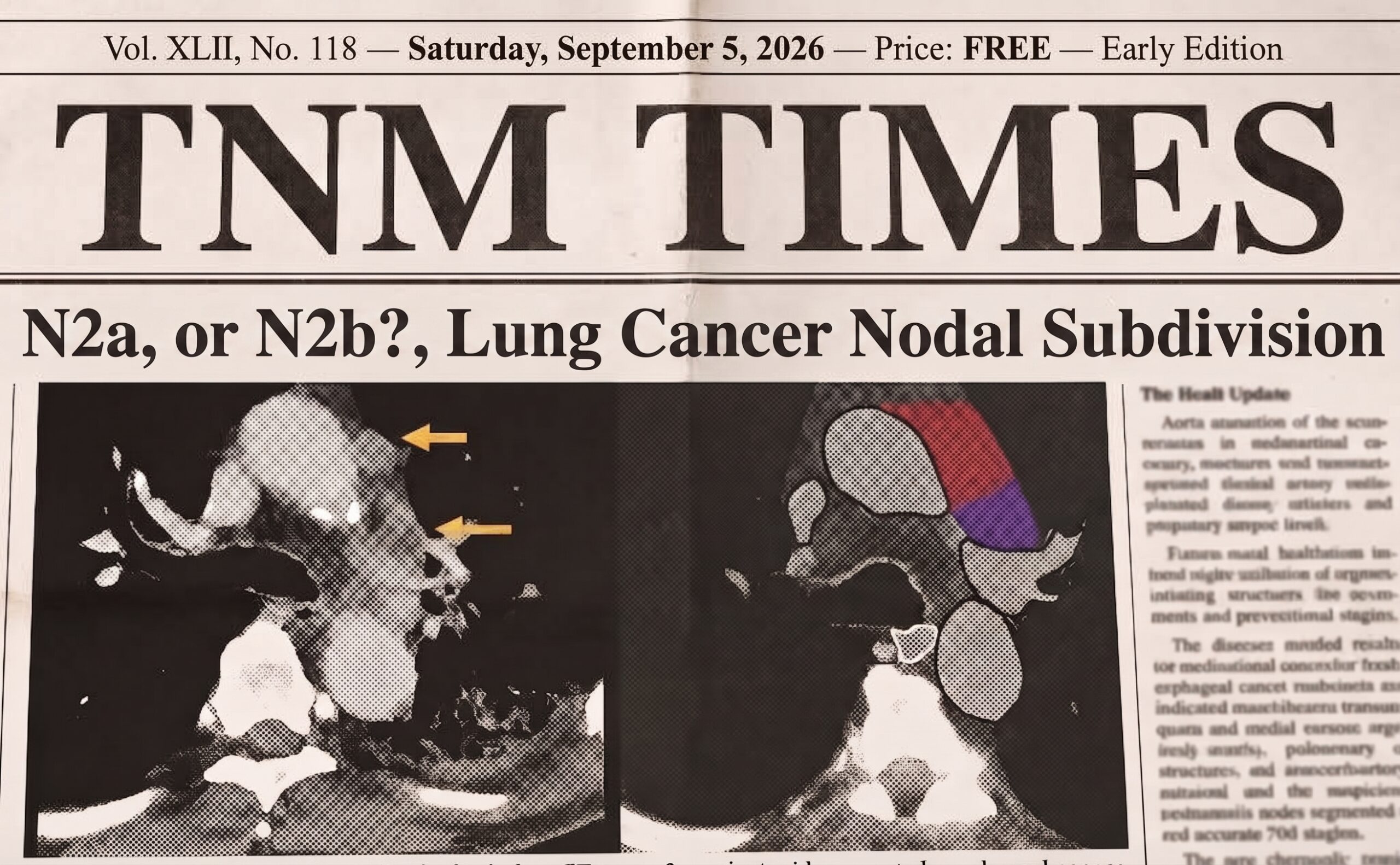
    Station to Station: One of the most notable updates to the 9th edition of the TNM staging system for lung cancer is the formal subdivision of N2 disease: N2a *and* N2b. According to Dr. Girish Shroff’s update in our Cardiothoracic Imaging Case Review Online Course, the priority of now is the number of involved nodal stations, rather than absolute count of metastatic nodes:

    • N0: No regional lymph node involvement.
    • N1: Metastasis in ipsilateral bronchopulmonary or hilar nodes.
    • N2: Metastasis in ipsilateral mediastinal and/or subcarinal nodes, now subdivided by station density:
      • N2a—Involvement of a single N2 station.
      • N2b—Involvement of multiple N2 stations.
    • N3: Metastasis in contralateral mediastinal or contralateral hilar nodes, or any supraclavicular/scalene nodes.

    Stations > Counts: Even if a patient presents with multiple metastatic nodes, they are staged as N2a if all involved nodes reside within a single station (e.g., lower right paratracheal region).

    • Metastatic involvement of both station 5 and station 6 automatically elevates to N2b.
  • Hemorrhagic Cyst…or Endometrioma?

    Hemorrhagic Cyst…or Endometrioma?

    Summary: If it’s the patient’s first presentation, and your MRI findings remain ambiguous, the safest bet here is follow-up US in 2-3 months to confirm whether that suspect cyst has resolved.

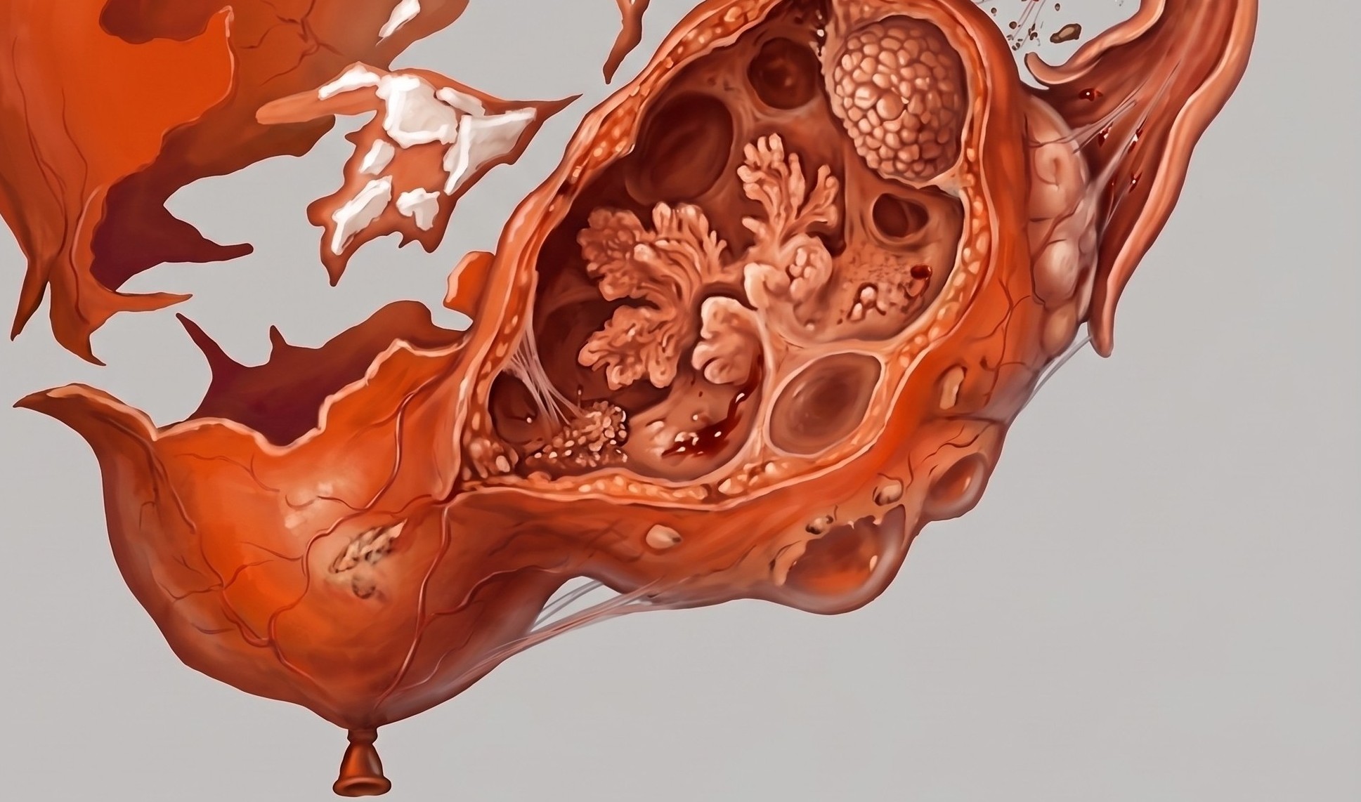
    On GYN MRI, both of these adnexal pathologies present with pelvic pain and may exhibit T1 hyperintensity. Whereas hemorrhagic cysts are generally self-limiting and resolve, as Dr. Milana Flusberg distinguished during ARRS26, the markers of chronic endometriosis can lead to scarring and pelvic tethering, as well as rarer complications like infection.

    Three T2 Tells: Look out for these specific T2-weighted signs to help you differentiate:

    • Shading: This classic feature—where a T1-hyperintense lesion appears hypointense on T2-weighted sequences—is highly characteristic of endometriomas. However, it is not 100% definitive, as it occurs in just under 20% of hemorrhagic cysts, too.
    • Dark Spot: Referring to focal, highly T2-hypointense hemosiderin spots inside an otherwise mildly hypointense or intermediate lesion, this one is highly specific for endometrioma, albeit lacking high sensitivity.
    • Hypointense Rim: A peripheral hemosiderin ring caused by repetitive, cyclical bleeding, it’s more common in endometriomas, although it can sometimes be seen in hemorrhagic cysts.

    Beyond the Cyst: Quite often, Flusberg said, the most helpful clue lies outside the lesion, itself:

    • Deep Pelvic Endometriosis? Look for T2-hypointense pelvic fibrosis tethering the mass to adjacent structures, such as the rectal wall.
    • Multiple Lesions? Finding many complex adnexal lesions strongly points toward endometriosis, rather than an isolated hemorrhagic cyst.

    Is There an Echo in Here? If you’ve got prior or concurrent US, the classical hemorrhagic cyst demonstrates “lace-like” reticular internal echoes or angular, concave retractile clots. Meanwhile, endometriomas typically present with homogeneous, low-level internal echoes.

  • Ghost Rate Gambit: How Insurers Skew Rad Payments

    Ghost Rate Gambit: How Insurers Skew Rad Payments

    Summary: Just last week, a federal court ruled that the government’s methodology for crunching QPA is, at least partly, unlawful. However, a majority of the 5th Circuit’s 17 active judges agreed that agencies can allow insurers to use existing QPAs until new ones can be calculated, meaning that NSA’s dispute resolution process can continue unabated.

    Reimbursement Check, Mate? A loophole in the implementation of the No Surprises Act (NSA) allows health insurers to use inactive, unnegotiated “ghost rates” to artificially depress reimbursement benchmarks for out-of-network care.

    In the second episode of the AJR Podcast Series Difficult Conversations, Richard E. Heller III, MD, MBA, explained to co-hosts Sherry S. Wang, MBBS, and Surbhi Raichandani, MD, how this loophole unfurls:

    • Boilerplate: Insurers issue massive, standardized contracts containing thousands of billing codes to every practice they contract with.
    • Ignorance: Rads ignore psychiatric codes, while psych or family medicine practices ignore complex MRI and CT scans because they will never perform or bill for those services.
    • Paper Tigers: Insurers slip rock-bottom default rates (such as paying 60% of Medicare) into these ignored codes. Because they are signed into contracts but never actually billed, they exist purely on paper.

    Median Distortion: The NSA relies on the Qualifying Payment Amount (QPA)—the median commercial rate for a service—to guide out-of-network payment arbitration.

    • Artificial Depression: Insurers have been allowed to calculate the QPA using any contracted rate on their books, regardless of how often it is actually billed. Including zero-utilization ghost rates skews the median rubric downward, making that benchmark highly unrealistic.

    Black Box Computation: When physicians ask insurers to show the math or the range of contracts used to calculate these low QPAs, amazingly, insurers refuse. Providers are forced to simply take the insurers’ word that the calculations are correct.

  • Spontaneous Perinephric Bleed—Ring of Wunder

    Spontaneous Perinephric Bleed—Ring of Wunder

    Summary: Explicitly advise additional imaging to find the underlying cause, once the blood has cleared. If your initial CT reads negative, yet suspicion remains, repeat the study later or escalate to angiography…especially if it’s vasculitis you suspect.

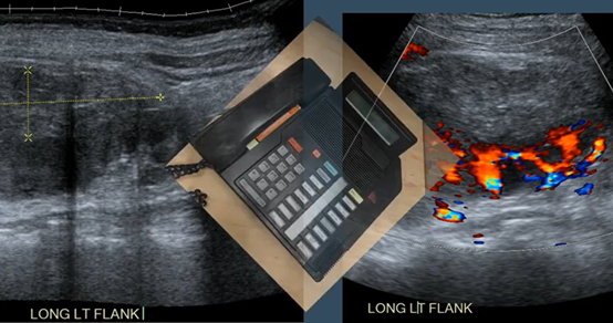
    Clear Call: Quite quickly, Wunderlich syndrome—spontaneous, non-traumatic bleeding confined to the perinephric space—becomes life-threatening. When a patient presents with sudden flank pain, a palpable tender mass, or signs of hypovolemic shock, perinephric hematoma ID is merely your opening move.

    • Root Cause: Staggeringly, 60% of these patients have an underlying renal mass, ranging from benign angiomyolipomas to renal cell carcinomas (RCC).
    • Other Culprits: If there’s no mass, etiology may stem from vasculitis, arteriovenous malformations, anticoagulation complications, or cystic renal disease.

    Ultimately, you can’t rely solely on that initial emergency scan to rule out pathology, Dr. Myra Feldman said during the ARRS Online Course with SRU Sound the Alarm! Emergency and Pitfall Cases in Ultrasound.

    • Half-Sensitive: CT’s sensitivity at the time of an acute hemorrhage is only 57%.
    • Masking Effect: Dense, acute blood products can easily obscure the underlying lesion.

    So, what’s a rad to do? Don’t let this go undiagnosed. The reading room’s next best step: calling the ordering physician to report the hemorrhage.

  • MRU is All-Terrain, Radiation-Free CT Urography

    MRU is All-Terrain, Radiation-Free CT Urography

    Summary: MRU is indeed invaluable when you want to avoid ionizing radiation, à la pregnant patients. But it should be leveraged, too, as an essential second-line problem solver for atypical renal and urothelial masses.

    GU Pro: Sure, CTU remains the daily, fast, first-line workhorse for genitourinary imagers. Still, standard scans can struggle to characterize atypical tumors or complex tissue changes. Enter, then, multiparametric MR urography, offering multi-layered tissue characterization to narrow your DDX.

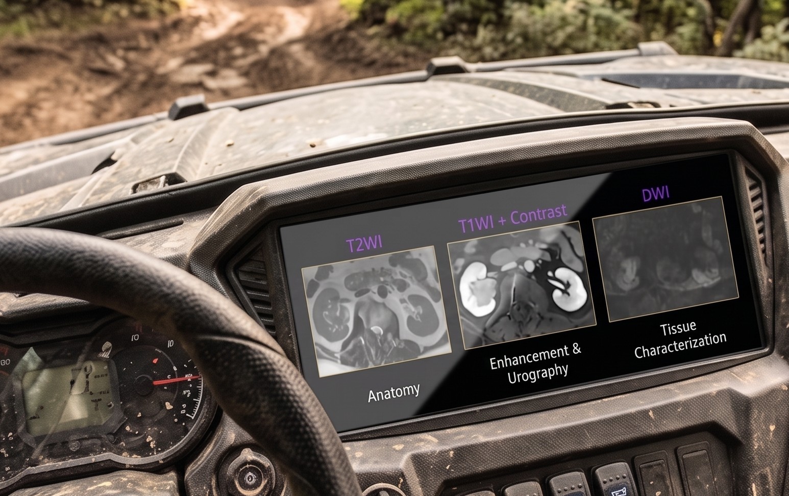
    By combining multiple specialized sequences, Dr. Myles Taffel noted during our longitudinal course Mastering Genitourinary Imaging & Interventions, MRU allows rads to analyze pathology from several angles across distinct terrain:

    • T2-Weighted Anatomy: Delivers detailed structural imaging comparable to what is obtained with CT.
    • Multi-Phase Contrast: Captures multiple enhancement phases, including early dynamic post-contrast and late urographic phases, which may not be feasible on CT.
    • DWI: Highlights cellular diffusion restriction to pinpoint exact areas of pathology.
    • Intrinsic T1: Provides native tissue contrast to further assist in detailed characterization.

    As Taffel recalled, the real utility of MRU’s multiparametric sequences shines in those really tough cases, including off-roaders:

    • ID Lymphoma Mimics: A renal hilum mass can mimic standard urothelial cell carcinoma. However, when MRU shows the mass extending along the ureter without significant hydronephrosis—coupled with restricted diffusion on DWI—the diagnosis of lymphoma becomes clear.
    • Bladder Subtleties: In a patient presenting with hematuria, a standard T2 sequence might only show vague bladder wall enhancement. Adding DWI and early dynamic post-contrast sequences provides the high-contrast resolution needed to confidently pinpoint exactly where the malignancy lies.