
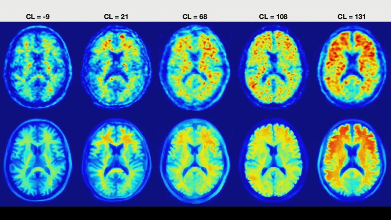
Summary: Ultimately, the positive/negative dichotomy just isn’t enough anymore. The Centiloid scale nurtures the nuance necessary for personalized treatment, as well as therapeutic response over time.
At Scale: Visually reading amyloid PET is too qualitative. Enter, then, the Centiloid scale, a quantitative tool that provides the precision necessary for selecting and monitoring patients on new disease-modifying therapies.
Standard Yardstick: With Alzheimer’s diagnostics bracing for more biomarker-based frameworks, amyloid PET has been dubbed “core 1,” capable of detecting pathology long before symptoms emerge. And as Dr. Lawrence Tanenbaum trumpeted during Imaging of Dementia and Alzheimer’s Disease: What You Need to Know, the Centiloid scale harmonizes results across different PET tracers and imaging centers—creating a single, site-agnostic measure of brain β-amyloid burden.
- 0: Mean uptake of healthy, amyloid-negative controls.
- < 10: Reliably negative.
- 10–30: Intermediate zone, where this “sweet target” population may represent the earliest detectable amyloid changes.
- 21–24: Threshold often used to maximize agreement with traditional visual interpretations.
- > 30: Reliably positive.
Moreover, immunotherapies like Lecanemab and Donanemab require longitudinal tracking to prove target engagement and confirm amyloid clearance. Tanenbaum reminds us, too, that some therapies were paused once a patient’s Centiloid score dropped below a specific threshold.
The Catch? Sure, quantification helps less-experienced readers identify ambiguous or borderline cases. Nevertheless, visual confirmation remains essential. Automated Centiloid scores can be “fictitious” or inaccurate if there is motion, significant brain atrophy, or atypical tracer uptake in the cerebellar reference region.
Leave a Reply