
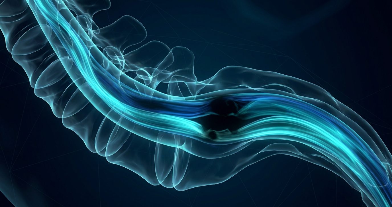
Summary: Clocking the flow void sign on T2 or STIR offers localization early in the workup—streamlining myelography and focusing treatment right there at the tear.
Spinal Gap: IDing spinal specifics is critical for classifying the cause of SIH and guiding myelography. As Dr. Joshua Russell Fage noted in this ARRS Web Lecture, the goal here is to establish the presence, or absence, of a spinal longitudinal extradural fluid collection. Immediate reporting directs the next best step, determining how subsequent myelography should be performed:
- Extradural Fluid Collections: Caused by dural tears, leaking meningeal diverticula, or arachnoid herniation near nerve roots.
- Fistulas won’t exhibit extradural collection.
- T2 Tips: Eval relies heavily on T2-weighted and T2 fat-saturated sequences to clearly delineate extradural fluid.
- Flow Void Sign: Seen on 2D T2-weighted or STIR images directly at the tear site.
- 3D Trade: Alas, Dr. Fage’s flow void sign is not visible on 3D acquisitions. (However, higher-res 3D sequences can sometimes visualize the dural defect directly.)
Leave a Reply