
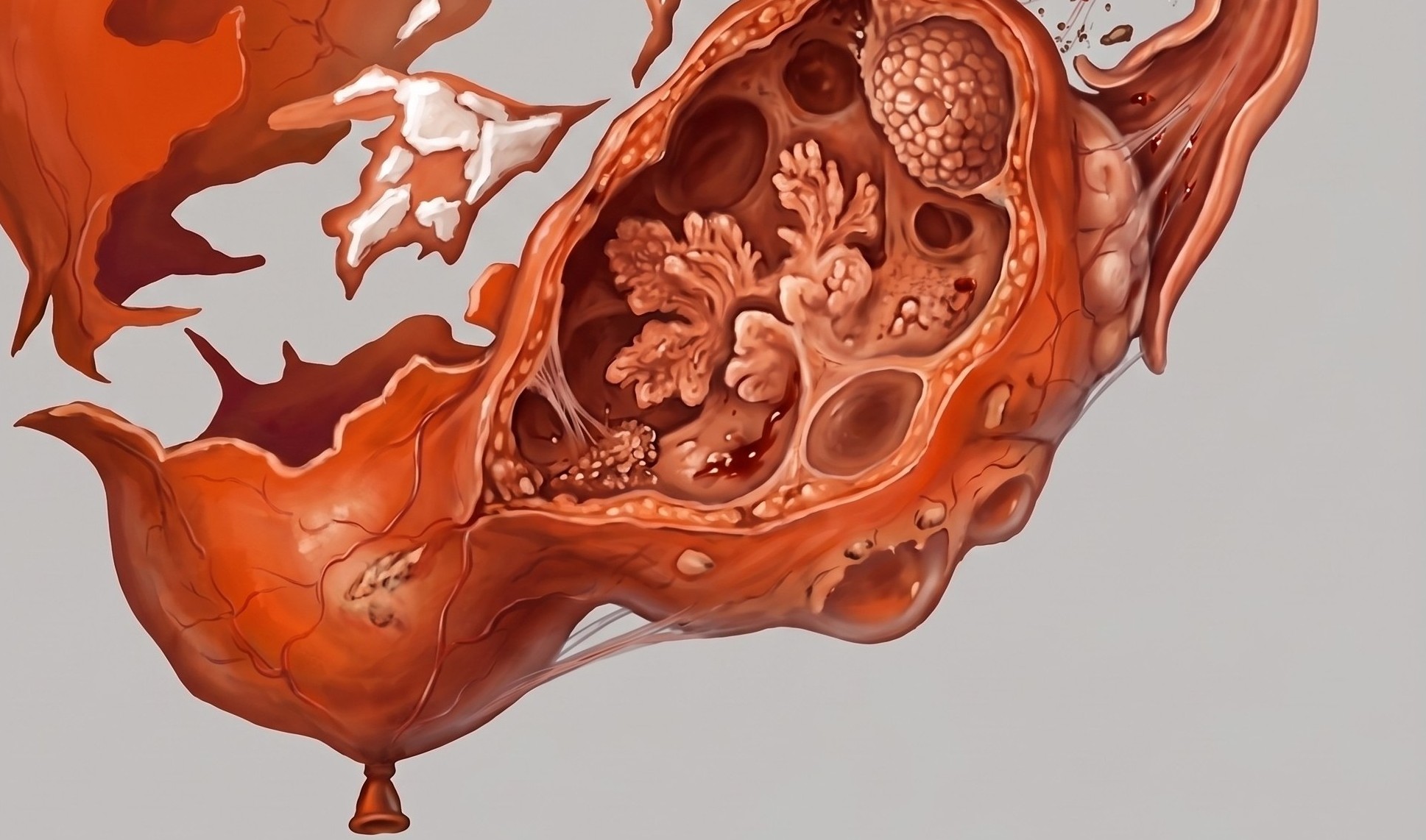
Summary: If it’s the patient’s first presentation, and your MRI findings remain ambiguous, the safest bet here is follow-up US in 2-3 months to confirm whether that suspect cyst has resolved.
On GYN MRI, both of these adnexal pathologies present with pelvic pain and may exhibit T1 hyperintensity. Whereas hemorrhagic cysts are generally self-limiting and resolve, as Dr. Milana Flusberg distinguished during ARRS26, the markers of chronic endometriosis can lead to scarring and pelvic tethering, as well as rarer complications like infection.
Three T2 Tells: Look out for these specific T2-weighted signs to help you differentiate:
- Shading: This classic feature—where a T1-hyperintense lesion appears hypointense on T2-weighted sequences—is highly characteristic of endometriomas. However, it is not 100% definitive, as it occurs in just under 20% of hemorrhagic cysts, too.
- Dark Spot: Referring to focal, highly T2-hypointense hemosiderin spots inside an otherwise mildly hypointense or intermediate lesion, this one is highly specific for endometrioma, albeit lacking high sensitivity.
- Hypointense Rim: A peripheral hemosiderin ring caused by repetitive, cyclical bleeding, it’s more common in endometriomas, although it can sometimes be seen in hemorrhagic cysts.
Beyond the Cyst: Quite often, Flusberg said, the most helpful clue lies outside the lesion, itself:
- Deep Pelvic Endometriosis? Look for T2-hypointense pelvic fibrosis tethering the mass to adjacent structures, such as the rectal wall.
- Multiple Lesions? Finding many complex adnexal lesions strongly points toward endometriosis, rather than an isolated hemorrhagic cyst.
Is There an Echo in Here? If you’ve got prior or concurrent US, the classical hemorrhagic cyst demonstrates “lace-like” reticular internal echoes or angular, concave retractile clots. Meanwhile, endometriomas typically present with homogeneous, low-level internal echoes.
Leave a Reply