
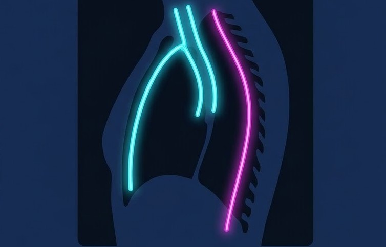
Summary: By marrying your findings with patient demographics, you can construct a highly focused DDX before ever sending the patient for cross-sectional imaging.
Full Felson: Evaluating the mediastinum is one of the most challenging aspects of reading chest radiographs because, well, masses are relatively rare. One of the biggest interpretive pitfalls? Glancing at a featureless mediastinum, seeing clear lungs, and calling the exam normal. Erroneously, of course.
During the ARRS26 honorary lecture celebrating Diane C. Strollo, MD, Melissa Rosado de Christenson, MD, detailed how rads can systematically characterize lesions and navigate classic compartments:


- Focal, or Diffuse, Lesion? Determine if a mass is focal, diffuse, or even multifocal.
- Diffuse, bilateral mediastinal enlargement that obscures normal anatomical landmarks typically indicates an aggressive lesion or lymphadenopathy.
- Unilateral, focal masses suggest a primary lesion, à la neurogenic neoplasm.
- Recognize the Landmarks: You gotta know your normal lines, stripes, and interfaces—right paratracheal stripe, anterior junction line, azygo-esophageal recess, etc.—to definitively ID if a mass is present.
- Read the Classics: Leverage signs like the hilum overlay and cervicothoracic signs to accurately locate abnormalities.
- Paging Dr. Felson! Though modern CT uses classifications like prevascular and visceral compartments, Felson’s anterior, middle, posterior compartments remain highly effective for narrowing differentials.
Leave a Reply