
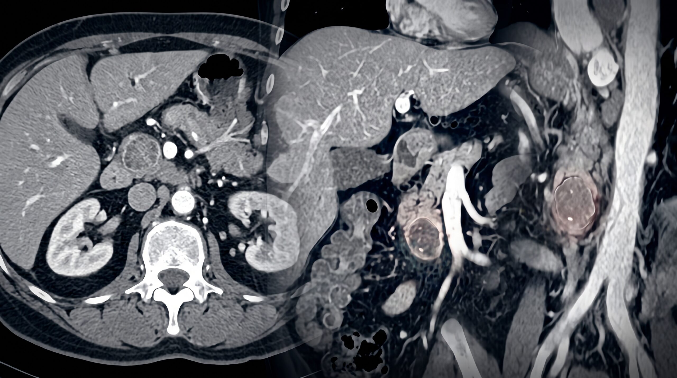
Summary: Effective evaluation requires reliance on structured imaging that’s razor-focused on cyst size, count, location, and calcification.
Nothing But PNET: Diversity is on full display with cystic pancreatic neuroendocrine tumors (PNETs), requiring a structured approach to calcification and enhancement, as well as clinical presentations. Detailing these distinctions during our online course Radiology Case Review–Abdominal Imaging, Mark Sugi, MD, noted that, typically, functioning tumors present early vis à vis clinical symptoms. Meanwhile, non-functioning tumors show up later and larger and undergo necrotic degeneration.
- Intratumoral: Punctate, mural, or non-mural calcifications should serve as signs for PNETs.
- Obstructionist: Even if a lesion obstructs the pancreatic duct, the presence of intratumoral calcification should arouse suspicion for PNET.
- Pattern Profile: PNETs characteristically demonstrate post-contrast enhancement, frequently exhibiting eccentric or rim-enhancing patterns.
Beware of Imitations: A circumscribed rim-enhancing lesion with subtle punctate calcification can closely mimic duodenal diverticulitis. Alternatively, ductal adenocarcinoma developing in the setting of pancreatitis with calcifications presents a similar kind of overlap.
- Insulinomas? In patients with multiple endocrine neoplasia type 1, insulinomas display as rim-enhancing, low-attenuation masses in the pancreatic body or tail with high uptake on ⁶⁸Ga-DOTATATE PET.
Leave a Reply