
With therapies evolving and technology updating, radiologists the world over are ready to rise to the challenge of delivering more precise staging and follow-up for rectal cancer.
On Sunday, April 12 in Pittsburgh, PA, abdominal imaging experts from both hemispheres will convene at the David L. Lawrence Convention Center and online for Rectal MRI, the 2026 ARRS Annual Meeting Global Exchange course featuring faculty from the Royal Australian and New Zealand College of Radiologists (RANZCR).
Codirectors Aliya Qayyum (ARRS) and Kirsten Gormly (RANZCR) are bringing together the field’s finest to share evidence-based insight and technical pearls designed for immediate clinical application. For radiologists seeking to refine staging, elevate posttreatment evaluations, and stay ahead of emerging imaging biomarkers, this expertly curated course offers globally relevant guidance.
Rectal MRI: A Cornerstone of Modern Care
Once an emerging tool, rectal MRI is now the gold standard for staging, enabling assessment of the T stage and detecting key prognostic features such as mesorectal fascia involvement (MRF) and extramural venous invasion (EMVI) (Fig. 1).
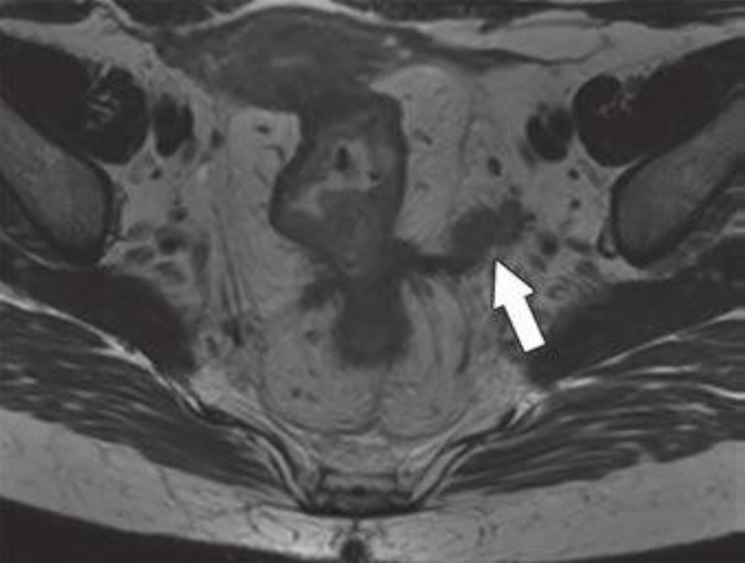
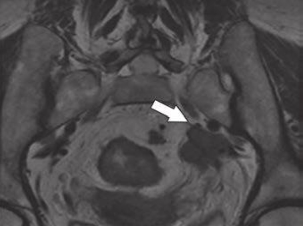
Fig. 1—59-year-old patient with rectal cancer with extramesorectal
vessel involvement, consistent with T category of T4b. Axial (top) and
axial oblique (bottom) T2-weighted images show rectal tumor that extends
through extramesorectal vein (arrow), which according to expert opinion
warrants classification as T4b.
Dr. James Costello (ARRS) will lead a session on foundational principles of T staging, MRF, and EMVI, emphasizing actionable strategies to refine reports and guide multidisciplinary teams. Building upon Dr. Costello’s foundation, Dr. Verity Wood (RANZCR) addresses the myriad nuances of assessing lymph nodes and how to identify poor prognostic tumor deposits. Pointing out pitfalls left and right, her lecture during the 2026 ARRS Annual Meeting Global Exchange with RANZCR will provide the latest imaging criteria to help radiologists render more confident interpretations.
From Early Detection to Posttreatment Decision-Making
As screening programs detect more early-stage tumors, MRI also has the ability to evaluate these lesions. Dr. Gormly will discuss how to assess early cancers and why acquisition technique for high-resolution T2-weighted images directly impacts interpretive accuracy for all rectal MRI parameters (Fig. 2).
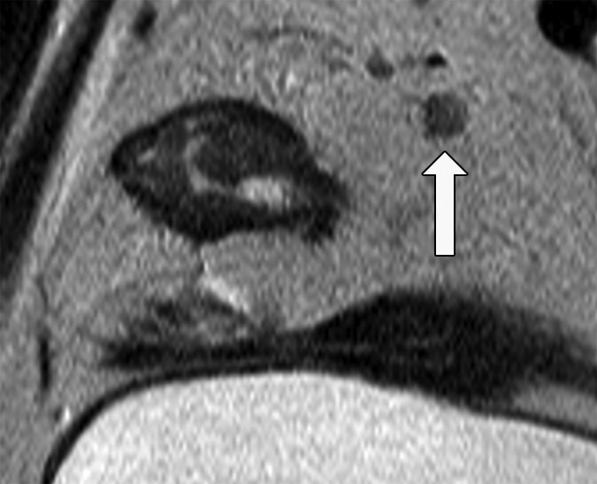
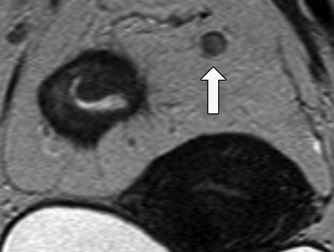
Fig. 2—Morphologic features of metastatic mesorectal nodes on MRI
and potential pitfalls in assessment. 58-year-old woman with rectal
adenocarcinoma. Oblique axial T2-weighted MRI (left) shows apparently
spiculated node (arrow). Graininess of image is related to poor signal-tonoise
ratio (SNR). Coronal T2-weighted MRI (right) shows that same node is
homogeneously T2 hyperintense with dark capsule (arrow), which is typical
of reactive mesorectal node. This image has superior SNR. Suboptimal images
can lead to erroneous assessment of nodal morphologic features. This
patient proceeded directly to surgery. Total of 38 lymph nodes (0.3–1.3 cm)
were harvested. Eleven of larger lymph nodes were serially sectioned before
submission for histologic processing. Final pathology revealed T3N0 disease.
Of course, staging is only part of the story. With the emergence of total neoadjuvant therapy (TNT) and “watch and wait” (W&W) protocols, radiologists play an increasingly vital role in posttreatment imaging. Dr. Raj Mohan Paspulati (ARRS) will outline W&W strategies in the United States, comparing tumor regression grading (TRG) systems and illustrating how MRI supports individualized care plans that may spare patients surgery.
The ability of MRI to predict patient outcomes can be considered more important than direct pathological correlation. Dr. Gormly will close the 2026 ARRS Annual Meeting Global Exchange course with her experiences in assessing emerging imaging biomarkers such as the “split scar sign,” aiming to improve patient selection for W&W, following a detailed explanation of how to assess residual tumor on high-resolution T2-weighted images. This is a key part of any posttreatment assessment system and is the cornerstone of the mrTRG system—the most validated MRI-based grading method globally.
Alliances Advancing Imaging
The mission of the ARRS Global Partner Society Program is to build long-standing relationships with key leaders and organizations in the worldwide imaging community—increasing awareness of our society’s services in specific nations, while raising the stature of Global Partner Societies among ARRS members. Every year, the ARRS Annual Meeting Global Exchange incorporates one partner society into the educational and social fabric of our meeting. ARRS members then reciprocate at the partner society’s meeting that same year.
Founded in 1949, RANZCR promotes and continuously improves the standards of training and practice in radiology and radiation oncology for the betterment of the people of Australia and New Zealand.
Rectal MRI not only reflects the robust collaboration between ARRS and RANZCR but also celebrates the global standardization of rectal MRI protocols.
And as Dr. Gormly told InPractice, “Australia and New Zealand’s early and ongoing partnerships with global leaders like Professor Gina Brown have positioned our region at the forefront of rectal MRI innovation. And this course is about sharing those insights with the world.”