
Lymphoma is the most common malignant tumor of the mesentery, occurring in 30% to 50% of patients with non-Hodgkin’s lymphoma. Because it often mimics sclerosing mesenteritis, identifying specific imaging clues is critical for an accurate diagnosis.
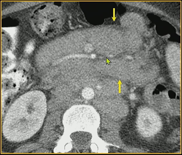
Bread & Butter Tip: The “sandwich sign“occurs when mesenteric masses involve both leaves of the mesentery while relatively preserving the central fat and vasculature. This creates a layered appearance, kinda resembling a sammy.
Key Differentiators for Lymphoma:
- Lack of Calcification: Untreated lymphoma typically doesn’t calcify. If calcification is present within mesenteric masses, lymphoma can generally be eliminated from the differential.
- Multifocal Disease: Lymphoma often presents with involvement beyond the mesentery, frequently extending into the retroperitoneum.
- Node Size: A short-axis dimension of 10 mm or more is a major red flag that requires further workup.
💯 Percent: Recent research indicates that the presence of just one of these worrisome features, significant node size or extra-mesenteric lymphadenopathy, yields 100% sensitivity and 100% NPV for malignancy.
Bottom Line: Whereas lymphoma can share features with mesenteric panniculitis (e.g., relative mass effect, tumoral pseudocapsule delineation, the fat ring sign), the latter is dominated by fat necrosis and inflammation—rather than the bulky, “sandwiching” masses seen in malignant disease.
Leave a Reply