Author: Logan Young

  • Better Days Ahead: A Briefing from Brazil’s COVID-19 Outbreak

    Better Days Ahead: A Briefing from Brazil’s COVID-19 Outbreak

    Published May 11, 2021

    avatar

    Rubens Chojniak

    Head of Diagnostic Imaging Department, A. C. Camargo Cancer Center São Paulo, Brazil
    Treasurer, Brazilian College of Radiology (2021–2022)

    avatar

    Valdair Muglia

    Associate Professor, Ribeirao Preto School of Medicine University of São Paulo
    President, Brazilian College of Radiology (2021–2022)

    For valiant service selflessly rendered on the frontlines of the fight against COVID-19, the American Roentgen Ray Society symbolically awarded each and every one of our members the 2021 ARRS Gold Medal. The ARRS Gold Medal Story Series shares perspectives of imaging professionals who conquered the day-to-day challenges of battling COVID-19.

    One of the countries most severely hit by the coronavirus disease (COVID-19) pandemic, cases and deaths are spreading in Brazil.

    The Brazilian health care system, “Sistema Único de Saúde,” is a universal, comprehensive, and free-of-charge public service assisting more than 120 million people. During previous public health emergencies, like the HIV pandemic of the 1990s, the system worked efficiently. Therefore, it was expected our system would place the country in an excellent position to mitigate the COVID-19 pandemic.

    That was not the case.

    Public and private hospitals had been reorganized to treat COVID-19 patients, but the country began to see a rapid uptick in new cases. Each region of the country—a country of continental dimensions—had peaked at different times, and several cities had their health care systems exhausted at different times.  

    The limited number of reverse transcriptase polymerase chain reaction tests available in many centers, and the prolonged time to process their results, have led to the early use of CT as an auxiliary method for screening suspected cases. This use of CT in suspected cases had been described in many countries experiencing similar scenarios and was mentioned in the guidelines of several international radiological societies.

    At our hospital, a public university hospital in São Paulo State with more than 1,000 beds, the scenario was no different. To help cope with the adaptations required to overcome the myriad challenges imposed by this pandemic, a Crisis Committee was created, gathering the major players engaging with COVID-19: medical specialists like radiologists, nursing and physical therapy staff, as well as other professionals providing all kinds of goods and services in a hospital. Since March 2020, our crisis counselors have convened every day (except from September to November), including weekends and holidays. Their daily briefings offer quick, practical solutions to dynamic problems.

    In treating COVID-19-positive patients, our radiological services have experienced a significant increase in chest CTs and a marked reduction in imaging tests performed for other indications. To help limit the spread of infection, many departments had to turn their focus toward fighting COVID-19.

    In September, due to general improvement of the overall situation in Brazil, some health services began returning appointments for care that had been postponed, particularly resuming treatment for chronic diseases and cancer.

    This scenario lasted for a short time, until January 2021, when the pandemic’s second wave hit Brazil even more severely with the more virulent variant, P1, accounting for more than 80% of new cases. At this time, the scene is even more stark, quickly draining the resources of both public and private systems. Even centers that offered adequate assistance in the first wave had their capacities depleted during this second wave. Currently, the major issues are lack of ICU beds dedicated to COVID-19 patients and a shortage of specific medical supplies, such as medications for intubation and sedation. Some parts of the country are experiencing an even worse scenario: limited oxygen supplies, for instance.

    Brazil’s national vaccination program against COVID-19 began in February 2021. The elderly and health professionals remain priorities. Although more than 25 million people have been vaccinated, that is merely 12% of our population.

    We are still facing an overloaded health system and a high number of cases and deaths, but we’re starting to see a significant reduction in the number of new COVID-19 cases. And as the vaccination campaign moves forward, we are hoping for better days soon.

  • True Team Efforts

    True Team Efforts

    Published May 10, 2021

    avatar

    Ali Gholamrezanezhad

    Assistant Professor of Clinical Radiology
    Keck School of Medicine, University of Southern California

    For valiant service selflessly rendered on the frontlines of the fight against COVID-19, the American Roentgen Ray Society symbolically awarded each and every one of our members the 2021 ARRS Gold Medal. The ARRS Gold Medal Story Series shares perspectives of imaging professionals who conquered the day-to-day challenges of battling COVID-19.

    As an emergency radiologist, my research projects have focused on emergency radiology and critical medicine. Through these research initiatives, I have identified aspects of my field that I find most rewarding. Focused on exploring new horizons in critical situations, I have found collaboration to be among the most valuable takeaways for my work. Reason being, it complements my hands-on practice and teaching, allowing me to have both an immediate and long-term impact on patient care, especially during emergency situations, such as trauma cases, burn victims, and urgent situations related to coronavirus disease (COVID-19).

    I am currently leading a multinational research team investigating the clinical and nonclinical features of COVID-19. Being able to have a personal impact in response to this pandemic has been a thrilling feeling for me. To date, our team has produced more than 45 journal articles, with over a dozen additional submissions under consideration. Our areas of focus have included: radiologic presentations of COVID-19, clinical factors predisposing patients to complications of COVID-19 (e.g., ICU admission, intubation, or death), long-term pulmonary consequences of COVID-19, the pandemic’s impact on health care workers and medical students, radiology department preparedness for surge potential, factors influencing differential case-fatality rates worldwide, and the best approach transitioning to the post-COVID-19 era. Over the past several months, my research team’s COVID-19 publications have been cited more than 2,500 times, accumulating more than 300,000 downloads by scientific and medical communities across the world. My colleagues’ work is considered a leading source of clinical information about SARS-CoV-2 infection.

    While I have taken great pride in our ability to produce a significant number of research articles on COVID-19, I also feel that our development of a major repository of COVID-19 imaging in such a short period of time, given the significant limitations of social distancing, is just as notable. One of the core things I have learned about meaningful research during this pandemic has been how to accommodate the critical factor of time sensitivity. Having hosted more than 200 videoconferencing sessions, we demonstrated our ability to plan, organize, and lead a team, showcasing my own project management and multi-tasking skills. I look forward to utilizing this amalgamation seamlessly when approaching oversight of future research projects. Specifically, I intend to apply these cross-departmental collaboration skills to extend my impact ability beyond my given area.

    During the outbreak of COVID-19, my team of researchers and medical students set out to educate the radiology community and broader health care system worldwide how to prepare for unusually high patient volumes, publishing several reference articles in various journals, including the AJR and Journal of the American College of Radiology. These articles were published in early February, when minimal COVID-19 cases had been reported in the United States. Our radiology research group formulated several critical recommendations for radiology departments to approach COVID-19 patients in the safest, most efficient manner. It was clear that if a flood of patients were to inundate even the most well-organized departments, that rush would be near impossible to accommodate. As a result, my team and I developed a mass casualty incident (MCI) plan, which consisted of several steps expressly geared toward viral outbreaks like COVID-19. We came up with a clear roadmap for preparation, resource mobilization, imaging chain, adjusting imaging protocols, and education. Specific to education, this plan included MCI simulation and in-service training. The core benefits to having an MCI plan in place include increased patient and staff safety, as well as a decrease in COVID-19 transmission.

    At the height of the pandemic, we conducted a national survey to evaluate the impact of COVID-19 on imaging practices. More than 800 radiologists across the country participated. According to our findings, a large portion of surveyed radiologists, 61%, rated their COVID-19-related anxiety a 7 or higher on a 1–10 scale. Upon further examination, we found that the higher the number of reported cases in a respondent’s respective state, the higher their reported score. Another key finding was that concern regarding personal health was the strongest connector to a higher anxiety score. Therefore, we determined that additional attention must be given to radiologists working in drastically altered practice environments and in remote settings.

  • Microaggressions: How to Be an Ally

    Microaggressions: How to Be an Ally

    Published April 27, 2021

    avatar

    Patrick Young

    Student Admissions Ambassador, Midwestern University Arizona College of Osteopathic Medicine
    President, Asian Pacific American Medical Student Association

    avatar

    Carolynn DeBenedectis

    Associate Professor (Breast Imaging), Vice Chair for Education, Radiology Residency Program Director University of Massachusetts Medical School/UMass Memorial Medical Center

    avatar

    Ann Jay

    Associate Professor (Clinical Radiology and Otolaryngology), Director of Head and Neck Imaging,
    Vice Chair of Education, Radiology Residency Program Director MedStar Georgetown University Hospital

    avatar

    Lori Deitte

    Professor of Radiology and Radiological Services, Radiology
    Vice Chair of Education Vanderbilt University Medical Center

    avatar

    Daniel Chonde

    Resident Physician, Radiology
    Harvard Medical School/Massachusetts General Hospital
    Chair, ARRS Diversity, Equity, and Inclusion Committee

    avatar

    Nolan Kagetsu

    Associate Clinical Professor (Neuroradiology)
    Icahn School of Medicine at Mount Sinai/Mount Sinai West Hospital
    Advisor, ACGME Office of Diversity and Inclusion

    Proper communication in a health care setting is vital to delivering quality care to patients. Without it, the quality of health care would be compromised, leading to greater overhead costs and, ultimately, negative patient outcomes. It is well-established that good communication requires basic health care literacy, intercultural competence, and language translation, when needed. But what about communication between providers? Towards nurses? Medical technicians? Medical students? It is easy to forget that patient care is a team effort, which entails cooperativity. While direct aggressive behavior is seldomly seen nowadays, subtle negative attitudes are often projected into biased mannerisms and come across as indignant, derogatory comments. Both these behaviors are unprofessional, but the latter is witnessed much more—to which it seems many prefer to turn a blind eye. Eventually, it becomes the status quo. Such comments sting for a moment but can be ignored; however, repetitive comments are damaging and lead to self-confidence issues and mental health conditions, such as anxiety and depression. These are microaggressions. It is imperative that microaggressions are addressed promptly and professionally to avoid escalating tension in the health care team.

    A microaggression is a comment or action that subtly and often unconsciously or unintentionally expresses a prejudiced attitude toward a member of a marginalized group. These types of comments are usually due to underlying implicit bias. Microaggressions are not just harmless side comments; they have significant psychological and physical consequences to the recipient. Microaggressions can be both verbal or nonverbal. Examples of verbal microaggressions include one attending saying to another attending, who is Asian in appearance (but is actually Korean): “We have a Chinese patient and need an interpreter. You speak Chinese, right?” Or a male saying to a female radiologist: “You are too pretty to be a radiologist and sit in the dark. You should be in pediatrics.” Nonverbal microaggressions could be a store owner following a black customer around the store, or a manager ignoring an idea when a female employee presents it, then praising a male employee for saying the same thing. When such examples are experienced as isolated events, they can cause the recipient to become angry or frustrated. When someone is the recipient of microaggressions repeatedly, these events become dehumanizing and can lead to anxiety, lack of self-worth, depression, as well as physical distress.

    Difficult conversations at work have additional complexities because of factors such as rank, seniority, perceptions of power within the organization, and perceived threats to work identity, which is often more deliberately crafted than the identity of our private lives. Difficult conversations can be unsuccessful because we bring assumptions and narratives about the intentions of others to the table, without being mindful of the fact that these assumptions are fabricated from our experiences in the world.

    Mindfulness is the practice of bringing your attention to the present moment without judgment. Mindfulness is a skill that, when learned, will hopefully lead to equanimity and the ability to respond, rather than reactHarris D. 10% Happier: How I Tamed the Voice in My Head, Reduced Stress Without Losing My Edge, and Found Self-Help That Actually Works—A True Story. It Books, 2014. Mindfulness is a key element in using the Most Respectful Interpretation (MRI) method of responding to others. Instead of automatic negative assumptions about someone else’s actions or intentions, you are deliberately mindful, assuming the most generous intentions for that person. Bringing mindfulness to a difficult conversation allows you to arrive with compassion and empathy, but without judgment. Doing this will make the other person less defensive and more open to deeper and richer conversation. The threats to identity and ego are diminished, and you allow space for someone else’s perspective to be true.

    A difficult conversation involves anything that is uncomfortable to talk about. Examples include confronting a supervisor making suggestive comments, a colleague unaware of their microaggressions, or coworkers with a conflict. Three questions to ask when contemplating a difficult conversation are:

    1. What do I really want?
    2. What do I want for others?
    3. What do I want for the relationship?Stone D, Patton B, Heen S. Difficult Conversations: How to Discuss What Matters Most. Illustrated, Penguin Books, 2010

    There is a tendency to avoid difficult conversations because they can make us feel uncomfortable, vulnerable, and anxious about challenging responses. However, unaddressed issues often simmer and can eventually erupt into an emotionally charged confrontation focused on blame and assumed intentions. Approaches to handling a difficult conversation well include shifting to a learning/curiosity stance, disentangling impact from intention, and moving from a blame frame to understanding contributions to the problem from both sides. Effective conversation skills include inquiry, active listening, paraphrasing, acknowledgement, reframing, and contrastingPatterson K, Grenny J, McMillan R, Switzler A. Crucial Conversations: Tools for Talking When Stakes Are High, 2nd ed. McGraw-Hill Education, 2011

    Ho CP, Chong A, Narayan A, et al. Mitigating Asian American bias and xenophobia in response to the coronavirus pandemic: how you can be an upstander. J Am Coll Radiol 2020; 17:1692–1694

    DeBenedectis CM, Jay AK, Milburn J, Yee J, Kagetsu NJ. Microaggression in radiology. J Am Coll Radiol 2019; 16:1218–1219
    . The goal is to move from a difficult conversation to a learning conversation with mutual understanding and purpose.

    Microaggressions can often be addressed with curiosity. For example, one could say, “I’m sorry, could you repeat what you just said? I’m not sure I understood what you said.”

    The timing of one’s intervention should be considered. We should consider “calling in” in private rather than “calling out” in public.

    New or renewed attention on how workplace and institutional culture and behaviors impact marginalized communities can be challenging. Most people do not receive training throughout their careers on these topics, and the cultural or societal implications they may bring up can be challenging. As education is a central pillar to the ARRS, it was determined necessary to establish a Diversity, Equity, and Inclusion (DEI) committee to help provide teaching and resources to members and the public on relevant topics.

  • The Dual Pandemics: Dismantling Systemic Injustices Through Intentional DEI Strategies and Inclusive Team-Building

    The Dual Pandemics: Dismantling Systemic Injustices Through Intentional DEI Strategies and Inclusive Team-Building

    avatar

    Jonathan Kruskal

    2021–2022 ARRS President

    Those of you I have connected with virtually over the past year may recall that, in addition to family photos, my office (and thus my zoom background) is adorned with my old cricket bat, indigenous South African art, Khoisan necklaces, hummingbird photographs, and Shona stone sculptures. These are just a few artefacts that represent my cultural identity, on which I’ve been reflecting a lot these days.

    One of the reasons I emigrated from South Africa after completing my medical and basic science training was to escape the abhorrent system of apartheid that I witnessed up close from a young age. My wife and I touched down in the U.S. in 1987 filled with hope and much anticipation. The days of watching fellow human beings suffer at the hands of systemic racism, marginalization, violence, and oppression were behind us, or so we thought. Perhaps our departure was one way of social distancing from that awful pandemic, though much guilt persists knowing that “running away” would not contribute to a solution in any lasting or meaningful way.

    Demolishing Normalcy

    Fast forward to the year 2020, and we find ourselves grappling with the factors that contributed to George Floyd’s death. Along with the outbreak of COVID-19, more than 15 long months ago, and the ubiquitous opioid addiction crisis, the America that we chose to move to is experiencing more than a single pervasive pandemic and finds itself in desperate and urgent need of a reckoning with structural racism.

    The last year has exposed centuries-long inequities, disparities, and ignorance, which impact our employees, peers, patients, loved ones, and communities in ways big and small, seen and unseen, told and untold. Absent diversity, equity, and inclusion (DEI) strategies, combined with social distancing protocols, full-time remote work, technology and commitment overload, and skyrocketing mental health concerns have rightfully demolished what we once believed were the tenets of effective teams; the trademarks of normalcy. To return to what we as radiologists do best—providing top-quality, safe, timely, and evidence-based care—we must work together to dismantle, then to rebuild the status quo. How can we do this?

    We Must Row as One

    Whether based in a hospital, private practice, or academia, we need to develop and implement DEI strategies that will build high-performing teams through intentional inclusion practices. It’s the only way we can ensure the highest-quality care for our patients, eliminate care and outcome injustices, and begin to narrow the health disparity gaps. We must acknowledge that, yes, we all have biases, many of which are unconscious.

    Consider the myriad of players and moving parts in our ecosystems: our technologists acquiring and managing images; our IT colleagues facilitating image interpretation, data management, and report communication; and our nurses providing compassionate, patient-centered care during minimally invasive procedures. We also have the essential contributions of our translators, transporters, schedulers, nurse navigators, medical assistants, advanced practice providers, administrators, and image repository staff. To effectively serve our patients, we must understand, respect, trust, and listen to one another. Simply put, we must row as one.

    Doing the Work

    As a first step, I encourage you to take Harvard University’s Implicit Aptitude Test to better understand some of your own biases. Set aside uninterrupted time, and take the test with an open and honest mind. You can also ask your employees or colleagues to do the same. Take time to discuss what everyone learned, and listen to each participant. Sit with them, either in person or virtually, and truly hear their experiences and perspectives. Make sure to create an environment of safety, compassion, and open-mindedness for each gathering. You can also consider designing a DEI survey for your team to receive anonymous or attributed feedback. In the spring of 2019, Harvard University created a three-minute “pulse survey” for its community. The executive summary, final report, and data charts and tables are available here.    

    In these discussions and surveys, you can also delve deeper into topics such as cultural humility, microaggressions, and the difference between bystanders and “upstanders.” The emerging practice of cultural humility, a commitment to lifelong learning about global cultural differences, encourages us to inquire and learn about the experiences and identities of others. Ignorance can lead to an intended or unintended microaggression, which Medical News Today defines as “a comment or action that negatively targets a marginalized group of people.” Another important term to learn and practice is upstanders, or people who speak or act in support of an individual or cause, particularly on behalf of a person being attacked or bullied.

    The Concept of Ubuntu

    The Zulu and Xhosa concept of Ubuntu emphasizes the importance of “being oneself through others,” a form of humanism best expressed by the phrase, “I am because of who we all are.” Imagine if we realized that our best personal function was dependent on the function of our entire team?

    To sustain and elevate team functionality, we must adopt this philosophy in a way that resonates with you. Perhaps it’s by remembering the Golden Rule, which instructs us to treat others the way we would like to be treated ourselves. Maybe it’s by thinking about Aristotle’s historic quote: “The whole is greater than the sum of its parts.”

    At the core of our impact as imagers is a broad swath of races, cultures, ideologies, genders, religions, age groups, and much more. Over the next year, we will continue to share DEI resources and invite members of our ARRS family to volunteer, as we develop educational materials that are the building blocks for individual members and practices to rebuild their teams. To submit ideas and feedback, please email me directly at jkruskal@bidmc.harvard.edu.

  • Untitled post 2205

  • Health Care Hacking—A System Update

    Health Care Hacking—A System Update

    Published March 16, 2021

    avatar

    Benoit Desjardins

    Associate Professor of Radiology, Hospital of the University of Pennsylvania

    With our April 2020 AJR paper, “DICOM Images Have Been Hacked! Now What?,” well received by the ARRS membership, we were asked to record both an accompanying podcast and live webinar. We also presented our results at the Cybersecurity Refresher Course during the Radiological Society of North America 2020 Scientific Assembly, as well as at many other venues. Due to so many new developments in the worlds of cybersecurity and health care since the publication of our original AJR review, I have been invited to provide an overview summarizing a few of those latest developments.

    Recent Hacks

    The number of cyberattacks targeting medical institutions continue to increase. In 2020, 79% of all cyberbreaches affected the health care sector, so our sector has become a prime target. The breach portal of the U.S. Department of Health and Human Services Office for Civil Rights has reported a total of 620 new breaches of medical records in 2020. Trinity Health in Livonia, MI took the largest hit, when more than 3.3 million records with patient information were compromised as a result of a ransomware attack on Blackbaud, a cloud computing provider selling a fundraising database software.

    Other recent breaches and attacks have received lots of media coverage, for example:

    • In September 2020, Universal Health Services (UHS) was forced to shut down all computer systems at its facilities around the U.S. after a cyberattack by the Ryuk ransomware. This attack was likely triggered by a phishing email. UHS operates more than 400 health care facilities across the U.S. and U.K. After the shutdown, many of their facilities were left without access to computer and phone systems. Access to anything computer-based—from old labs and ECGs to radiology studies—was lost. UHS was forced to redirect ambulances and relocate patients in need of surgery to other hospitals nearby. Following the incident, four deaths were reportedly caused by delays in lab results arriving via courier. However, it is unclear whether or not these deaths were directly related to the cyberattack.
    • In Düsseldorf, Germany, also in September 2020, a ransomware attack against Heinrich Heine University gravely affected its University Hospital. Cybercriminals encrypted about 30 hospital servers, preventing access to important medical information for patient care. Doctors had no access to this information, so patients had to be redirected to other hospitals. As a result, a female patient in transit to the emergency department in critical condition was rerouted to a hospital 20 miles away. With the detour causing a one-hour delay in her care, she died in transit—known to be one of the first cases of proven death from ransomware.
    • A study by Greenbone Networks in September 2019 revealed that, worldwide, billions of confidential medical images on DICOM servers were freely accessible on the internet. This study headlined the news and even caught the attention of Congress, where Senator Mark Warner of Virginia became a strong supporter of improving the security of medical servers and images. In October 2020, New Net Technologies directed a follow-up study and discovered that, in the U.S. alone, millions of unprotected medical images were still exposed on the internet.
    • In March 2020, Russian hackers (APT29), who were also responsible for the Democratic National Committee hack in 2016, inserted malicious code within an update of SolarWinds’ Orion software, which monitors the computer networks of governments and businesses to detect problems. Once the hacked update was downloaded by users, the perpetrators were secretly granted remote access to all the networks monitored by Orion, allowing for complete control and the ability to easily steal information. Hundreds of government institutions and private companies have been affected, including the Departments of Homeland Security, Treasury, Commerce, and Justice, as well as the Pentagon, Postal Service, and National Institutes of Health. As this article is being written, investigation continues to reveal the full extent of the damages. So far, at least 250 federal agencies and businesses have been compromised by the hack.

    COVID-19 Vulnerabilities

    The outbreak of coronavirus disease (COVID-19) created a new set of problems for cybersecurity, producing a triple threat for health care systems:

    1. rapid expansion of networked devices and services, creating an expanded attack surface
    2. increase in the different types of cyberattacks
    3. fewer available resources to defend against cyberattacks

    The use of telehealth has surged during the COVID-19 pandemic. At my institution, the Hospital of the University of Pennsylvania, consultations by telehealth skyrocketed from 50 to 7,000 per day. Many radiologists continue to work remotely from home, creating additional vulnerabilities in security.

    A home radiology workstation connects to a home router, which connects via the internet to the hospital virtual private network device, which itself connects to the hospital servers. Each of these connection points are vulnerable. Radiologists reading remotely often forget to change the default administrator password on their home router. Hackers have performed domain name system hijacking on hundreds of thousands of home routers, redirecting links from legitimate institutions to hackers’ websites and intercepting data.

    As soon as COVID-19 became a pandemic, there was a massive surge in different kinds of cyberattacks. Whenever there is a social crisis, cybercriminals exploit the situation in full force, as people are more stressed out and, therefore, more prone to make mistakes. Phishing attacks pretending to originate from entities such as the World Health Organization spread across the globe like wildfire. These emails included fake links and malicious file attachments.

    Real websites, such as the Johns Hopkins Coronavirus Resource Center’s COVID-19 map, were quickly duplicated, becoming major sources of worldwide malware distribution. Upon visiting the fake websites, computers became infected by Trojans stealing crucial information. Information about those fake websites was spread via phishing emails, malicious online advertisements, social engineering, and search engines. Since the beginning of the pandemic, over 60,000 COVID-19-related fake websites have been created.

    Meanwhile, nation states, like Russia and China, have been attacking pharmaceutical companies and vaccine developers to steal intellectual property. They use phishing emails to obtain login credentials, and then use exploits to transmit files or execute code remotely. Two well-known groups of cybercriminals, APT29 from Russia and APT41 from China, have stolen COVID-19 research data by exploiting weaknesses in servers and routers.

    Some Solutions

    Late last year, the National Cybersecurity Center of Excellence, part of the National Institute of Standards and Technology (NIST), released its final practice guide on how to protect PACS technology and DICOM servers. NIST recommended a combination of strategies, including defense in depth, access control mechanisms, a holistic risk management approach, and the use of cloud storage. A defense in depth strategy involves multiple layers of defense at the level of the perimeter, the network, the workstation, the application, and the data; if one fails, data are still protected. NIST also recommended network segmentation into groups of devices sharing similar functionalities. This can be accomplished through virtual local area networks or by finer segmentation using software-defined networking—often used to secure legacy devices that lack inherent security features.

    Also in December 2020, the Health Sector Cybersecurity Coordination Center issued a sector alert regarding vulnerabilities in DICOM image servers, while adding a series of recommendations on how to better protect them. These suggestions included general ones (use secure passwords, close unused computer ports, apply the most recent patches), as well as using encryption of data at-rest and in-motion, restrictions on network access, and network segmentation.

    The U.S. government enacted into law the Internet of Things (IoT) Cybersecurity Improvement Act of 2020 in December, too. Responding to the significant increase in control of IoT devices by cybercriminals during the year, this law establishes new mandatory minimum-security standards for IoT devices purchased using government funds, including supply chain security.

    Early this year, the U.S. government enacted into law the HIPAA Safe Harbor Bill to amend HITECH (Health Information Technology for Economic and Clinical Health) to incentivize the use of cybersecurity best practices for meeting HIPAA requirements, especially those recommended by the NIST. The HITECH Act from 2009 was responsible for expanding the adoption of electronic health records (EHR) by health care providers— keeping one million U.S. physicians busy every evening, entering clinical data into the EHR.

    What can we expect in 2021 for cybersecurity in health care? The new administration will start by repairing the massive damages to the cybersecurity infrastructure caused by the previous administration. Nation states will continue cyberattacks on COVID-19 vaccine developers to steal trade secrets and gain a competitive advantage. Smaller health care institutions, barely surviving this pandemic economically, will face increased attacks by cybercriminals. They do not have the same cyberdefense budget and manpower as larger institutions, although they face the same cyberthreats. As health care organizations transition more and more of their data to the cloud, we should see increasing numbers of data breaches from patient data on cloud infrastructures. Phishing with a health care theme will be more prevalent, given the focus on all health issues during COVID-19. And IoT and wearable medical devices will remain targets of cyberattacks for the foreseeable future, until the industry fully implements the new IoT security standards imposed by the latest legislation.

  • Imaging Patients with COVID-19: Current Perspective

    Imaging Patients with COVID-19: Current Perspective

    Published March 16, 2021

    avatar

    Lawrence Goodman

    Emeritus Professor of Radiology, Pulmonary Medicine, and Intensive Care Medical College of Wisconsin, Milwaukee

    avatar

    Luis Antonio Sosa

    Assistant Professor of Radiology, Section of Cardiothoracic Imaging Medical College of Wisconsin, Milwaukee

    In early 2020, as the coronavirus disease (COVID-19) epidemic spread, numerous articles appeared on imaging for the diagnosis of COVID-19. Descriptions were often discordant and confusing because of small sample sizes and selection biases. Those discrepancies have been largely resolved. As clinical experience and testing improved, imaging’s role has largely switched from diagnosis to aiding prognosis and clinical management.

    Asymptomatic/mildly symptomatic patients: The Fleischner Society (Radiology: Cardiothoracic Imaging, Vol. 2, No. 3) and others see no role for chest x-ray (CXR) or CT for diagnosis, unless comorbidities exist or testing is otherwise limited.

    Moderately/severely symptomatic patients: CXR is often negative early, only to turn positive subsequently. The most characteristic findings are basilar and peripheral ground-glass opacities (GGOs) (Fig. 1).

    Unfortunately, only a minority of patients have this typical COVID-19 pattern. In most patients, the disease is located more diffusely or elsewhere. Consolidation may be present initially with more severe illness or duration (Fig. 2).

    Early on, CT may be positive when the CXR is negative. The typical GGO distribution is similar, but usually more extensive than on CXR (Fig. 3).

    Adenopathy, cavitation, and effusion are uncommon early on. Compared to other viral pneumonias, peripheral lower-lobe GGOs are more common in COVID-19, whereas other pneumonias tend to have more diffuse disease. There is considerable overlap, however.

    Reporting guidelines: Many templates have been proposed. Radiological Society of North America guidelines (Radiology: Cardiothoracic Imaging, Vol. 2, No. 2) define:

    • CXR = negative, COVID-19-like; regular pneumonia, other disease
    • CT = negative, typical of COVID-19; indeterminant, not COVID-19

    Both show moderate interobserver reproducibility.

    Follow-up of established disease: On CXR and CT, GGOs and focal area of consolidation often progress and may evolve into a bilateral acute respiratory distress syndrome-like pattern when cytokine storm develops. Less common CT findings include dilated peripheral pulmonary vessels, adenopathy, rounded infiltrates, and signs of bronchial inflammation. Hospital-acquired bacterial pneumonia may complicate COVID-19 and vice versa. Preexisting lung disease (e.g., chronic obstructive pulmonary disease, interstitial lung disease, etc.) further complicate interpretation. Clearing usually starts after 2 weeks.

    COVID-19 may cause hypercoagulability, leading to an increased incidence of both emboli and in situ thrombi and deep vein thrombosis.

    Pleural effusions may appear late, and barotrauma causing pneumothorax appears to be more common than in other viral pneumonias.

    Beyond the thorax: COVID-19 affects more than the lung. It is now clear that this is a systemic disorder affecting many organs.

    As with lung involvement, patients with comorbidities (e.g., diabetes, cardiopulmonary disease, etc.) appear more likely to develop extrapulmonary diseases—summarized briefly below.

    Cardiac disease: A significant minority of patients develop cardiac disease, evidenced by elevated troponins and other cardiac markers. Imaging may show evidence of heart failure, myocarditis, coronary vasculitis, mural thrombi, and pericardial effusions. Cardiac MRI has an important role, since it can identify myocardial inflammation. Myocarditis has been associated with poor outcomes, including cardiac dysfunction and mortality (either related to COVID-19 or other cause).

    Neurological disease: Approximately 15–20% of hospitalized patients develop altered mental status or more focal symptoms. Imaging is positive in a minority, however. Ischemic infarcts are the most common imaging findings, probably related to coagulopathy. Reported infrequently are hemorrhagic stroke, cranial nerve inflammation, encephalopathy, and worsening of multiple sclerosis plaques.

    Renal disease: Elevated renal function tests are not uncommon, and acute renal failure has been reported in some cytokine storm patients. Ultrasound may show increased renal echogenicity.

    Gastrointestinal disease: Diarrhea and other gastrointestinal symptoms are not uncommon. Imaging may show ileus, dilatation, bowel loops (diffuse/focal), and CT may reveal contrast-enhanced bowel wall. Liver function tests are often abnormal, but failure is uncommon.

    Long haulers: A significant minority of patients have residual vague or specific symptoms. Cough and dyspnea are frequent and often clear with supportive therapy within 30 days. Imaging is suggested for symptoms lasting beyond 30 days. Approximately a third of post-hospital patients will have residual signs of “fibrosis” and other residual ground-glass patches. Signs and symptoms in other organ systems may also linger. Clinical and imaging understanding are still evolving and may change as second- and third-wave infections hit, mutations occur, and vaccinations alter immunity. Stay tuned.

    This piece first appeared in ARRS’ SRS Notes.

  • Imaging the Past in Situ: Paleoradiology and the Saqqara Necropolis Excavation

    Imaging the Past in Situ: Paleoradiology and the Saqqara Necropolis Excavation

    Published in InPractice, Spring 2021: Volume 15, Issue 2

    avatar

    Sahar N. Saleem

    Department of Radiology
    Kasr Al Ainy Faculty of Medicine
    Cairo University Egypt

     
    avatar

    Zahi Hawass

    Former Minister of Antiquities of Egypt, Cairo


    Ancient Egyptians believed in an afterlife, that it was necessary to preserve the body after death. They perfected the process of mummifying the dead and buried them with their treasures in deep underground burial shafts, as in the Saqqara necropolis, or in expertly crafted mountain tombs, such as The Valley of Kings.

    Paleoradiology: Imaging the Insides

    In 1896, only a few months after the discovery of radiography, prior to its use on living humans, the first tests of the mysterious form of radiation were done on Egyptian mummies: one of a human and one of a cat. For the first time, x-ray film showed what was inside a mummy—without the need to dissect or even unwrap it. This event marked the birth of paleoradiology. The term refers to the use of non-invasive medical imaging modalities to study ancient human and animal remains or objects. Paleoradiology has evolved alongside medical imaging methods since its first uses. Today, a wide array of imaging modalities can be applied in archaeological work: conventional radiography, computed radiography, direct digital radiography, CT, micro-CT, endoscope, and MRI.

    Latest Findings in Saqqara

    The Saqqara necropolis had been an ancient burial site since the Old Kingdom (ca. 2686–2181 B.C.) and was in use through much later periods for royalty, noblemen, priests, and even common people. Located about 30 kilometers south of present-day Cairo, Saqqara is a United Nations Educational, Scientific, and Cultural Organization (UNESCO) World Heritage Site, hosting several archaeological excavations. Our recent mission at Saqqara, near the pyramid of King Teti (2323–2291 B.C.), unearthed great discoveries, including the temple of Queen Naeret, wife of King Teti. We also discovered 52 burial shafts (1522–1069 B.C.), containing coffi ns and a large number of rare artifacts: statues, board games, jewelry, amulets, stellas, as well as a papyrus 3 meters long by 1 meter wide with inscriptions from The Book of the Dead.

    Field Paleoradiology: Imaging With Archaeological Context

    Paleoradiology can give us important information regarding the condition of a mummy or other ancient artifacts. Imaging mummies in a proper facility provides the optimum technical conditions. However, during excavations, it is often impractical to transport mummies to an imaging facility; it may even be harmful to vulnerable mummies. Instead, we use dedicated mobile imaging equipment to reach the mummies inside their tombs, a nondestructive option by which we can accomplish the same goals.

    Traditional radiography has been used in field paleoradiology, but it is a cumbersome process to develop x-ray film in situ. Therefore, we use computed radiography and direct digital radiography technologies that have the advantage of being filmless systems, allowing for instant review and further manipulation of displayed images.

    Conquering Technical Challenges

    Careful site inspection is crucial preparation for the use of these field paleoradiology techniques. First, we determined the topography of the site—type, position, and relationship of the burial objects—and the data we intended to collect. We then identified any safety concerns, like applying radiation protection measures to conduct responsible field paleoradiology. The burial shaft in Saqqara was narrow, located 10 meters underground, leading to a room jammed with more than 50 coffins stacked atop one other. To better navigate this architecture, we designed detachable, lightweight radiography accessories that we could bring down and mount inside the burial shaft. These included a tube holder, a framed mummy bed with a plexiglass top, as well as a cassette holder for lateral viewing.

    We x-rayed the closed coffins, altering radiographic exposure settings and positioning according to the mummification style and the structures we aimed to visualize: bony skeletons, dense amulets, or soft tissues.

    Interpretation Issues

    Unlike patients, mummified remains do not have clinical history, and their historical context is often questionable. In fact, we identified a female mummy via radiograph that had been placed inside a coffin bearing inscriptions of a male’s name. The interpretation of mummy radiography also requires awareness of the ways in which the desiccation of human tissue affects its morphology, as well as the changes that resulted from different mummification practices. Additionally, some mummified bodies could be covered by artifacts that might be misinterpreted as pathology.

    Thus far, our field paleoradiology study has been successful, resulting in valuable details regarding the sex, age at death, and possible cause of death for the mummies in the Saqqara burial shaft. We were also able to identify jewelry, amulets, and embalming materials inside many of the mummies that reflected their socioeconomic status. Moreover, we gained new perspectives in to the health status and diseases of the studied mummies, which can provide a better understanding of the natural history of disease, itself.

    Field paleoradiology allowed us to determine the mummies’ levels of preservation—to avoid moving vulnerable mummies unnecessarily. This enabled us to carry out a type of triage; mummies whose images suggested were in good condition and needed further investigations could be moved to a nearby medical facility for a CT scan, while those in more delicate condition were left in situ.

    An invaluable tool to unlock the mysteries of mummification, paleoradiology puts us face-to-face with the men and women of ancient Egypt. Even thousands of years after their deaths, we’re still gaining remarkable insights into their lives simply from imaging their remains. Our work at the excavation site at Saqqara has not ended, of course, as what we have unearthed to date is merely a fraction of what we expect to exist.

  • Siege or Soar?

    Siege or Soar?

    avatar

    Alexander Norbash

    2020–2021 ARRS President

    This is my fourth and final column in an intense and unforgettable year. I have heard many opine that, somehow, we are going to be stronger and smarter and more evolved as a result of the many trials and tribulations we have faced. I don’t know so, but I do certainly hope so—hoping that something positive comes out of fear, pain, suffering, and uncertainty. In a certain way, I suppose we as physicians must possess such an optimistic mindset to survive and keep moving forward, despite the human misery and pain which we as radiologists literally see on a daily basis. As I think about the year, I can’t help but generate a list of questions for every challenging and engrossing topic, questions that I believe are intuitive, and questions that command my attention and test my optimism.

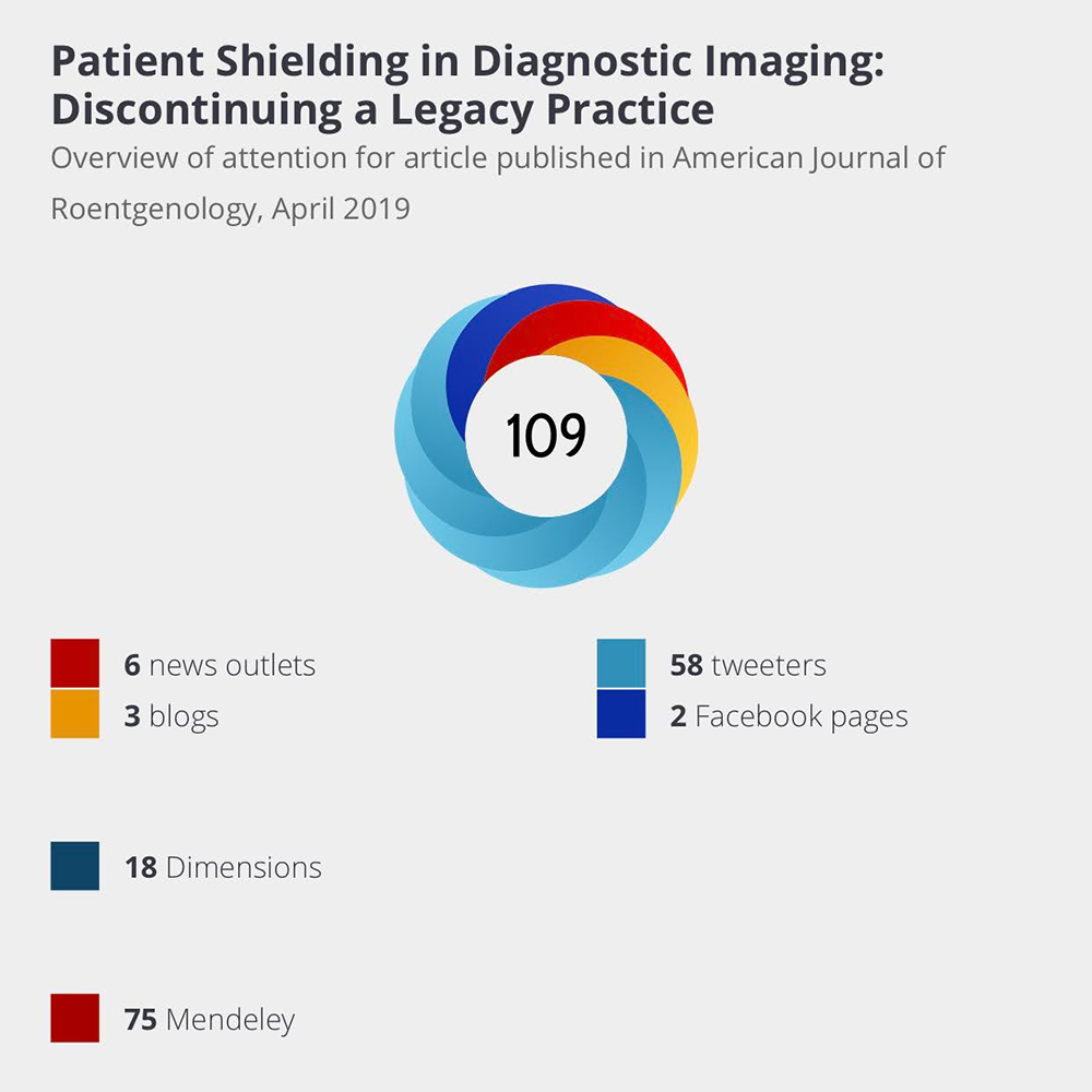
    In this year, there has been no dearth of challenges, large and small, that affect and threaten to derail our daily lives and, therefore, the practice of our specialty. Our year opened with COVID-19, affecting our lives like an unpredictable and interrupted set of explosions. It affected the financial security of our department, our salaries, our personal sense of security (affecting ourselves and our families), and our ability to serve our patients, who were either sheltering at home or suffering from a disease which we incompletely understood. Just when we thought we had turned a corner, a larger wave comes crashing on top of us. We followed a handful of models telling us what to expect for a wave, insofar as magnitude and timing were concerned, realizing these predictions may as well have been science fiction. We congratulated ourselves for having several vaccinations, then realized that we could not effectively manage distributing and administering these vaccinations at anything faster than a trickle. As I write this column, we have administered 20 million vaccinations. By some estimates, we have to give 229 million doses to reach 70% of our population of 328 million for effective immunity, if conventional models hold. That means we are 8% of the way there, in the one year and five days since the first U.S. case was reported, on January 19, 2020. Are we committed to social distancing, as I drive up the Pacific coast and see numerous outdoor restaurants open, including some with maskless servers? Have we understood how we will ensure sufficient supply chain management to optimize vaccination production, delivery, and inoculation? Do we know what the longterm effects will be on our businesses and our children’s education, and have we established plans to address the fallout?

    Before we could even gain our bearings in dealing with this new pandemic reality, we had to deal with the reality of race riots burning our streets, evidencing centuries of outrage and anger, threatening to tear us apart. As an occasional visitor who picks up fragmented and incomplete impressions of communities I have visited over the years, I never thought I would see Minneapolis in flames, or Seattle. I never imagined Portland as a city under siege. Did we divert to a better place after the Watts riots, or have we learned nothing? Did we resolve any of the contributory factors? Do we have a plan? Did we agree on a course of action that would address divergences and prevent future race riots?

    Perhaps all of our problems are really only psychosocial, since we are clearly masters of the natural world, so we may have thought as we debated the validity or existence of global warming. Then, we blinked, and life and earth reminded us of our hubris and fallibility: massive, unprecedented forest fires choked our newly darkened ochre skies in California, we wiped ash off of our windshields in the mornings before going to work, burning flames took away every material possession our friends and families cherished. Homes and property disappeared with a snap of Thanos’ finger. What are we going to do during next year’s fire season? Have we done anything to better address those seemingly inevitable forest fires? Did we solve this problem? Are we ready to take on the next series of forest fires with greater effectiveness, confidence, and less loss of property and life?

    And then, after a pandemic, race riots, and forest fires, the fourth horseman of the apocalypse turned out to be an angry mob, numbering in the tens of thousands, breaking into our seat of government while it was in session, intending to kill or physically harm our vice president, our senators, and our representatives. Despite the historic siege of the U.S. Capitol and all that it implied, the sun rose the following morning. In one fortnight, a peaceful transfer of power took place, and both the relevance and power of our systems and processes were reinforced. Are we now committed as a society to respectful disagreement? Will we need 20,000- plus troops in Washington, D.C., for inaugurations and legislative sessions to maintain law and order? Is the pendulum going to slow down and stop swinging way to the left, then way to the right?

    So, I can’t help but ask these questions. I would presume you, too, have at least one question: what relevance does any of this have to the ARRS and this column? I would respond as follows: we do not have control over many external factors in our daily life, and we may not be able to easily address external questions; however, we do have the power to mobilize our societal membership to create a better future for our patients and society through the improved delivery of health care—the scope and scale at which we operate, that is—and we have to aspire to work as a committed group of colleagues to positively influence our professional future, which is in our control. Oddly enough, we also have the opportunity to address some of those external big questions through our society, particularly the aspects that overlap with our professional lives.

    No matter what, we show up every day, and we do our best to help our fellow citizens. We are part of a group of compassionate, professional, and knowledgeable citizens. And even if the skies are on fire, if there is a literal plague among us, if there is revolution in the streets, if the future of our government is in question, we do our job to the best of our ability. Because we are hopeful, because we are committed, and because we have faith in our processes and our systems.

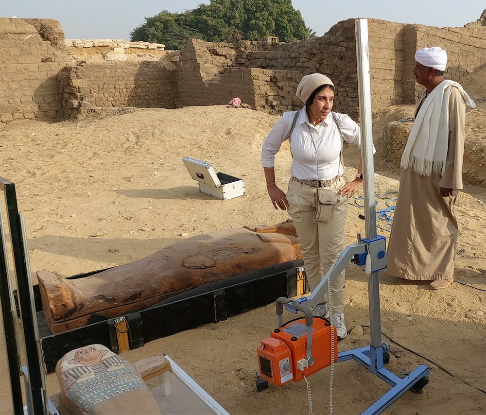
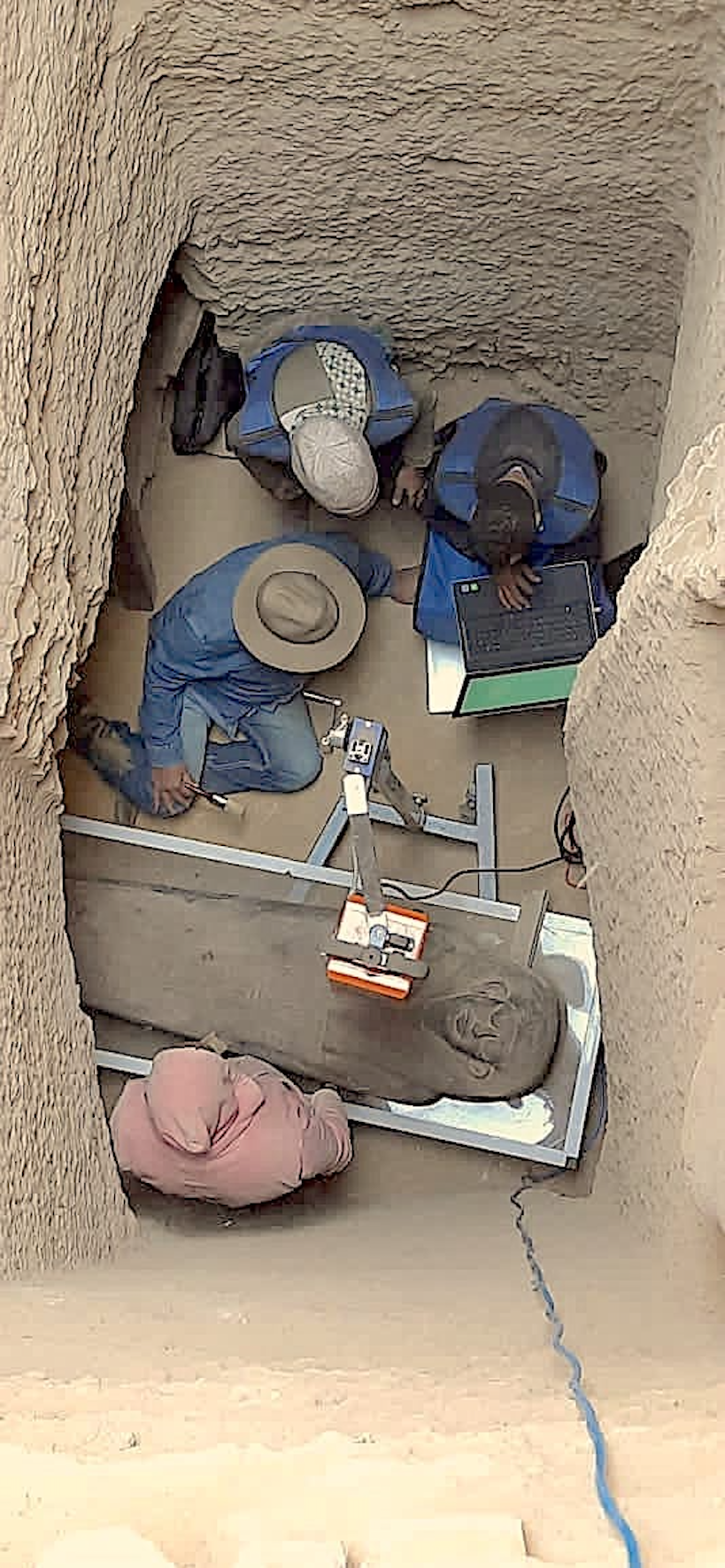
    Our professional society reflects the same collective ethic. Our central belief hinges on having faith in the power of our collective. We hope to educate each other, support each other, and facilitate our collective progress, so that we may become the very best that we can be at what we do. It doesn’t matter if we have to convert the in-person ARRS Annual Meeting to an all-virtual convening, if we must distribute our educational materials in a more effective way, if we need to focus our practice communications on the realities of pandemic management, if we need to share fast-breaking scientific communications regarding COVID-19 to help you work better and smarter, or if we utilize our platforms and publications as a bridge to timely and essential topics, such as diversity in health care. This professional society tirelessly does what it has to do to inspire and empower you.

    As I hand off this column to my dear successors, please allow me to ask three things of you, dear reader. I ask you to care, I ask you to engage, and I ask you to volunteer.

    I ask you to care because that will fuel your efforts. Caring creates motivational reserves that are virtually limitless. Caring provides purpose, and purpose allows us to muscle through uncertainty in the quest to find solutions. Everything that happens should matter to you, be it race riots or forest fires. Whether or not they are in your backyard, you should care, and you should want to care. Once you care, you will think of solutions, you will share these solutions, and, ultimately, we will collectively iterate and address our problems. It starts with caring. Great things are only possible when we care about the world around us and the events that affect our lives.

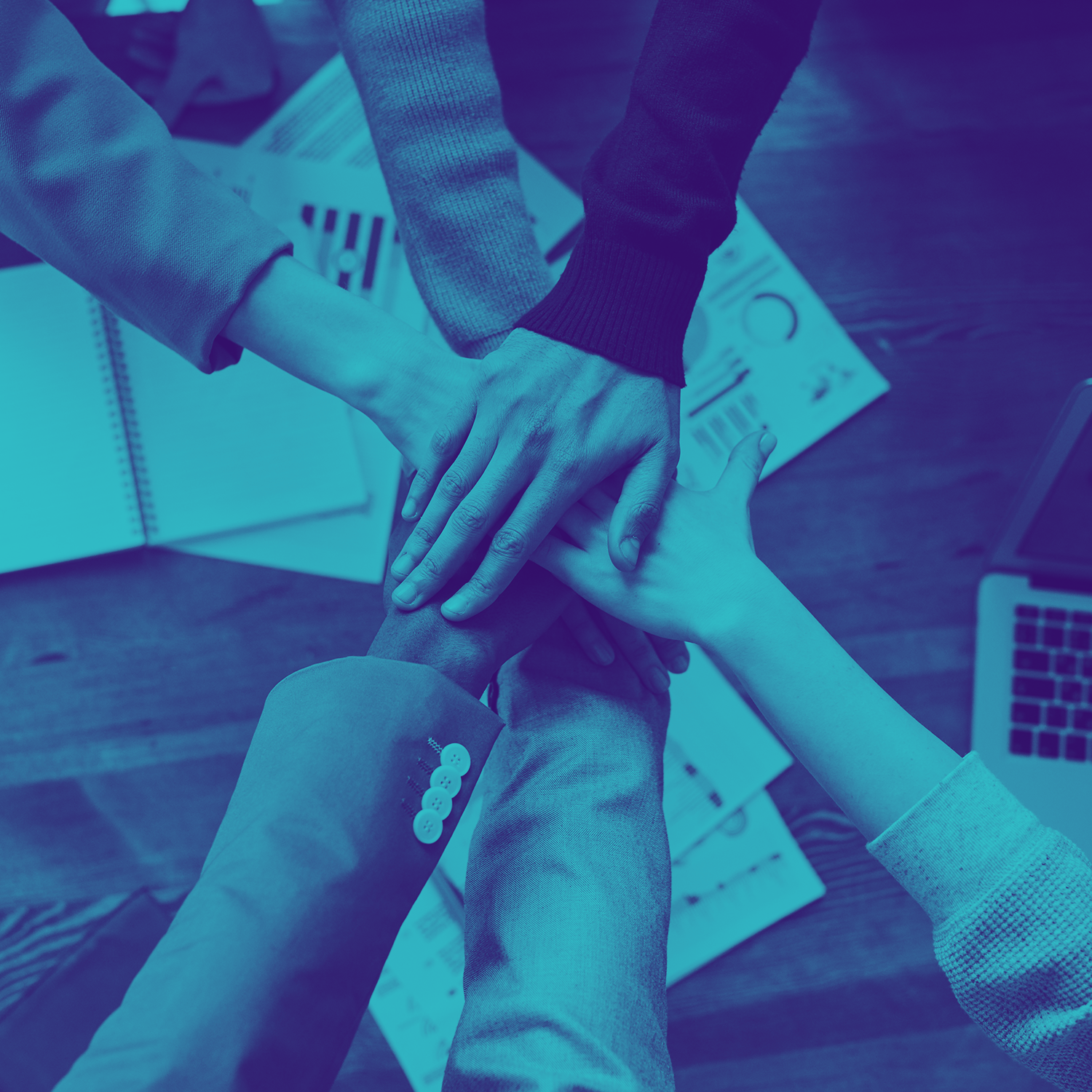
    I ask you to engage because not one of us is an island, and no individual one of us has the answers. Answers are generated by groups and teams. This means choosing to effectively establish dialogue, which is bidirectional information fl ow necessitating transmitting and receiving. If I’m only listening, then I’m not contributing to the conversation. If I’m only speaking, then I’m not listening suffi ciently to contribute to the conversation in a meaningful way. I have to train myself to listen, and I have to train how to express myself. When we connect with each other—and we translate our caring into effective communication—if we are prepared to both listen and speak, then we leverage the cooperative wisdom of the crowd into a powerful understanding. No matter how fascinating and experienced a single life may be, it cannot compare with the shared experiences and gained lessons and perspectives of a dozen lives. Great things are only possible when we communicate with each other.

    I ask you to volunteer, so we can all be part of something bigger, so we can align our efforts and energies, so we can not only imagine positive change, but we can realize it. Part of the beauty of professional societies is the remarkable power evidenced in likeminded individuals standing side-by-side with their shoulders to the wheel, pushing hard, and doing the best they can to create a better future. Sometimes, this testbed demonstrates remarkable returns. As one small example, I can see a brief line of succession where our societal executive leadership is concerned, and I am inspired by the depth of character, thoughtfulness, and energy I see for the next several cycles and years. We have great leaders moving into their ranks, who will serve our society exquisitely well. Great things are only possible when we work together.

    Please care, engage, and volunteer. You secure our brightest future, and you validate all that we do. And for that, I owe you all an unrepayable debt of gratitude.

  • New to AJR—Altmetric

    New to AJR—Altmetric

    Published March 16, 2020

    First used as a hashtag and codified more than a decade ago, altmetrics present a more comprehensive alternative to the traditional citation impact criteria of scientific publishing. Mainstream media, online-first publications, social and academic networks, public policy documents, post-publication forums, Wikipedia even—these complementary bibliometrics help to paint a truer picture of engagement with scholarly work, especially work that finds second, often third lives beyond the towers of academia.

    Altmetric.com is a data science company uniquely leveraged to capture this full array of online sources and collate all the disparate activity accordingly. In fact, just to the right of the abstract for every AJR article published on AJRonline.org, you will now see a multicolored “donut” badge visualizing a real-time summary of the attention that article is receiving (red for news outlets, orange for blogs, blues for social media, etc.) For numerical context, you will notice the data output gets assigned an Altmetric Attention Score, a high-level assessment of both the quality and the quantity of attention said AJR article is receiving, weighted according to the relative reach of each attention source, itself.

    Perhaps most importantly, if you click anywhere on the embedded donut, then you will be directed to a dedicated AJR Altmetric details page. Here, you, too, can follow the myriad conversations this specific AJR article is engendering—geographic and demographically—in one convenient, shareable URL.

    Want to be notified whenever someone shares or discusses the details for a certain AJR article? You can sign up for e-mail alerts under the Summary Tab of any AJR Altmetric details page. While you are free to sign up for notifications regarding multiple AJR Altmetric research outputs, you will only receive a single email alert, sent via once-a-day digest.

  • Continuing Radiologist Education—A New Paradigm Using Clinical Test Sets

    Continuing Radiologist Education—A New Paradigm Using Clinical Test Sets

    Published on January 22, 2021

    avatar

    Mo’ayyad Suleiman

    University of Sydney, Australia
    DetectED-X

    avatar

    Patrick C. Brennan

    University of Sydney, Australia
    DetectED-X

    avatar

    Ziba Gandomkar

    University of Sydney, Australia
    DetectED-X

    Although we have known for many decades that radiologists do make errors and the number of these can be significant, continuing educational solutions rarely recognize the practicalities facing modern-day clinicians. These practicalities include: proven effectiveness, methods of delivery that align to busy clinical workloads, modules that are compatible with any technological display, and tailoring education to reflect the unique errors each radiologist makes. Also, under the current climate of pandemic and travel restrictions, the tremendous difficulties of physically attending conferences and other educational venues must be recognized.

    The current article focuses on education using clinical test sets, which contain data sets on normal and abnormal cases with known truth that the clinician must diagnose. We aim to summarize what busy radiologists in 2020 require from continuing educational solutions, illustrating how effective, relevant, available, and tailored education can be delivered to all using the DetectED-X platform (Sydney, Australia). 

    So, What Is Needed?

    High Quality Data Sets Combined With Performance Algorithms

    Radiologists need effective and clinically relevant education that is available at all times. One solution that has been shown to work focuses on the provision of high-quality radiologic test sets that are specifically selected and designed to improve diagnostic efficacy. Via their local workstations, clinicians can view and judge sets of usually up to 60 DICOM cases at full native resolution, with higher than normal prevalence, and search for clinically relevant lesions validated by pathology findings using the DetectED-X platform. This way, it is possible to encounter in 1 hour the number of specific abnormalities only presented over periods of years. Radiologists should be able to judge these cases the same way they do clinically, so expert behaviours are relatively unaffected, and once all cases are judged and suspicious areas of interest are marked, clinicians should receive instant results on their performance using well-known and benchmarked metrics, such as sensitivity, specificity, lesion sensitivity, ROC, and jackknife alternative free-response ROC (JAFROC) (Fig. 1). Algorithms should then support these judgments by enabling the presentation of all errors made by each radiologist and identifying specific areas for immediate improvement (Fig. 2).   

    Accessible and Recognised Education

    To improve accessibility, it is essential that continuing medical education using test sets is available 24/7, regardless of clinicians’ location, must be completed in timeframes that align to busy clinical workloads, and should provide immediately much needed continuing medical education (CME) credits or continuing professional development (CPD) points. Also, to maximize ease of use, educational solutions should be compatible with all types of mobile devices, should involve test sets with smaller number of cases that can be completed in as little as 20 minutes, and must ensure that every activity is rewarded with CME or CPD—regardless of whether the clinician is located in the United States, Europe, or Asia.

    Tailored Solutions

    Tailoring to each clinician’s needs is required. No longer should clinicians receive test sets or other material that is common for everyone: Dr. Brady will make different mistakes from Dr. Cusack, so why should the two receive the same education? Artificial intelligence (AI) technology is now sufficiently mature to offer a robust educational solution. Elsewhere, in the entertainment industry for example, AI is now being utilized for the personalization of viewing platforms based on previously collected data to improve user experiences. Such advances can now be applied to online medical education, facilitating customized educational materials that accurately recognize each clinician’s strengths and weaknesses. By combining algorithms previously developed for the general-purpose recommender system, such as movie recommendation tools, with radiology-specific knowledge driven from research on factors defining case difficulty for specific clinicians, AI models can predict and access cases most suitable for each user.  

    One Potential Solution

    Test set technologies have been available over the last 3 decades, particularly in the United Kingdom, Australia, and New Zealand, and these have shown demonstrable mean improvements in diagnostic performance of 34%. However, these test sets can take up to 2 hours to complete and are usually only available via high-quality clinical workstations with images loaded locally. A new approach to address accessibility has been launched by DetectED-X, a University of Sydney startup, where high-quality cases are now available through modules (the smallest of which take only 20 minutes to complete), are available on any mobile device, and are immediately certified with CME credits or CPD points available for every activity. To ensure that this education is available at fast speeds, regardless of wherever in the world the radiologist is located, DetectED-X is working with GE Healthcare, Volpara Solutions, and Amazon Web Services to ensure robust and widespread distribution. Highly accurate tailoring based on tens of thousands of interactions with radiologic images is part of the DetectED-X approach, so that each clinician, regardless of training or experience, has an optimized educational experience. Its evidence-based AI algorithms for recognizing clinical error-making patterns have been tested using more than 600 clinicians reading mammograms or lung CT images, with an accuracy of 80% and 83% for predicting the positive and negative cases that each clinician has difficulty with, respectively. Once case difficulty for a clinician has been set, this is coupled to the clinician’s learning objective (e.g., whether they want to see a test data set, oversampled with calcifications or denser breasts), then a personalized test data set is presented to the clinician. Radiologists need a new approach so that effective, relevant, and tailored education is available everywhere and at all times. The COVID-19 pandemic has brought this need to the forefront.

  • What Lies Ahead for Radiology?

    What Lies Ahead for Radiology?

    Published on January 22, 2021

    avatar

    Alexander Norbash

    2020–2021 ARRS President

    I cannot get the image of the raging forest fires out of my head, even though I saw the carnage from the safety of my sofa on my iPad. Even so. And a close friend of mine lost his house and everything within it, leaving with only the clothes on his back and what little he and his family could carry. And he is grateful that his family lost no lives that day. Literally today, 12,600 firefighters are fighting 14 major wildfires in California, which have already claimed more than 4 million acres. Twelve-thousand firefighters in California alone. That is about half the number of radiologists in the entire U.S. And 4 million acres? Rhode Island has a surface area of less than 1 million acres, Delaware slightly under 1.6 million acres. Even Connecticut is slightly below 3.6 million acres. Our country is on fire, and we go about our daily lives…

    What do forest fires have to do with radiology? I submit that health care may be more akin to an uncontrolled forest fire, rather than the controlled and carefully harnessed fire in a foundry that allows us to forge steel and alloy.

    Natural questions include pondering the nature and distinction of solvable and unsolvable problems, as well as attribution. When do you stand by and watch a forest fire consume your house, and when do you fight it without endangering your life and health? Couldn’t anything have been done to prevent this? Can’t anything be done to fix this? If solvable, is the solution really cleaning forest floors, ridding them of combustible biomass before flames have a chance to perform the same function? Is this really attributable to global warming, and is it too late to do anything? Are forest fires even a necessary evil that allows renewal and rebirth, despite the massive sorrow accompanying the human experience?

    Although a raging forest fire is terrifying, especially when you are fighting the fire and sense it closing in on you from all sides, a single candle’s dancing flame is hypnotizing, and its generation of light and energy is magical. A collection of votive candles can be moving and sacred. A campfire can be comforting and protective. I recall many campfires I sat around as a boy scout, where we shared stories, enjoyed each other’s company, and felt both close to nature while safe and comfortable. Even though there was a dark silence surrounding us, inside that cylinder of light and warmth projected by the campfire was the illusion of safety. If you will forgive the analogy, although our health care ecosystem from a 30,000-foot perspective may be a forest fire, the single flame of an individual patient interaction or single health care encounter is sacred and at times life-affirming for those of us who are moved by the nature of trust patients place in our hands.

    For those who have not visited San Diego, we are arid and considered a coastal desert, and our terrain is varied, including steppes, buttes, mesas, and multiple valleys. As you would surmise, we are situated in close proximity to a number of mountain ranges. As you ascend on drives in these mountains, at higher elevations, you encounter beautiful forests. Whether driving to Mount Palomar, which houses a spectacular astronomical observatory on top of a peak, or the Anza-Borrego Desert 90 minutes northeast, the majesty of nature is undeniable. As you drive through our pine forests, spectacular vistas come into view, and off to the sides of the roads, you can’t help but notice groves with massive trees showing charred trunks. Where these charred trunks are found, the undergrowth is different. There is little brush, there is filtered and streaming light, and oddly enough, one finds fields of wildflowers in these clearings. Many decades ago, I recall reading Norman Maclean’s Young Men and Fire, regarding the 1949 Mann Gulch Fire. This book is a management mainstaple regarding tactics, leadership, communication, and learning systems. Several of the preventable fatalities were attributed to firefighters refusing to chuck their equipment and heavy packs in order to lighten and therefore save themselves. So many unnecessary casualties—a costly lesson for future literal and figurative firefighters. Do we learn from each other? Are we willing to jettison our heavy and cumbersome conventions to save ourselves?

    Perhaps health care is somewhat analogous to a forest fire raging out of control. Some of us feel a sense of anxiety about the unpredictable winds of the future and how they will affect us. There are many changes perched on our doorsteps, preparing to hit us hard. Whether planned evaluation and management code adjustments threatening us with over 10% of our income this coming year, Rad Partners and MEDNAX merging 10% of all U.S. radiologists into one consolidated supergroup redefining the structural nature of practice, or large-scale venture capitalist and hedge fund purchasing of radiologist practices affecting our autonomy, we lack control and fear for our safety. Similar to a clearing forest fire, perhaps we’re in for rupture of our status quo as an essential step in reordering the foundation and practice of radiology.

    Certainly, the COVID pandemic has significantly changed our academic practices, and who knows which of the changes we are seeing will be durable and which will be enthusiastically thrown out at the earliest possible opportunity. Have the COVID pandemic, civil unrest, and financial crisis all served as combustible agents where health care and, more specifically, imaging are concerned? The COVID pandemic has directly affected our financial practices and our procedural practices, underscoring inequities in care that have resulted in extraordinary mortality rates where the disenfranchised are concerned. Civil unrest has reminded us of these same persistent inequities in care. The financial crisis; more of the same. It is natural following a crisis, perhaps even more so following multiple simultaneous crises, to discuss and understand what we have learned. We are midstream and in no way past our crises, nevertheless, it helps to elevate our vision and see where we are heading as an essential exercise and to grant us temporary respite from today’s challenges. If nothing more, at least it allows us to lift our optimistic spirits, instead of suffocating under the weight of what is undeniably present in today. Perhaps in looking to tomorrow and moving toward action we should focus collectively on how we can provide the best possible care to the largest number of patients. I submit that the central reason for the forest fire which is health care could well be inequity in care delivery, misbalanced and at times exaggerated profitability, and a lack of effective population health. Does the public know about radiology, care about it, and fight to protect and elevate it? This repair work necessitates my personal engagement as a single radiologist. This demands my involvement in driving down costs, attempting to ensure the propriety and efficiency of imaging in each case, and eradicating inequities as much as possible. No one else is picking up the ball and confidently addressing the issues. Regrettably, these actions are neither natural, nor have they really been expected of me to date. My job to date, as an individual radiologist, has been to manage worklists, ensuring that individual dictations are correct. That said, if I focus on my work RVUs and bonus, and do not ensure the essential and full appreciated value of my contributions as a radiologist, will I be effectively valued?

    I’m sorry to end with a cliffhanger; a question to which I do not have a ready answer. What can I do, apart from drilling down on my worklist as an individual radiologist, to serve the largest number of patients most effectively, or has my campfire already contributed to the creation of a massive and clearing forest fire? I fear the response to this question will determine whether we become giant redwoods, or whether we will feed fields of flowers.