Category: Articles

  • The Journey to AI for COVID-19 Radiography

    The Journey to AI for COVID-19 Radiography

    Published September 29, 2020

    It all began with The Roentgen Fund® grant for deep learning cardiac MRI

    avatar

    Albert Hsiao

    Associate Professor of Radiology, University of California-San Diego
    2019 ARRS Scholar

    A specific form of artificial intelligence (AI), called a convolutional neural network (CNN), is rapidly becoming a standard tool for analysis of biomedical images, including radiography, CT, and MRI. Within only a few years, many laboratories have grown to adopt this technology to answer specific medical questions, make technical advances, or enhance clinical workflows. For example, CNNs have shown a remarkable ability to detect lung cancer on CT scans or diabetic retinopathy on fundoscopic photographs. One important reason for this is simplicity and ease of use, as CNNs are capable of learning characteristics or “features” of disease without being explicitly programmed. Another reason for rapid adoption of CNNs is remarkable generalizability, as they can be taught to be insensitive to artifacts, body habitus, technique, or even modality (CT or MRI). Just like humans.

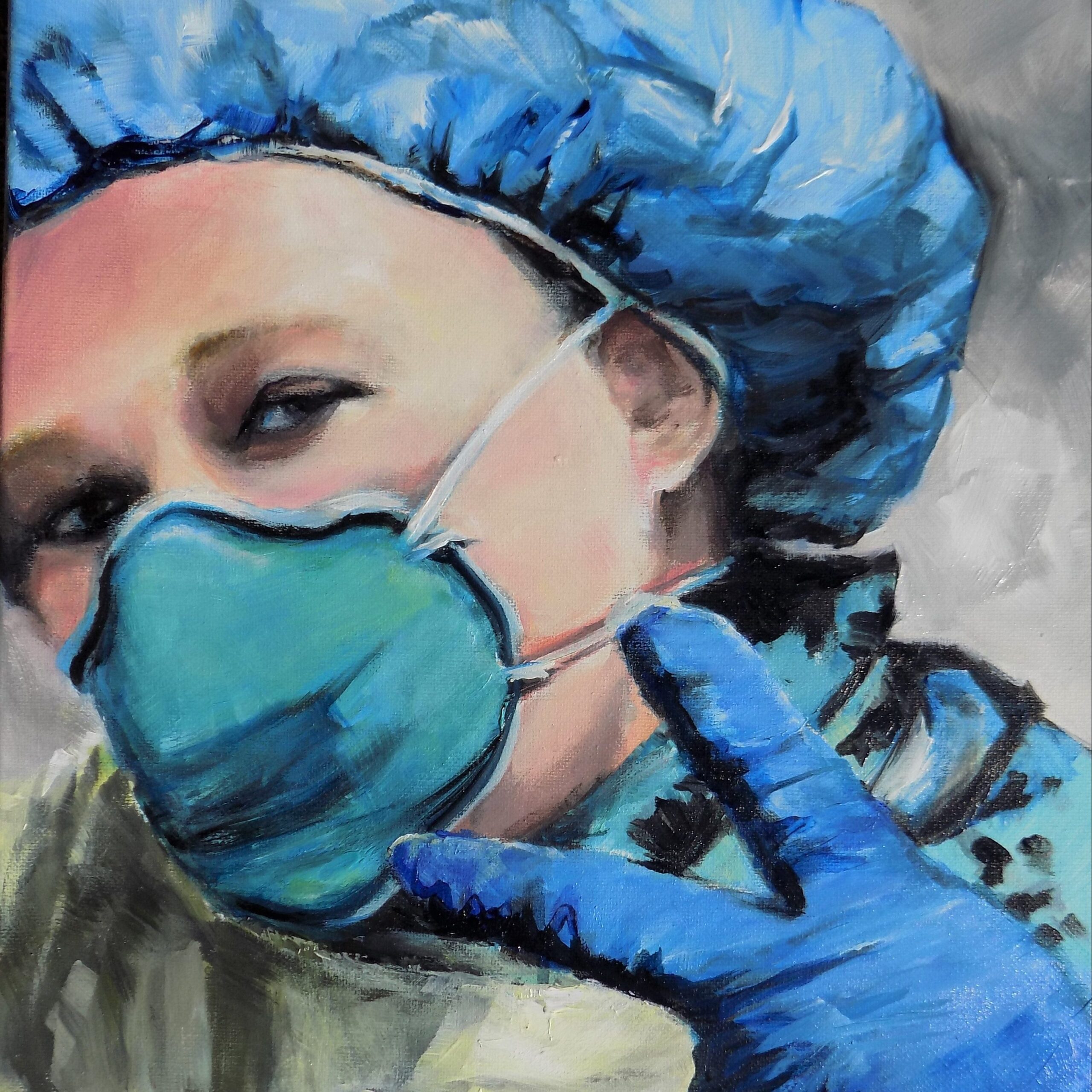
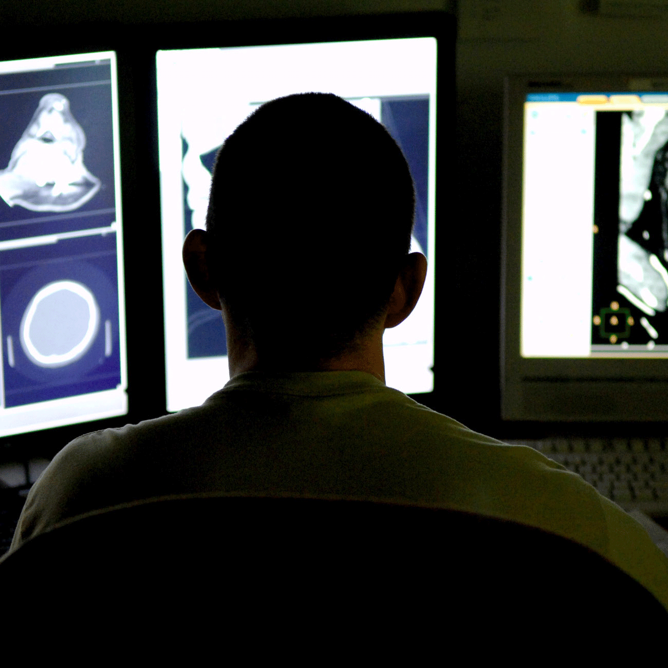
    The remarkable power of CNNs has led some physicians to conjure up mythical battles between man and machine, leading radiologists to reflect on how CNNs contribute and provide diagnostic value in medical imaging. With time, many of us now see this technology as a way to enhance our practices, reducing the clutter and labor that limit our ability to engage our higher cognitive skills to diagnose and manage our patients. There are few clinical arenas in diagnostic radiology that are more interesting but beset by more clutter and labor than cardiac imaging. That is, in fact, one of the reasons I was reluctant to pursue this field, initially choosing intervention instead. Fortunately, I was guided to take notice that where there are challenges, there are often extraordinary opportunities.

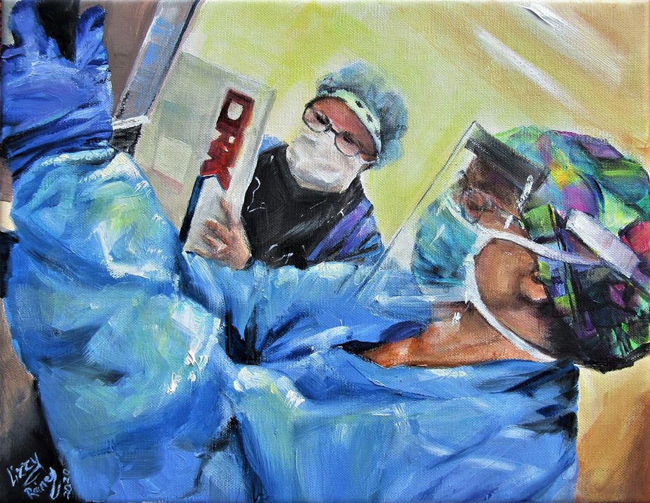
    Several years ago, we began exploring the potential of CNNs to simplify the process of performing and interpreting cardiac MRI. The idea was simple. There were too few technologists proficient in cardiac MRI, and the ability of radiologists to carefully supervise these examinations has been compromised by diminishing reimbursements and increasing clinical volumes. However, if we could use AI to automate the manual tasks on the scanner, we might be able to both improve image quality and reduce the amount of training needed for our technologists. AI could serve as our conduit of knowledge to improve the quality of the imaging we provide. Our efforts quickly showed promise, and we began integrating our approach into clinical software and the MRI scanner with research grants and partnership with GE Healthcare.

    The ARRS Scholarship provided me an opportunity to take this project in a new direction, considering it not just as a clinical end-product, but also exploring fundamental questions about how we bring AI to our practices. Specifically, in our proposal entitled “Adaptive Artificial Intelligence for the Acquisition and Analysis of Multiplanar MRI,” we asked: What tools do we have at our disposal to ensure that our algorithms would work well across multiple field strengths and technological improvements in MRI? Imaging equipment is constantly changing and improving, and AI algorithms need to grow and adapt, just as we do. We had some hints that CNNs could have the flexibility to continuously learn. With my brilliant students, we are looking at this from several angles, asking several key questions. Can we predict which images the AI algorithm will fail to process correctly? If so, can we selectively collect these images to teach our AI algorithm to learn from these cases and improve its performance? The results, so far, are promising, and we are preparing a manuscript on this topic for submission.

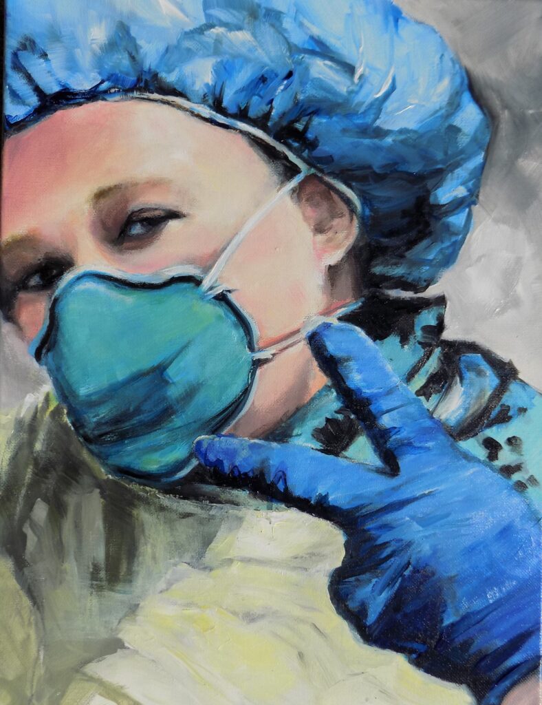
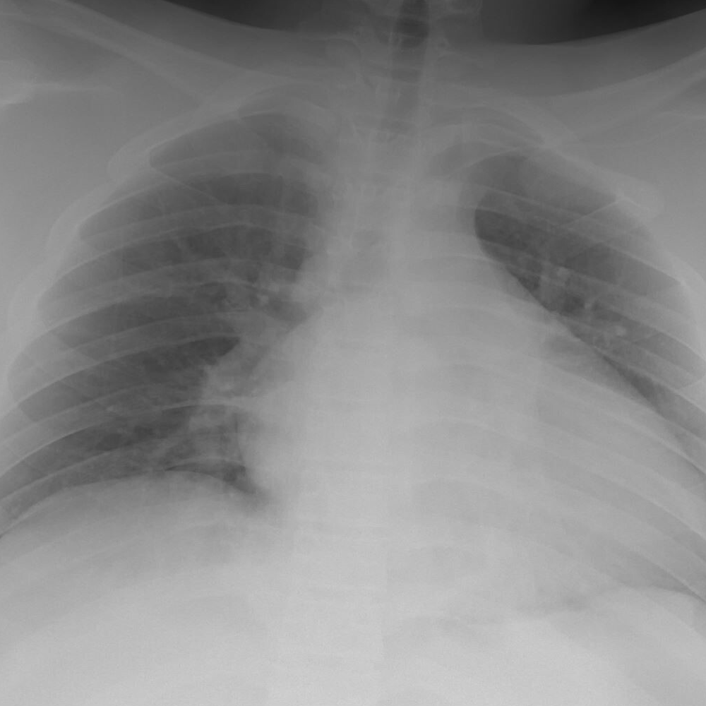
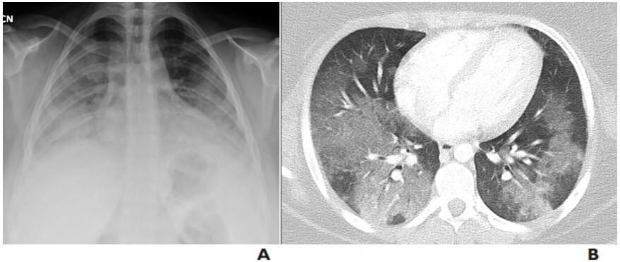
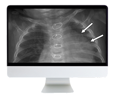
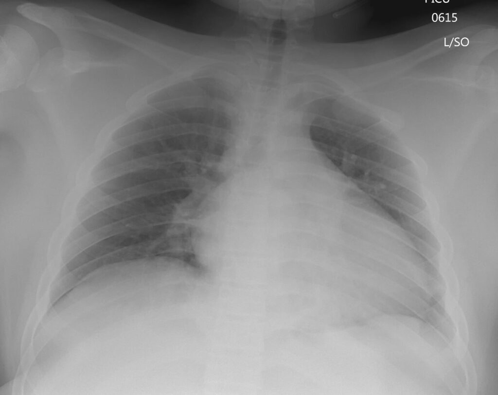
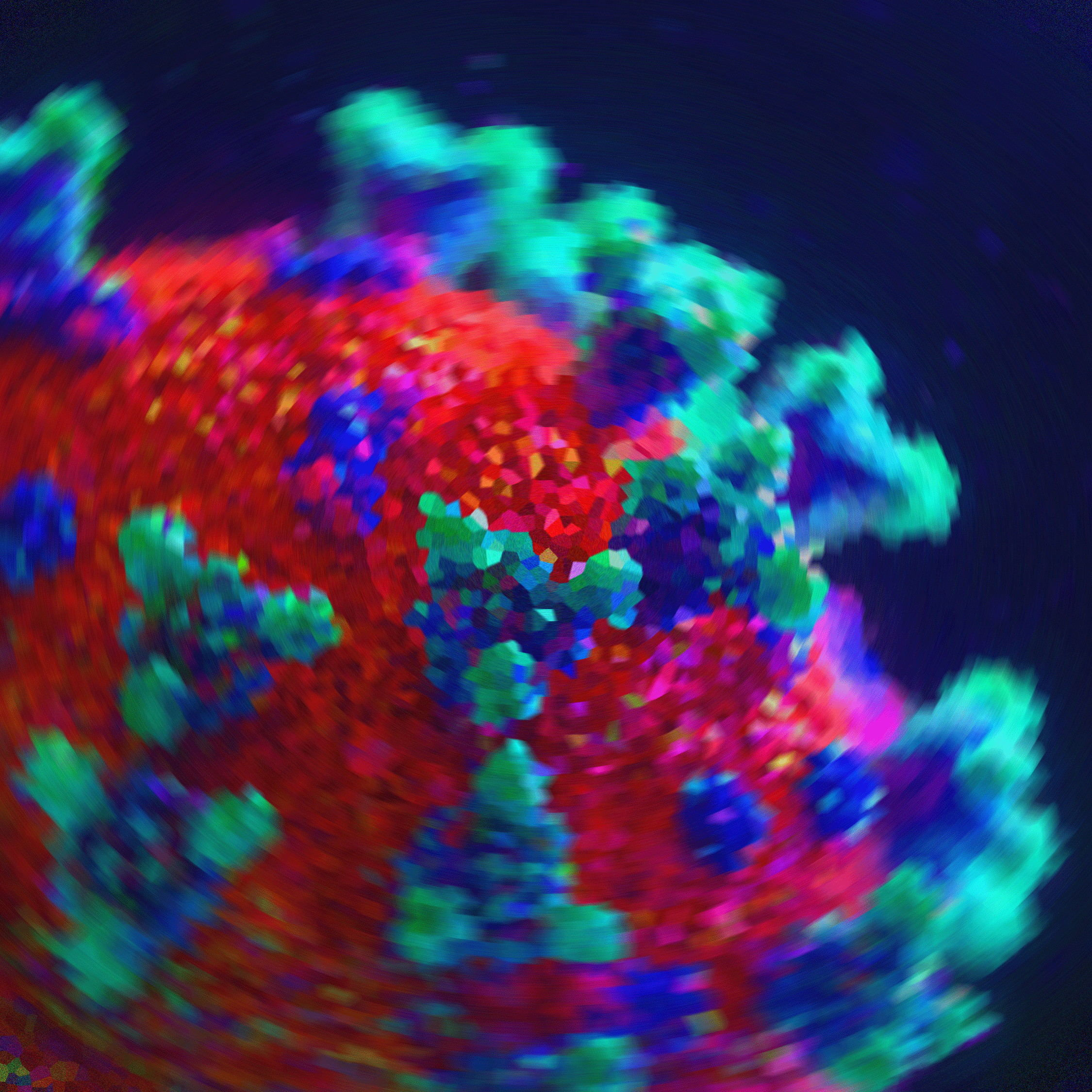
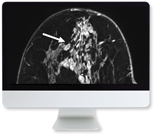
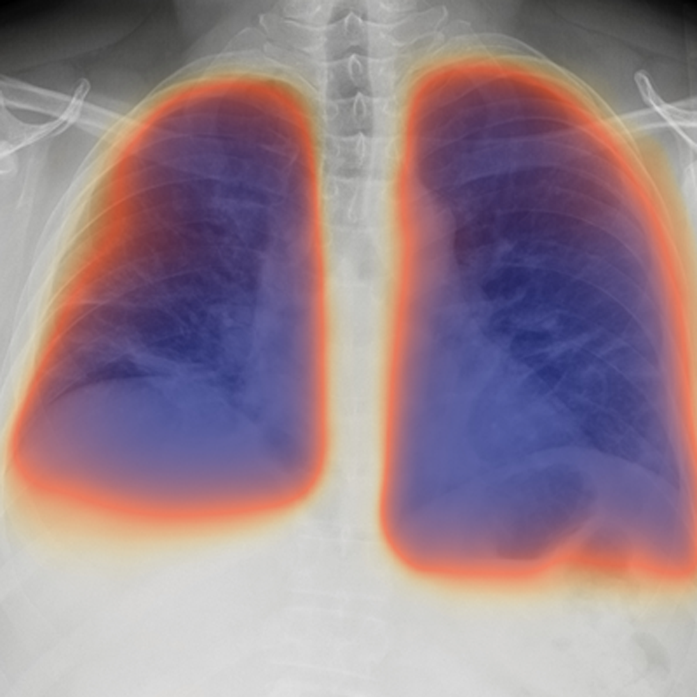
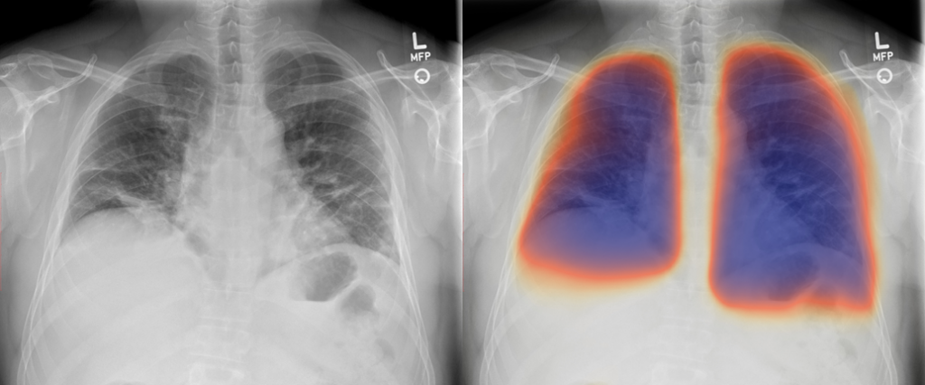
    We also looked at this from another perspective. There are far more x-rays in my practice than cardiac MRIs. Data are king when it comes to developing AI. Further, early and atypical pneumonias can be readily missed by trainees and even by experienced faculty. Could we use the same approach that we use to identify the mitral valve on cardiac MRI to find pneumonia on radiography? Could we enhance our x-rays with color, the same way we use 4D Flow to enhance cardiac MRI? Yes and yes.

    We did not anticipate that coronavirus disease (COVID-19) would become so prevalent and change our lives so dramatically, but as it emerged, we had the opportunity to test our AI algorithm on x-rays from some of the first patients diagnosed with COVID-19 pneumonia. Surprisingly, it tracked well with the severity of pneumonia. We have since received funding and support from the University of California Office of the President, National Science Foundation, NVIDIA/Groupware, Amazon, and Microsoft AI for Health to further explore this promising approach to AI.

    We did not seek initially to develop an AI algorithm to better diagnose and manage COVID-19 when exploring our strategy for cardiac MRI. However, it certainly goes to show that benefits from investments in research and education are not always linear and predictable. They create opportunities for young clinicians and researchers to tackle important questions that can ultimately shape the future of our field.


    Help The Roentgen Fund® provide support to talented young radiologists with your tax-deductible gift. 100% of your donation will go to funding scholarship and fellowship programs. Receive a special thank-you gift from ARRS when you contribute $200 or more, in addition to many other donor benefits.

  • Fear and Hope in Indiana: One Rad Tech’s Look at COVID-19

    Fear and Hope in Indiana: One Rad Tech’s Look at COVID-19

    Published August 25, 2020

    avatar

    Lizzy Rainey, R.T. (R)

    Franciscan Health Lafayette

    A radiologic technologist and respected painter in Lafayette, Indiana, Lizzy Rainey transforms complex medical images into what she calls “landscape anatomy.” Since 2008, her paintings have appeared on the covers of American Society of Radiologic Technologists scientific publications more than 21 times. Today, her work hangs in the Pentagon and in medical centers across the country.


    My story is about fear. It’s also about hope.

    I’ve been a radiologic technologist for nearly 40 years. At first, I felt confident we had a handle on this virus. I knew we could simply use our protective equipment, and it would keep us safe.

    I’ve survived working with devastating diseases before: TB, HIV, H1N1, meningitis, CDIFF, strep, influenza. In the past, we always had PPE, the proper training, and the knowledge of how to keep safe.

    This time, it didn’t take long to realize the world had changed overnight. Now, our protective tools are rare and guarded. For the first time in 40 years, I feel there is a chance some of us may not survive this one. The fear is very real.

    My coworkers and I continue to work with first-contact patients in the ER and patients on the floors, while using the best protective gear possible. But we often have to search for it, demand it, beg for it, and even attempt to create our own.

    Someone told me, “It’s like when you’re on vacation and run out of clean clothes, you wear your best dirty shirt.” But that statement offers little comfort when our lives depend on what we wear. As the days go by, more of the appropriate gear has started to become available. But not knowing when or if this situation will come to an end, or if our PPE will always be there, creates unrelenting stress for all of us.

    I’ve already seen my coworkers frightened, panicked, in tears. Yet everyone has somehow found their self-control and overcome the worst of their anxiety. So far, none of my colleagues have refused to do their job, even with their very real concerns.

    As a grandmother and technologist in my late fifties, working weekends in a busy hospital, I wonder when I will safely hug my kids and grandkids again. Even if they lift the social distancing order, I’m committed to keeping away from people for their safety.

    I find myself counting the days of the virus incubation period every week, wondering if I will have symptoms before the week ends and I return to the ER, just to begin counting the days again.

    There are no definite answers right now. My fear is real, but so is my hope. My hope comes from my faith in God, and I also paint. Yes, I paint my feelings and that brings me great hope. I’ve been painting portraits of my hospital colleagues and other technologists in their protective gear. Not necessarily portraits praising the individual but demonstrating the spirit of the job.

    The paintings show them in the midst of the crisis, working in Indianapolis, Indiana’s Methodist Hospital, draped in protective gear. One painting captures a moment after the successful completion of a dangerous procedure at my hospital, Franciscan Health in Lafayette, Indiana. My colleagues strike a pose, smile, and flash a peace sign. All this while enjoying the job. I hope, through my paintings, you can feel their compassion, purpose, and, yes, hope—even through the fear we feel every day.

    Lizzy Rainey’s essay and paintings are shared in collaboration with the American Society of Radiologic Technologists (ASRT), the premier professional association for the medical imaging and radiation therapy community. With more than 157,000 members, ASRT has a profound commitment to the ongoing support and advancement of radiologic technologists.n

  • A Commitment to Remain Engaged and Awoke

    A Commitment to Remain Engaged and Awoke

    Published August 18, 2020

    avatar

    Alexander Norbash

    2020–2021 ARRS President

    Do you believe that a radiologist deserves to comment on social inequities in a column such as this? Today, I believe that it is appropriate for me to do so. I do believe that, as a physician, I have a perspective that allows me to have an informed opinion where public health is concerned. And I believe that inequity, poverty, and racism are all public health problems. Whether or not they are solvable is beyond my understanding, given the human condition and history. If you feel that I have no right opining on such as this, I pray that you will forgive me this deviation.

    On Thursday, April 9, I sent my last editorial to our InPractice editor. At the time, the focus of my thoughts dealt with the COVID-19 crisis and all that we saw, anticipated, and feared at the time as a direct result. I thought we were facing a once-in-a-lifetime problem that would demand our full attention, cost us many lives and considerable hardships, and might take two or three years to address. I even assumed at the time that we would be facing some type of large-scale societal financial crisis, above and beyond the imminent financial crisis I thought our health system would be facing. We thought we would figure this out. I did not imagine I would see a bigger crisis than COVID in my time, and I certainly could not imagine my commenting on a bigger crisis in my three remaining editorials. I was mistaken. 

    That was a very long time ago. George Floyd was alive on that day, and he continued to walk, breathe, and live among us for the next six and one-half weeks. And then, everything changed for a brief time. We all stirred to awaken—and I fear we have rolled over and fallen asleep again—back into our highly factionalized and outraged dreams. Summed up, the consequences and impact of perpetual, persistent, and unrelenting racism subjugates the COVID crisis to pale by comparison.

    In this phase of my life, I believe that, as a healer, I should care about the health of individuals and society. Perhaps my span of interest should expand beyond just minding my business as an imager. Perhaps I should try to at least voice my thoughts about social inequities, and how those inequities erase multiple lifetimes of my professed service to society.

    Mr. Floyd was killed, and we witnessed his death. Many of us gathered around our understanding that over 400 years of oppression, we have been either beneficiaries, perpetrators, victims, or bystanders. If victims, we are rightfully outraged. If perpetrators, I have a difficult time characterizing or understanding the defensive sentiment. If bystanders and beneficiaries, then we are faced with the leaden and heavy realization of our complicity, and our contribution to the ongoing racial crisis. Collectively, the end result has been neglect and denial of a purely man-made evil, where we have literally failed to lift a finger to solve this crisis over the centuries. With Mr. Floyd’s killing, many of us were now embarrassed into some temporary form of consciousness, and sought to right 400-plus years of a wrong. Perhaps momentarily, we recognized that the overt outrage of human ownership had been allowed to transform into the covert insidiousness of multigenerational oppression, poverty, disempowerment, and irreversible disadvantage. And now, we thought ourselves awoken, and we almost mobilized to do something real. Closer than ever before. Then again, maybe not so close. I don’t know. Memory is brief and incomplete.

    Watts was big, with nearly a thousand buildings destroyed or burned over five days in 1965. Did anything substantive change after Watts? If so, why are there less black male medical students in 2020 than there were in 1973? Does that not shock us? Did Mr. Floyd also die in vain, as one of thousands? You would think that shock charges us to concrete and actionable charges to society. I don’t see the crisis seized upon, nor the corrective actions enabled. I saw large groups of committed individuals mobilized in the streets with my own eyes, propelled by the strength of their convictions and their fearlessness. And I don’t see where all that energy and passion was constructively directed.

    We are physicians and health care workers, which means that we not only care for the human physic, we realize the irreducible entanglements that the body has with society, spirit, and soul. Can we be radiologists and not care about lead levels in water supplies, intentional violence, and child hunger because we are only interested in the latent image? Can we be disinterested by social inequity? Can we be untroubled by injustice, when such injustice neutralizes multiple lifetimes of our collective best radiologist efforts?

    So, what are we supposed to do? Perhaps, at the least, we have to commit to not only work on equity in our immediate environment, we have to also recognize the insidious permanence of the condition. Yet there are also specific steps and actions that we can take.

    What are concrete steps we can take? Where our immediate environment is concerned, we are all in need of implicit bias training and bystander training. How else can we change our immediate environment unless we understand what is hurtful, and unless we each believe our duty includes stepping in and clearly addressing inequity? There are dozens of online implicit bias training modules that can remind us that each and every one of us suffers from implicit bias, and that this awareness of our implicit bias is the first step in controlling it.

    Bystander training is more complicated. It demands an understanding from each of us about when and how we would step in when we see something heinous, or even something just slightly hurtful. Alternatively, would we prefer to lower our heads and walk away to save ourselves? And then, following that, could we look at ourselves in a mirror? Learning about microaggressions and interventions is time consuming, and another ingredient that contributes to establishing a fair and equitable society.

    A higher level of commitment is that of outreach and pipeline creation, where we choose to go to such groups as disadvantaged elementary and high school students and tell them about radiology—taking along team members such as technologists and nurses, so these students can envision an alternative future for themselves. Even if we only touch a small number of lives, in and of itself, that has meaning and substance, and may result in further cycles of engagement and economic salvation. It takes so little additional effort to positively affect the lives of others.

    Only through vigilance and an understanding of the need for durable and enduring change can we hope for improvement. More than 400 years of inequity demand continuous and unrelenting pressure for change to occur. This is not a moment, or a project, or an initiative. It is a commitment for life to remain engaged and awoke, in order to better the condition of our brothers and sisters. For many of us, it is our understood duty.

  • Pediatric COVID-19 and MIS-C: What Lies Beyond Pneumonia?

    Pediatric COVID-19 and MIS-C: What Lies Beyond Pneumonia?

    Published August 10, 2020

    avatar

    Alexandra M. Foust

    Department of Radiology
    Boston Children’s Hospital, Harvard Medical School

    avatar

    Ricardo Restrepo

    Department of Radiology
    Nicklaus Children’s Hospital, Florida International University

    avatar

    Edward Y. Lee

    Department of Radiology
    Boston Children’s Hospital, Harvard Medical School

    Since the publication of our initial AJR article, “Pediatric SARS, H1N1, MERS, EVALI, and Now Coronavirus Disease (COVID-19) Pneumonia: What Radiologists Need to Know”, the coronavirus disease (COVID-19) pandemic has continued to grow—more than 15.7 million cases and 640,000 deaths worldwide, as of July 26, 2020. During this time, understanding of the imaging manifestations related to pediatric COVID-19 pneumonia, and the more newly defined COVID-19-related entity multisystem inflammatory syndrome in children (MIS-C), has continued to increase; however, substantial uncertainty regarding the imaging findings of pediatric COVID-19 and MIS-C still exist. Our article highlights a few key points regarding what is currently known about the imaging findings of pediatric COVID-19 and MIS-C for practicing radiologists.

    What is Typical Pediatric COVID-19?

    A recent meta-analysis of 7,780 pediatric patients positive for COVID-19 found that the mean age of patients was 8.9 years with a slight male predominance (55.6%). Underlying comorbid medical conditions were identified in 35.6% of patients. Overall, pediatric patients demonstrated a more mild clinical course than adults with 19.3% of patients completely asymptomatic, 3.3% requiring intensive care, and only 7 reported deaths (0.09%). The most commonly observed clinical complaints among symptomatic patients were cough and fever, and elevated inflammatory markers such as C-reactive protein, procalcitonin, and interleukin-6 were frequent.

    Radiographically, typical imaging findings of pediatric COVID-19 pneumonia have been characterized as bilateral peripheral and/or subpleural ground-glass opacities and/or consolidation in a lower-lobe predominant distribution.

    Fig. 1—13-year-old female with obesity and positive COVID-19 reverse transcription–polymerase chain reaction test who presented with fever and severe shortness of breath. (A) Frontal chest radiograph shows bilateral lower lung zone-predominant consolidation and ground-glass opacities, which are typical chest radiographic findings of pediatric COVID-19 pneumonia. (B) Axial lung window CT image demonstrates diffuse peripheral predominant ground-glass opacities in both lungs.

    Although a unilateral or bilateral distribution of parenchymal abnormality may be observed in pediatric COVID-19 pneumonia, the differential for unilateral disease is somewhat broader. Thus, a unilateral distribution has been defined as indeterminant. The halo sign, a rounded consolidation surrounded by a rim of ground-glass opacity, can be seen during the early phase of pediatric COVID-19. Therefore, the halo sign is also considered typical when present in an immunocompetent patient, as it has a narrow differential. Additional important considerations for radiologists are atypical imaging findings that raise concern for alternative diagnosis, including centrilobular nodules, focal segmental/lobar consolidation, cavitary lesions, pleural effusion, and lymphadenopathy.

    What is Multisystem Inflammatory Syndrome in Children (MIS-C)?

    As suggested by the name, MIS-C is a post-viral inflammatory syndrome observed in pediatric patients with prior COVID-19 infection (within the past 4 weeks) that results in injury to multiple organ systems, most frequently involving > 4 systems. Some researchers have called MIS-C a “Kawasaki-like” disease due to the potential overlap in clinical presentation, including fever, conjunctivitis/rash, cardiac dysfunction + coronary artery dilation, and hemodynamic instability. Unlike pediatric COVID-19 pneumonia, children who develop MIS-C demonstrate a more severe clinical course, requiring intensive care management in up to 85% of cases, and have a higher death rate of 4%. As might be expected, the imaging findings in MIS-C differ from those observed in typical pediatric COVID-19 pneumonia.

    What Are Important Differences Between Typical Pediatric COVID-19 and MIS-C?

    Thoracic Findings

    Chest imaging studies in pediatric patients with MIS-C may be normal or may demonstrate abnormalities, mainly related to underlying cardiac dysfunction. On chest radiograph, this may manifest as cardiomegaly, increased prominence of pulmonary vasculature, interstitial edema, or pleural effusions.

    Fig. 2—15-year-old male, reverse transcription–polymerase chain reaction test positive for COVID-19, who presented with MIS-C associated with COVID-19, with symptoms including fever, vomiting, and diarrhea. Frontal chest radiograph shows cardiomegaly. Subsequently obtained echocardiogram demonstrated dilated left ventricle and moderate systolic dysfunction.

    Chest CT may show similar findings and, in some cases, may demonstrate pericardial effusions, coronary artery dilation, and/or pulmonary embolism. Echocardiograms often demonstrate left ventricular dysfunction and/or reduced ejection fraction, as well as pericardial effusion, and/or coronary artery dilation.

    Whereas typical COVID-19 pneumonia presents with bilateral peripheral and lower-lobe predominant ground-glass opacities and consolidation, the distribution of pulmonary parenchymal abnormality in MIS-C tends to be central and perihilar in distribution and more frequently presents as increased pulmonary vascularity, although airspace consolidation may be seen in advanced stages of cardiac failure. Additionally, cardiomegaly and pleural/pericardial effusions are often observed in MIS-C, but they are rare in pediatric COVID-19 pneumonia.

    Extra-thoracic Findings

    Extra-thoracic manifestations are not generally observed in pediatric COVID-19 pneumonia. However, as may be expected in an inflammatory disorder involving multiple organ systems, extra-thoracic findings are not uncommon in MIS-C—especially in the abdomen. Reported intra-abdominal abnormalities in MIS-C include bowel wall thickening, ascites, right lower quadrant fat stranding and/or lymphadenopathy, hepatomegaly, gallbladder sludge and/or pericholicystic fluid, and increased renal cortical echogenicity.

    Fig. 3—7-year-old girl, reverse transcription–polymerase chain reaction test positive for COVID-19, who presented with MIS-C associated with COVID-19, with symptoms including fever, vomiting, abdominal pain, and hypotension. Transverse grayscale ultrasound image of the right lower quadrant shows thickened bowel loops (arrows) and ascites (asterisk).  

    Five Take-Home Points for Diagnostic Radiologists

    1. “Typical” pediatric COVID-19 pneumonia presents as bilateral peripheral and lower-lobe predominant ground-glass opacities/consolidation + halo sign.
    2. Centrilobular nodules, parenchymal cavitation, focal lobar/segmental consolidation, pleural effusion, and lymphadenopathy are atypical in pediatric COVID-19 pneumonia.
    3. MIS-C has a more severe clinical course than pediatric COVID-19 pneumonia, often involving > 4 organ systems, with up to 85% of patients requiring intensive care.
    4. Thoracic imaging findings observed in MIS-C, including cardiomegaly, pleural/pericardial effusion, coronary artery dilation, or pulmonary embolism, differ from typical findings in pediatric COVID-19 pneumonia.
    5. Extra-thoracic manifestations are not uncommon in MIS-C and generally are a manifestation of inflammatory change (bowel wall thickening, lymphadenopathy, fat stranding, pericholicystic fluid, ascites) and/or organ dysfunction (hepatomegaly, increased renal cortical echogenicity).

    As our understanding of pediatric COVID-19-related disease continues to grow, it is essential for practicing radiologists to be aware of the imaging findings in this patient population. Additionally, as the imaging features are quite different, awareness of the differences between pediatric COVID-19 pneumonia and MIS-C are critical to accurate diagnosis and optimal management of pediatric patients.

  • New Rituals for Radiology Residents During COVID-19

    New Rituals for Radiology Residents During COVID-19

    avatar

    Lekui Xiao

    Mayo Clinic

    The COVID-19 pandemic began to impact our program (Rochester, MN) in mid-March, requiring rapid and near constant changes to the entire curriculum. Our chief residents divided us into teams—with one team responsible for call coverage and mandatory rotations, and others on back-up and remote learning. Our program organized daily case-based teaching sessions with staff and distance curricula. This time felt eerily reminiscent of the first two years of medical school, due to being somewhat removed from significant clinical duties! Regular communication with program leadership and co-residents helped alleviate a lot of the stress brought forth by COVID-19.

    Around mid-May, we began transitioning back into clinical rotations. Although it is terrific to be back in the reading rooms, it is clear that we’re in a “new normal” for at least the immediate future. The number of people in attendance at noon conference is limited to accommodate social distancing, with many residents watching remotely. Masks, face shields, and meticulous sanitation have become a ritual.

    The month of July has been uniquely different because our new first-year residents just started. They would usually interact with the upperclassmen and staff through informal gatherings and welcome parties outside of work. However, because of institutional COVID-19 precautions, social opportunities to welcome them have been limited.

    Now is a memorable and unique time to be a radiology resident, as we have faced many challenges. I am eager to resume pre-COVID traditions and social activities with a new appreciation for my residency’s supportive learning environment.

  • Radiology Residency Looks Different with COVID-19

    Radiology Residency Looks Different with COVID-19

    avatar

    Sean Golden

    The Johns Hopkins Hospital

    The Johns Hopkins Radiology Residency, like most training programs across the country, has been deeply affected by the COVID-19 pandemic.

    In late March, our program temporarily transitioned to a skeleton staffing model, where the majority of residents stayed home and were unable to read studies remotely. Key rotations were delayed or cancelled, and two residents were called upon to staff the ICU. To mitigate the diminished case volumes, faculty and fellows created daily virtual didactic sessions to supplement our regular morning conferences and independent study. Special emphasis was placed on preparing the first-year residents for independent overnight call, which we begin in July of our second year at Hopkins. An innovative online call-prep curriculum was created by our associate program director, where first-years independently reviewed DICOM images of previous overnight studies, submitted an impression to an online dropbox, and then reviewed the pertinent findings with an attending radiologist.

    In May and June, COVID-19 numbers in Maryland began to decline, and our program gradually returned to a relative state of normalcy (albeit socially distanced). Now, as numbers in Maryland once again climb, the hospital leadership has asked for another group of volunteers to assist in the ICU and on the floors.

    The COVID-19 pandemic has created unprecedented challenges for our faculty and residents alike, but it has also highlighted the department’s ability to adapt. Our reading rooms and conferences may look a little different than last year, but our commitment to education, research, and patient care carries on.

  • New Words for Radiology Residents During COVID-19

    New Words for Radiology Residents During COVID-19

    avatar

    Joshua T. Olson

    Mayo Clinic

    The coronavirus disease (COVID-19) pandemic brought with it a new lexicon of phrases to describe daily resident life. At our radiology residency program at the Mayo Clinic in Rochester, Minnesota, we don appropriate “personal protective equipment,” or “PPE,” for clinical encounters and at the reading station. We also practiced “distance learning” for a month, rotating between essential rotations and studying from home as our institution prepared for COVID-19. We came away with a renewed appreciation for the value of one-on-one teaching and the irreplaceable learning that comes from the patients we are humbled to serve. We understand the concept of “hyper-locality” and how our local COVID-19 environment is radically different from others.

    Similarly, we have been presented with new challenges. The rescheduling of the American Board of Radiology Core Exam has necessitated multiple redesigned call schedules. With the necessity for safe distancing practices, residents have found creative strategies to collaborate and celebrate milestone personal and professional achievements. Attentiveness and flexibility in meeting the ongoing and changing educational and personal needs during this stressful time have promoted cohesiveness and trust between residents and our program’s leadership. 

    Integrating these adaptations will allow us to grow stronger as we look toward the post-COVID-19 resumption of a “new normal” residency environment. The Mayo Clinic’s history is one of perseverance and innovation, borne in the wake of a devastating tornado which nearly destroyed the town almost 140 years ago. Together, I am confident our residency will persevere through the present pandemic—maintaining our strong tradition of innovation to ensure a world-class training environment, prioritizing resident wellness and the old adage that “the needs of the patient come first.”

  • An Action-Based Radiology Residency for COVID-19

    An Action-Based Radiology Residency for COVID-19

    avatar

    Lei Yu

    University of Nebraska Medical Center

    Unprecedented times call for unprecedented actions. The past few months have been the most uncertain and challenging time in our lives, and we are all doing the best to adapt.

    In mid-February, Nebraska Medicine quarantined 13 passengers of the Diamond Princess cruise ship in our National Quarantine and Biocontainment Units. The awareness and preparedness for COVID-19 started early among our institution, including focused refresher training on PPE use for all the residents to ensure safety during patient care. 

    Our department and the residency program reacted to the pandemic with a proactive mindset, including postponing non-urgent outpatient imaging studies and procedures, adopting structured virtual didactics, and altering our reading room layout to follow social distancing guidelines. As COVID-19 cases rapidly climbed nationwide in late March, rotational changes were made based on the waves of teams approach. We divided our residents into two teams: one working in the hospital to fulfill clinical duties, the other working from home to ensure a healthy workforce as a backup. Our schedule was very flexible and gave special considerations to residents who were in need of childcare due to closure of schools and daycares, as well as for family emergencies. In order to facilitate our educational program during these trying times, many online educational resources were made available. Our department always kept our wellness in mind—arranging for snacks and beverages to be brought into the reading room every afternoon, which was something we all looked forward to.

    Lack of information can be unsettling in times of uncertainty. To keep us informed, our program sent out timely COVID-19 communications to provide updates and policy changes at both local and national levels, along with wellness resources to help cope with both internal and external stresses. 

    A silver lining from this pandemic is that it allowed us to pause and appreciate our life, our health, and our families. In the wise words of Marcus Aurelius, “The impediment to action advances action. What stands in the way becomes the way.” Look forward to the post-pandemic time, when we shall become stronger.

  • Radiology Residency in the Midst of COVID-19

    Radiology Residency in the Midst of COVID-19

    Published July 15, 2020

    avatar

    Ian Y.M. Chan

    Chief Radiology Resident, Western University
    Chair, ARRS Resident Advisory Subcommittee

    Published July 15, 2020

    Like many facets of life, radiology residency has not been immune to a new reality wrought by the coronavirus disease (COVID-19) pandemic. As one of the first cities in Canada to have a confirmed case of COVID-19, the novel coronavirus was certainly top of mind among our residents, fellows, faculty, and staff at Western University in London, Ontario, Canada. As residents, we worked closely with our program director to advocate for necessary changes to our program to ensure our safety while continuing our education during this pandemic.

    As our government mandated physical distancing and temporary business closures, our department also acted in concert and canceled most outpatient imaging. As a result, we noticed a sudden drop-off in cases for residents to report during the day. While after-hours resident call duties remained, we modified our daily work routine for two months by implementing a schedule of alternating a week of working in hospital and a week of self-studying at home. Importantly, we continued to receive dedicated radiology teaching as our “hot seat” case conferences and academic half-day lectures proceeded as scheduled via video conferencing.

    There was also the looming anticipation of our redeployment to other clinical services. This has not yet transpired, largely due to public health policies that have slowed community transmission for the time being. Fortunately, we have had access to appropriate personal protective equipment when caring for patients. Stress management has indeed been especially crucial to our wellness during this trying time. I believe that constant, albeit distanced, social interaction with colleagues and friends has helped emphasize our shared experiences and that “we are all in this together.”

    The Greek philosopher Heraclitus once said, “change is the only constant in life.” During this harrowing time, this axiom still rings true with our residency training as we adapt to this pandemic.

    Stay safe and be well!

  • Breast Imaging: One Size Does Not Fit All

    Breast Imaging: One Size Does Not Fit All

    avatar

    Linda Moy

    Professor of Radiology NYU School of Medicine Center for Advanced Imaging Innovation and Research
    Laura and Isaac Perlmutter Cancer Center

    Published June 22, 2020

    Breast cancer is the most common cancer in women worldwide with approximately 2 million cases diagnosed each year. In the United States, breast cancer is the second leading cause of cancer-related mortality among women. Multiple studies showed that regular screening mammography reduced breast cancer mortality by 40% or more. However, this year, we learned that men at high risk of developing breast cancer may benefit from mammography.

    Screening of Men at High Risk for Breast Cancer

    Researchers at NYU School of Medicine conducted the largest review in the United States of the medical records of men who have had a screening mammogram. The study involved 1,869 men, ages 18 to 96, who had a mammogram between 2005 and 2017. Some men sought testing because they felt a mass in their breast, while others had no symptoms and wanted to be screened because a family member had recently received a breast cancer diagnosis. In total, 41 men were found to have breast cancer, as confirmed by breast tissue biopsy. Among the 271 men who had screening exams, 5 had the disease. All those with breast cancer had surgery (mastectomy) to remove their tumor. A key finding was that mammography was more effective at detecting cancer in men with high risk than is the norm for women with average risk of breast cancer. For every 1,000 exams in these men, 18 had breast cancer. By contrast, the detection rate for women is roughly 5 for every 1,000 exams.

    Among the study’s other main findings was that men who had already had breast cancer were 84 times more likely to get it again than men who had no personal history of the disease. Men with an immediate relative who had breast cancer, such as a sister or mother, were three times more likely to develop the disease. Other men with elevated risk of breast cancer included those of Ashkenazi descent, an ethnic group widely known for high rates of some cancers (who were 13 times more likely to get breast cancer than non-Ashkenazi men) and those who had genetic mutations, such as BRCA1 or BRCA2 (up to 7 times more likely than men with no genetic risk). Current National Comprehensive Cancer Network Guidelines only recommend checking for breast cancer as part of annual physical exams, not using more sensitive imaging tests like a mammogram, for men age 35 and older with BRCA mutations. The take-home point is that men need to be more aware of their risk factors for breast cancer and that they, too, can develop the disease.

    Contrast Enhanced Spectral Mammography

    Another exciting development is that the armamentarium that radiologists have to detect breast cancer continues to expand. The latest imaging tools focus on functional imaging tests that reveal physiological activities within the breasts. Functional imaging includes measuring changes in metabolism (e.g., PET/CT or PET/ MRI), changes in the tumor cellularity (diffusion weighted imaging on MRI), regional chemical composition (MR spectroscopy, sodium MRI) and changes in blood flow. The increase in the number of blood vessels (neoangiogenesis) and the increased permeability of blood vessels that feed a tumor are the two main factors that account for uptake of gadolinium that leads to the enhancement of breast cancers on MRI. But MRI is expensive, and we know that iodine based contrast may also be used to detect breast cancer. As a result, contrast enhanced spectral mammography (CESM) is being used in both the screening and diagnostic setting. Similar to breast MRI, CESM identifies the increased blood flow associated with breast cancer and is largely independent of breast density.

    Compared with MRI, clinical implementation of CESM is much easier and at much lower costs. CESM requires an intravenous injection of iodinated contrast (dose 1.5 ml/kg) and a dual energy mammography system. A “low-energy” acquisition image resembles a normal mammogram, whereas a “high-energy” image, using a keV above the k-edge of iodine, will enhance with the contrast agent signal. Images are performed in the standard craniocaudal and mediolateral mammographic views. The postprocessing, recombined iodine-only mammograms will identify enhancing lesions. Studies show that CESM has good diagnostic accuracy when used to evaluate the extent of disease in women newly diagnosed with breast cancer. Like breast MRI, CESM outperforms combined mammography and ultrasound in the detection of additional disease and in assessment of tumor size, compared with pathology. The literature reports that CESM has a small reduction of sensitivity, while providing a higher specificity compared with breast MRI in the evaluation of tumor extent.

    In addition, CESM works well as a screening exam with an additional cancer detection rate 6.6 – 13.1/1,000 over conventional mammography. Therefore, it may be feasible to screen a larger population of women (e.g., women at moderately increased risk for breast cancer), and it may be advantageous to use CESM over breast MRI in this subset of women. Overall, CESM is a safe technique with a modest increase in the radiation dose compared to conventional mammography. Also, serious adverse contrast reactions are infrequent. Currently, the lack of a CESM-compatible biopsy device for lesions exclusively seen on CESM is a limitation of this new technique.

    Abbreviated Breast MRI and Ultrafast MRI

    Recently, many authors have evaluated the potential of an abbreviated breast MRI to increase the accessibility of breast MRI, especially for the screening of women at above-average risk for breast cancer. The conventional breast MRI exam is a 30-minute examination that is expensive and not well tolerated by some patients. These factors, along with the limited availability of MRI scanners, preclude population-wide screening with breast MRI. Abbreviated MRI, with shorter image acquisition and interpretation times, may increase the availability of breast MRI and reduce the costs. The basic abbreviated breast MRI protocol includes a pre-contrast and one post-contrast T1-weighted imaging, along with subtraction images and maximum intensity projection images. Multiple variations on this basic protocol have been evaluated. These protocols exploit the high sensitivity of MRI, while reducing acquisition and interpretation times. A recent review of 21 studies on abbreviated breast MRI, performed in eight different countries and in over 4,500 women, confirmed the diagnostic accuracy was similar to the full breast MRI protocol.

    With stronger magnets and improvements in breast coils and MRI software, ultrafast sequences have been developed to measure the rapid arterial perfusion and the rapid venous drainage of breast cancers. The temporal resolution of ultrafast protocol is typically less than 10 seconds/frame and may be incorporated into abbreviated or full breast MRI protocols. The hope is that imaging faster may allow radiologists to better distinguish between benign enhancing lesions and background parenchymal enhancement from breast cancers. Ideally, ultrafast MRI sequences may allow for increasing the specificity of abbreviated breast MRI, without increasing the scan time.

    Artificial Intelligence

    Without a doubt, artificial intelligence (AI) is the most talked about new diagnostic development in the field of radiology. Breast imaging is at the forefront of this research because we have decades of experience using computer-aided detection (CAD). Further, similar to chest radiography, large numbers of screening mammograms are available to be converted into datasets to train these AI algorithms. These new deep learning–based CAD models are proliferating due to recent breakthroughs in computer technology, data science, and algorithm development. Computer processing speed and memory have increased exponentially, owing to faster graphics processing units and parallel processing. Simultaneously, there have been mathematical advances that enabled the use of complex and multilayered neural networks, which led to a markedly improved performance of machine interpretation of highly standardized imaging tasks (e.g., predictions of cancer or no cancer).

    New CAD platforms will differ from traditional CAD in several important ways. Some of these deep learning models no longer require manual feature design and minimize training with humans (i.e., radiologists). These AI algorithms learn discerning features that are best predictive of outcomes independently and may identify novel imaging features that are imperceptible to the human eye. The capacity for continuous feedback and learning will allow deep learning–based CAD to improve over time. In theory, deep learning algorithms can be trained for pattern recognition of image data (pixel-related information), correlate that data to tumor registry data (the truth), and assess risk when it recognizes a similar pattern (predict likelihood of cancer). Further feedback into the AI algorithm of whether that prediction is correct and truth-based will improve its performance in the future. New CAD systems may eventually be able to identify novel features associated with more relevant cancers by incorporating patient- and tumor-level variables—a task that is now performed in small groups of patients, usually in the research setting. This design has the potential to maximize the mortality benefit of breast cancer screening and to address the issues of overdiagnosis and overtreatment. Therefore, there is hope that these deep learning algorithms may hold real potential to improve clinical care.

    Clinical Case-Based Review of Breast Imaging

    Focus on honing your breast imaging diagnostic skills. This special course combines didactic lectures and rapid fire case review sessions to provide deeper insights into breast imaging.

    The literature on these AI algorithms for mammography shows that their performance for lesion detection and classification are approaching that of radiologists. A preliminary study showed a similar diagnostic accuracy for an AI algorithm that evaluated screening digital breast tomosynthesis exams. It is anticipated that these AI algorithms will play a major role in screening in the near future, both to improve the quality of the screening programs and to assist with the increasing workload of interpreting screening mammograms. Furthermore, early studies suggest that when AI models predict a very low likelihood of malignancy, these mammograms may be triaged and interpreted by the algorithm alone, saving time and resources.

    Other potential applications for screening mammography deep learning models beyond lesion detection and classification include assessment of mammographic breast density. This quantitative analysis of breast density is important because supplemental screening is recommended in women with mammographically dense breasts. A more recent development is utilizing these AI algorithms to predict a woman’s risk for developing breast cancer in the future by incorporating the normal mammographic parenchymal pattern (density, texture, etc.). Risk assessment may be further personalized when information from the electronic health record is included in deep learning risk models. The addition of radiogenomics, which combines radiologic phenotypes with underlying genetic signatures, has the potential to add relevant tumor and patient predictive and prognostic information using information extracted from images.

    Although there is a lot of excitement about AI, many experts urge caution because these AI tools haven’t been evaluated in a wide variety of clinical settings. Most studies on AI and breast imaging are retrospective enriched reader studies. To increase the generalizability of the results, prospective studies in different patient populations should be performed.

    It is clear that the “one size fits all” approach may no longer be relevant. Instead, the standard mammograms, ultrasounds, and breast MRI exams are being tailored for specific clinical indications, often augmented with AI tools.

  • Radiologists, Now More than Ever

    Radiologists, Now More than Ever

    avatar

    Alexander Norbash

    2020–2021 ARRS President

    Published June 22, 2020

    On Thursday, March 19, the governor of California issued a mandate for state citizens to stay at home and not congregate, except for essential and emergency needs. I manage the radiology department at the University of California-San Diego, and needless to say, this mandate was therefore of particular and great interest to me. At the time, I saw this as a brave and decisive move on the governor’s part. Given the lack of other governors showing the wisdom of consensus in such an action, I also felt this instance of decisiveness demonstrated a high degree of confidence in his advisors and his own perspective. Many of us were paying attention to a number of news stories that suggested our lives were about to be significantly disrupted.

    Ever since December 2019, as with many of my acquaintances and family members, I had been following at a distance and with one eye the epidemic in Wuhan. My attention and interest increased exponentially as December proceeded and Wuhan locked down tighter and tighter, then as December turned to January with mounting casualties, and more so as various reports regarding the impact of the epidemic and control measures on citizens came to light. Perhaps many of us even preceding this pandemic have a personal story regarding the invisible erstwhile masters of the universe, or something related. I certainly did. My personal story dealt with the swine flu vaccination of 1976. I was very close to my maternal grandparents. My maternal grandfather was on a ventilator for two weeks, and at one point, he could only blink due to Guillain-Barré syndrome. We held our own breaths as he very slowly improved. It took him two long years to fully recover. As a consequence, I was fascinated by virology in medical school as a result of my grandfather’s misadventure, which regrettably was about 34 years in the past. Perhaps I paid just a little more attention to epidemics and cures than the average citizen.

    Somehow, seeing two hospitals erected in one week—the first with 1,000 beds and the second with 1,300 beds—was a firm nudge in the ribs for me. Firstly, I felt that was an impossibility for any of the environments that I have worked, meaning if we ever had to add a thousand hospital beds in a week, we couldn’t possibly do it. Second, it underscored the seriousness of the situation and that the epidemic may well be headed to our shores.

    So, I read up on the popular culture sounding boards dealing with the 1918 influenza epidemic, SARS in 2002, H1N1 in 2009, and MERS in 2012. Naturally, I also went back to the bubonic plague. Reading did not build up my confidence. 

    As the shelter edict was issued in California, immediate and rapid changes in our delivery of health care took place. These changes influenced and affected our faculty, our residents and trainees, our patients, our clinical operations, our research, and our staff. Research came to a near grinding halt, and clinical volume dropped by 75% overnight, levelling off at 45% down. We wrestled with a span of consequences, from “how do we teach remotely?” to “what do we do with idle staff?” 

    Dr. Norbash explains how The Roentgen Fund® supports innovation in the radiology field.

    Our elective cases nearly instantaneously and precipitously dropped in number as patients sheltered at home, and we realized how much of our volume was truly nonurgent and nonemergent. Our residents and technologists and faculty bravely and selflessly provided the same exceptional level of service they had always provided, only now with the understanding and belief that there is increased risk of harm to self. We watched the calendar and counted the days, preparing to the best of our abilities for the inevitable tsunami to hit. Our leadership assembled emergency plans with exceptional sophistication and creativity, doubling our ICU beds and creating standard operating procedures overnight in droves. Nearly three weeks later, the tsunami hasn’t hit us. We have, however, watched closely and day by day as our worst nightmares unfolded across the country, in New York City, where unparalleled compassion, ingenuity, and creativity have been demonstrated by our remarkable colleagues. The rest of us are petrified that what New York City is experiencing could be our future, and we convince ourselves that our lesser population density and lack of success with mass transit somehow protects us.

    We really don’t know when and how this is going to end. Maybe, by the time you read this, the whole thing is over and solved. Maybe we are seeing multiple tsunamis scattered across the country battering us on a daily basis. As I write this, we certainly don’t know if San Diego is relegated to a gentle slope for an infinity, necessitating masks and social distancing forever, rather than a bump or a tsunami with an implied and potential resolution of sorts. In the meantime, we are relieved to have been spared massive carnage up until now, although there still are lost lives with the accompanying scarring, sorrow, and regret one would expect. In the meantime, we are trying to understand how to provide our faculty and staff some semblance of a paycheck when revenue is down by 40%, giving them a heightened sense of purpose with a sprint that is turning into a marathon, which will be progressively more difficult if and as this crisis stretches into the summer, fall, and possibly beyond. Not knowing is the most difficult part. We need to salvage the critical, scientific, and teaching missions that distinguish us, in preventing irreparable damage to what keeps us distinct and gives us unique value: teaching and discovery.

    Many predict that the lockdown will be extended to the end of May, and some predict the end of June. Just today, a newsfeed quoted an extremely influential billionaire stating we won’t be over COVID-19 until Fall 2021. There’s much discussion regarding how much unemployment and financial instability we collectively will tolerate. We, as a people, are not in all instances risk averse. After all, we have states where motorcycle riders can ride without helmets, and legions of humans still smoke cigarettes. We also have too many people who suspect the value of immunizations. There is also much discussion regarding how we will deal with multiple recurrent waves of COVID-19 rising proportionately with our inevitable societal lapses in vigilance and awareness, if COVID-19 becomes an annual affair. 

    In the moment, I am inspired by my colleagues throughout the health system. My fellow radiologists are optimistic and creative, perpetually showing their innovative and flexible spirit. Every day there are new solutions and new approaches percolating among them, as they do their best to ensure optimal deployment of our tripartite mission. All this in graceful partnership with technologists, nurses, front desk staff, and trainees. Our brilliant departmental resident AI scientists have even deployed an intelligent tool in our PACS that will catch pneumonias, which may be too subtle for the naked eye to see. In the middle of this crisis, there are blinking flashes of creativity going off like lightbulbs. Everywhere under this roof, radiologists as tinkerers and creative spirits who are solving problems, as we elevate and illuminate each other’s vision. 

    I can’t imagine a better group of compatriots to have in my lifeboat.

  • Andrew Rosenkrantz Named 13th Editor in Chief of AJR

    Andrew Rosenkrantz Named 13th Editor in Chief of AJR

    Published May 10, 2020

    Andrew B. Rosenkrantz

    Come July, the future of the 113-year-old American Journal of Roentgenology (AJR) will rest in the hands of “one of the most widely published researchers in academic radiology” (Radiology Business Journal).

    As prolific as his city is sleepless, Andrew B. Rosenkrantz of New York University has edited the textbook MRI of the Prostate: A Practical Approach and authored or co-authored more than 350 peer-reviewed publications, all while training some 40 clinical fellows and mentoring over 80 residents resulting in publication. As NYU Grossman’s Professor of Radiology and Urology, Director of Prostate Imaging, Director of Health Policy, and Section Chief of Abdominal Imaging, he thrives in every last one of those professional titles, too.

    For this professional society, in particular, Rosenkrantz remains so much more.

    An ARRS member since 2004, he has received both the 2014 Melvin M. Figley Fellowship in Radiology Journalism and the 2017 Leonard Berlin Scholarship in Medical Professionalism. In addition to starring roles with ARRS’ Publications and Practice Improvement Committees, Rosenkrantz serves on the Scientific Program Subcommittees for Genitourinary Imaging, Efficacy, Administration, and Informatics.

    Speaking on his appointment to AJR’s chief chair, Deborah Baumgarten, ARRS Publications Committee chair, said, “It became clear during the selection process that Andy Rosenkrantz is visionary, dedicated, proactive, and really quite brilliant.”

    InPractice spoke with AJR’s soon-to-be editor in chief—a creative and affable man who, despite being aged much closer to the left side of 40, was named AuntMinnie’s Most Influential Radiology Researcher of 2018 and can already measure his CV in plain-text kilobytes.

    InPractice: You will be just the 13th chief editorial officer of the world’s oldest continuously-operating radiological journal. For context, when did you first encounter “the yellow journal?” And what does taking the reins from someone like Thomas Berquist mean to you now?

    Andrew B. Rosenkrantz: I began reading AJR early in residency, around the time that Robert Stanley began as Editor in Chief. At the time, given their educational value, I was drawn to the journal’s clinically-oriented research and image-rich review articles. Indeed, it was quickly clear that radiologists could rely on each issue to provide a wealth of practical content and that staying abreast of the journal’s latest articles would help in learning to be a clinical radiologist. I’ve remained an avid reader since that time, including throughout Berquist’s tenure. During his many years at the helm, Berquist has worked tirelessly on behalf of the quality and integrity of the journal’s content and launched a staggering array of pilots and new initiatives to the benefit of the journal’s authors, reviewers, and readers. It is an enormous privilege, though also humbling, to now have this opportunity to follow Berquist in this role.

    IP: From 2012–2015, you were AJR’s CME Consulting Editor for Genitourinary Imaging; currently, you’re one of the journal’s five Genitourinary Imaging Assistant Editors, a position you’ve held with distinction since 2014. To what do you attribute your success at AJR?

    ABR: I’ve benefitted greatly from the AJR as a practicing radiologist, and I have felt that it’s been important to give back and serve the journal as opportunities to do so have arisen. Over the years, I’ve been fortunate to have been provided chances to support the journal in these various editorial board capacities, and I have sought to make the most of these roles. I’ve also come to recognize the importance of the entire editorial team in enabling the journal to thrive, and I look forward to empowering a new generation of editorial board members to continue to shape the journal.

    IP: You published your first article in AJR in 2010, and since that February issue, you’ve authored and co-authored some 60 articles, letter-to-editor replies, and guest editorials for the journal. Given your wide-ranging interests, as well as that “Most Influential Radiology Researcher of 2018” laurel from AuntMinnie, what is it about AJR, specifically, that’s drawn and kept your attention?

    ABR: Even as my own research interest have evolved, the AJR has remained a primary journal in which to try and publish. AJR publishes articles on a wide range of topics, covering all areas of radiology practice. Despite this breadth of the journal’s content, it has maintained a compelling track record of publishing articles that are clinically impactful and will make a difference in radiologists’ practice. The journal’s editorial board has done an impressive job of staying in touch with its readership and knowing what articles its readers will find interesting and relevant to their day-to-day work.

    IP: Meanwhile, you’ve been “Rocking the Review” for AJR for more than a decade, receiving the Top, Outstanding, and two Distinguished Reviewer Awards. How does the implied dichotomy here (author vs reviewer) influence your overall approaching to medical publishing?

    ABR: Authors and reviewers need to work together to produce the highest-quality final accepted manuscripts. Reviewers must recognize their role as not just advising whether to accept or reject a submitted paper, but to provide the critical feedback that will fundamentally improve the paper. Authors must take the reviewer feedback seriously and be as responsive as possible in revising their work. The AJR will focus on strategies for best engaging and serving both of these important groups.

    IP: Similarly, as the recipient of ARRS’ Figley Fellowship and Berlin Scholarship, how have these two Roentgen Fund® accolades— the first for journalism, a second for professionalism—informed your subsequent research and practice?

    ABR: The Figley Fellowship provided a unique opportunity to learn the inner workings of the journal and its operations. I was invited to spend time at ARRS headquarters in Leesburg, Virginia and work closely with the journal staff—observing all the steps in the review and production pathway, from manuscript submission to publication. That experience laid a key foundation for an even deeper level of involvement with the journal in the following years. I dedicated the Berlin Scholarship to exploring issues relating to diversity among radiologists pursuing research and publication, encompassing projects seeking to not only understand challenges and barriers, but also strategies and opportunities for change. Likewise, this work will be important in guiding the journal in the coming years.

    IP: As the incoming Editor in Chief, do you foresee a more equitable union of, say, the types of informatics research you’ve been pursuing at the Neiman Health Policy Institute with the more diagnostic content for which AJR has long been heralded?

    ABR: No question, AJR has been a home for outstanding research and reviews in health policy, along with the journal’s more traditional diagnostic content. A large part of the journal’s appeal has been the inclusion in each issue of articles addressing policy, quality, informatics, and other aspects of modern radiology practice management. More recently, the journal has introduced “Best Practices” articles that provide an evidence-based assessment to guide radiologists in addressing focused clinical questions. These articles have quickly become very popular with the journal’s readership and will become an even more frequent component of the journal in the coming years.

    IP: These days (and especially with AJR), a scientific journal’s impact factor is a lot more than just a number. Can you explain your philosophy concerning impact factor at large?

    ABR: The impact factor reflects the number of citations in a given year to the journal’s contents in the prior two years, divided by the total number of citable items in the journal in those two prior years. As citations by subsequent investigators indicate that an article is influencing future researchers, the AJR will seek to publish high-quality, innovative articles that will contribute to a growth in its impact factor. At the same time, this metric is only one component of a journal’s overall reach, not necessarily reflecting interest by broader audiences. Thus, the journal will need to complement impact factor with other measures, including those relating to social media and online communication platforms, in tracking its influence.: These days (and especially with AJR), a scientific journal’s impact factor is a lot more than just a number. Can you explain your philosophy concerning impact factor at large?

    IP: An abdominal imaging specialist yourself, what would be on the not-too-distant horizon for AJR regarding your primary research focus: prostate MRI? Relatedly, how close are researchers to something like an optimal MRI for targeted prostate biopsy and risk assessment?

    ABR: As it turns out, a good number of the landmark papers in prostate MRI were published in AJR over the past decade, a testament to authors’ recognition of the journal’s role as a leader in clinically-oriented radiological research. While I’ve largely pulled back on my own research efforts in prostate MRI, I continue to be amazed by the tremendous work being pursued in this area by numerous research teams across the globe. In the next few years, I anticipate that we’ll see research in this field seeking to validate shorter and more streamlined prostate MRI protocols, establish paradigms that leverage prostate MRI results to reduce the overall number of biopsies performed, and support wider adoption of MRI-guided minimally invasive therapies for prostate cancer.

    IP: Given all that has happened in medical imaging since AJR was established—and particularly what’s happening in the field right now—what would you mark as the biggest challenges to and opportunities for radiology here in the 21st century?

    ABR: Radiology is inherently a technology-driven specialty, and radiologists have always been leaders in embracing new technologies and quickly translating these to clinical practice. A critical challenge now facing radiology is to continually ensure the value of such technologies—beyond, say, incremental improvements in image quality. As a specialty, we must be prepared to address deeper questions, such as how our latest technological advances alter care pathways and improve outcomes that are meaningful to patients. Patients, payers, and policy makers are expecting us to provide a strong evidence basis to support the clinical adoption of the new imaging methodologies that we develop. This creates an exciting opportunity for researchers in the field to take the lead and pursue the kind of novel, high-quality work that will provide this important evidence to support our clinical practices.