
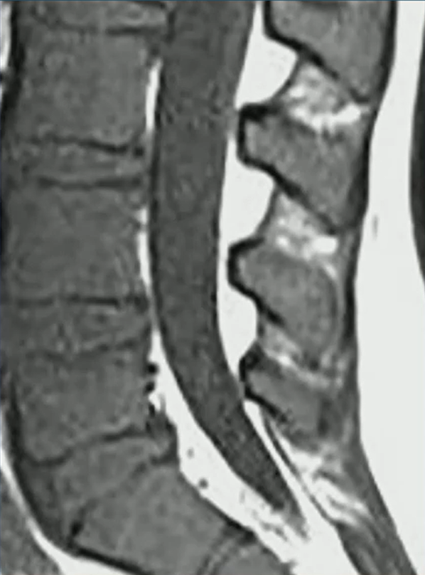
When a pediatric patient presents with back pain, MRI of the spine is a powerful tool to distinguish between benign and malignant processes. In this case of a 7-year-old girl from a Quick Byte presentation by Laura M. Fayad, MD, the imaging reveals systemic replacement of the normal bone marrow.
Primary Observations
- Darkness: On T1-weighted imaging, the marrow signal is abnormally dark.
- Vertebral Compression: Multiple areas of the spine show vertebral body height loss and fractures.
- Diagnostic Worry: Indeed, this combination of diffuse signal abnormality and pathologic fractures is highly concerning for leukemia.
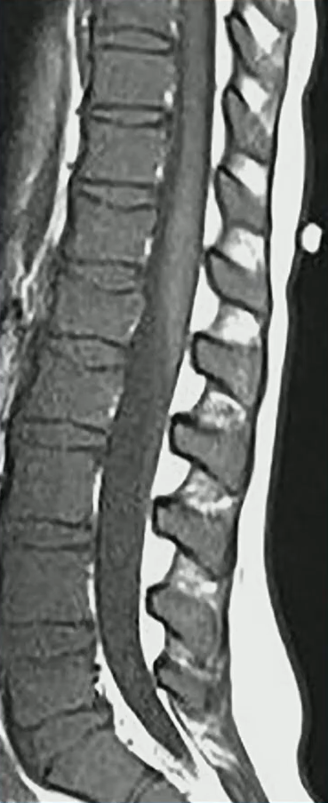
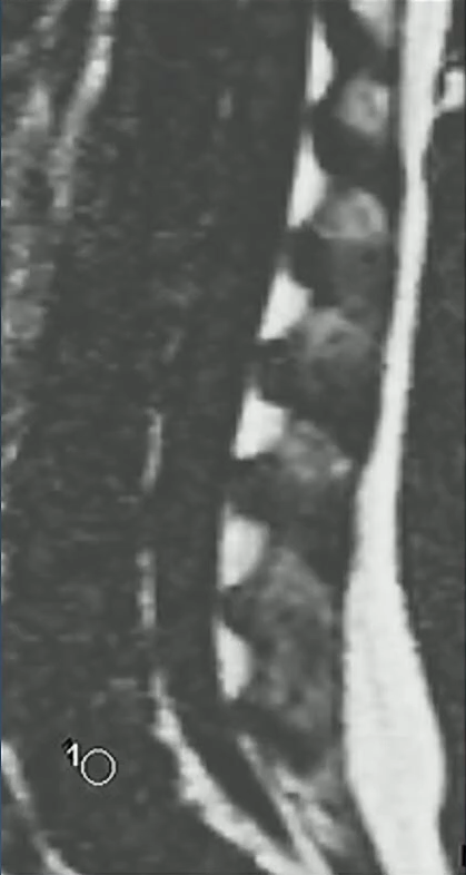
Dixon for Clarification: To confirm if the marrow has been replaced by tumor, a variation of the in-and-out-of-phase gradient echo, the Dixon technique, can be used. Dixon provides fat-only and water-only images to isolate specific tissues.
- Fat-Only Imaging: In a normal patient, fat should be visible within the marrow.
- The Dropout Test: In this patient, while fat is clearly visible in the subcutaneous and epidural spaces, there is a complete absence of fat signal within the bone marrow.
- Conclusion: The total lack of marrow fat indicates that the space has been entirely replaced by malignant cells.
Fat Fraction Analysis: Beyond visual inspection, we can measure the MRI fat fraction to provide an objective data point.
- 10% Rule: In pediatric patients, a fat fraction of less than 10% is a significant indicator of malignant bone marrow.
- Comparison: Normal control marrow typically maintains a much higher fat fraction, often ranging from 20% to over 50%.
| Marrow Category | MRI Fat Fraction (%) | Clinical Significance |
| Malignant | < 10% | Highly concerning for leukemia or tumor infiltration. |
| Normal (Controls) | > 20% | Indicates healthy, fat-containing marrow for age. |
Leave a Reply