
Presented during the ARRS Annual Meeting’s Musculoskeletal Radiology Review, this case utilizes CT (and an arboreal aid) to highlight a benign lipoma with cortical attachment, emphasizing key differentiators from aggressive lesions.
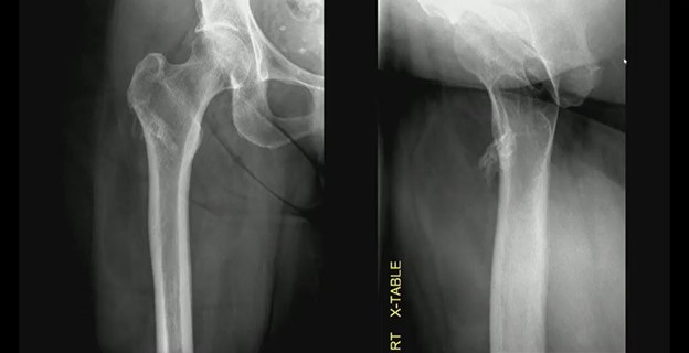
Imaging: Two views of an unusual surface lesion coming from the femur. Bone exostosis is evident. On the right, it doesn’t appear there’s any communication with the marrow space. Looking at soft tissues, you can make out a large, low-density mass—sort of draped below that little piece of bone coming off of the cortex.
CT shows the bone exostosis corresponds with this lesion coming from the surface of the cortex. It does *not* involve the medullary space; it doesn’t extend through the cortex. That low-density mass corresponds with this very fatty mass atop the bone.
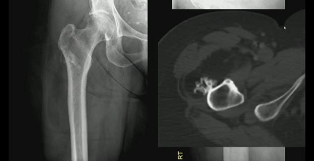
Sure, there are a few septations, but no other suspicions.
DDX? This combination of a benign fatty mass attached to the underlying bone surface is a parosteal lipoma.
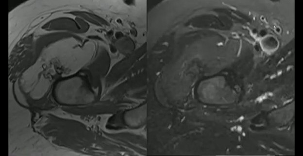
- “Trunk”—The bony outgrowth emerging from the femoral cortex.
- “Branches”—That large, low-density soft-tissue mass draped over the bone.
Bottom Line: Differentiating benign surface lesions from aggressive bone tumors is critical for avoiding unnecessary biopsies and patient anxiety. And if the “trunk” (i.e., bone) and “branches” (i.e., fat) are present sans marrow involvement, it’s likely just a lipoma.
Leave a Reply