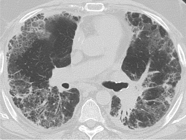
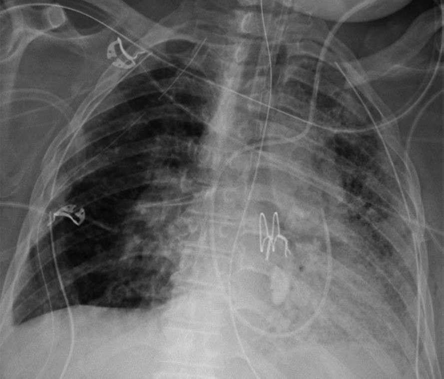
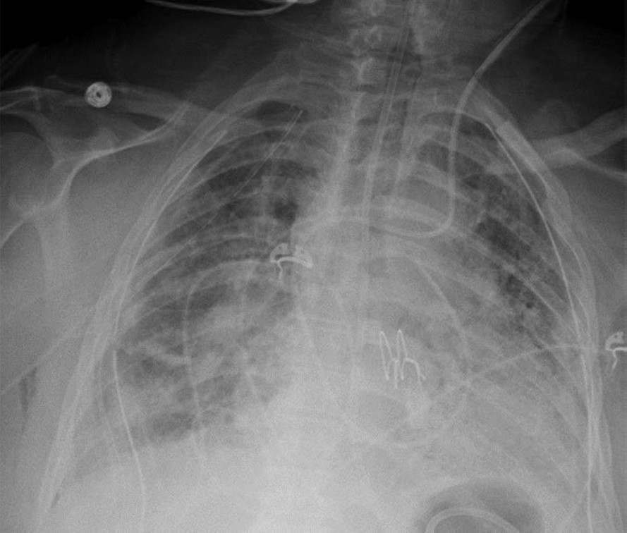
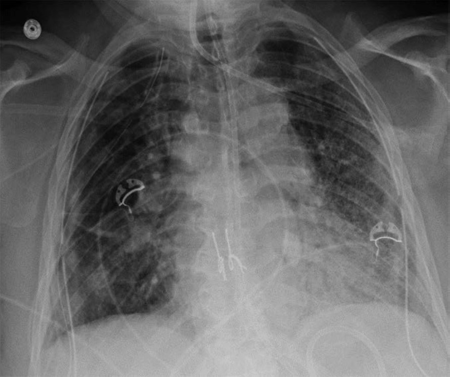
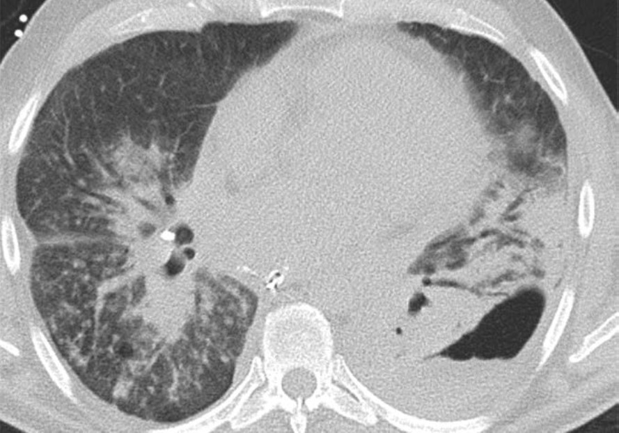
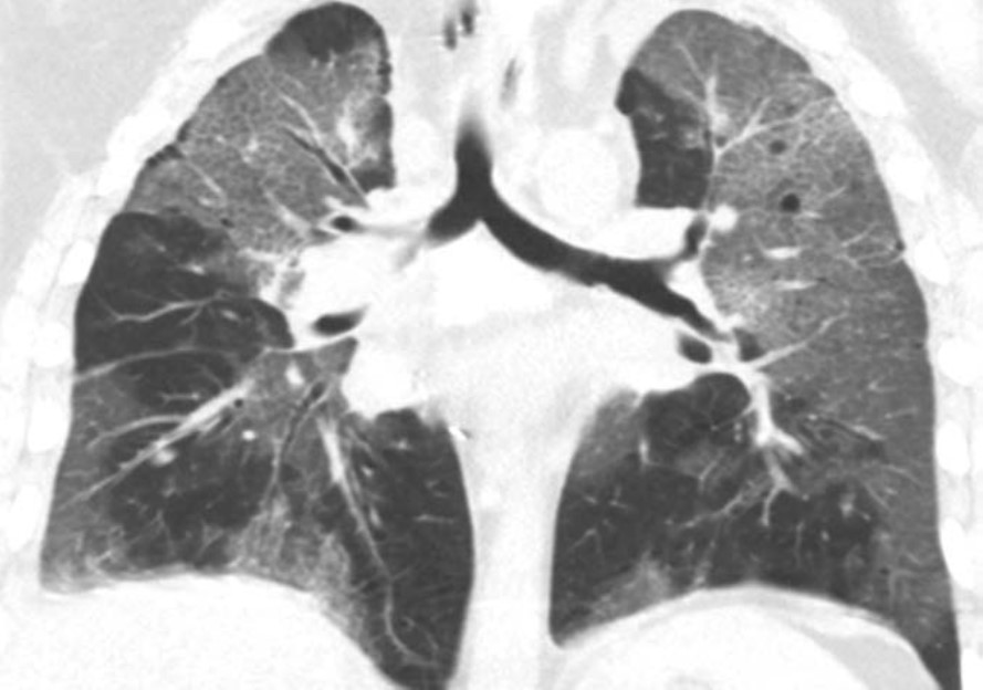
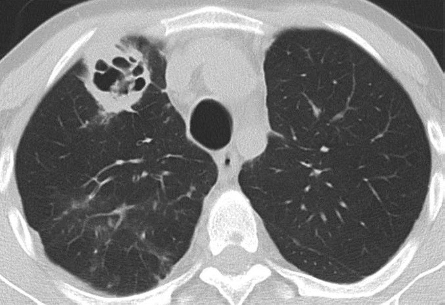
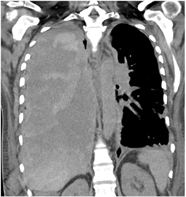
The American Roentgen Ray Society (ARRS) proudly announces publication of the inaugural issue of its brand-new, peer-reviewed radiology journal: Roentgen Ray Review.
Marking the 125th anniversary of North America’s very first radiological society, ARRS’ Roentgen Ray Review (R3 for short) will continue to publish image-rich, clinically relevant content each week at R3journal.org.
Edited by John R. Leyendecker, MD, R3’s Pictorial Essays, Clinical Practice Challenges, Case-Based Reviews, Case Reports, and unique article formats like “Don’t Miss This” enable today’s working radiologists to integrate critical knowledge into imaging practices that continue to grow in both volume and complexity. Already, R3 is keeping busy ARRS members up to date on the latest developments in the specialty without compromising their demanding workflow routines.
Among the Volume 1, Issue 1 Roentgen Ray Review articles that our membership can access any time and anywhere are a few that exemplify ARRS’ mission:
Overview of Imaging Intracranial Infections
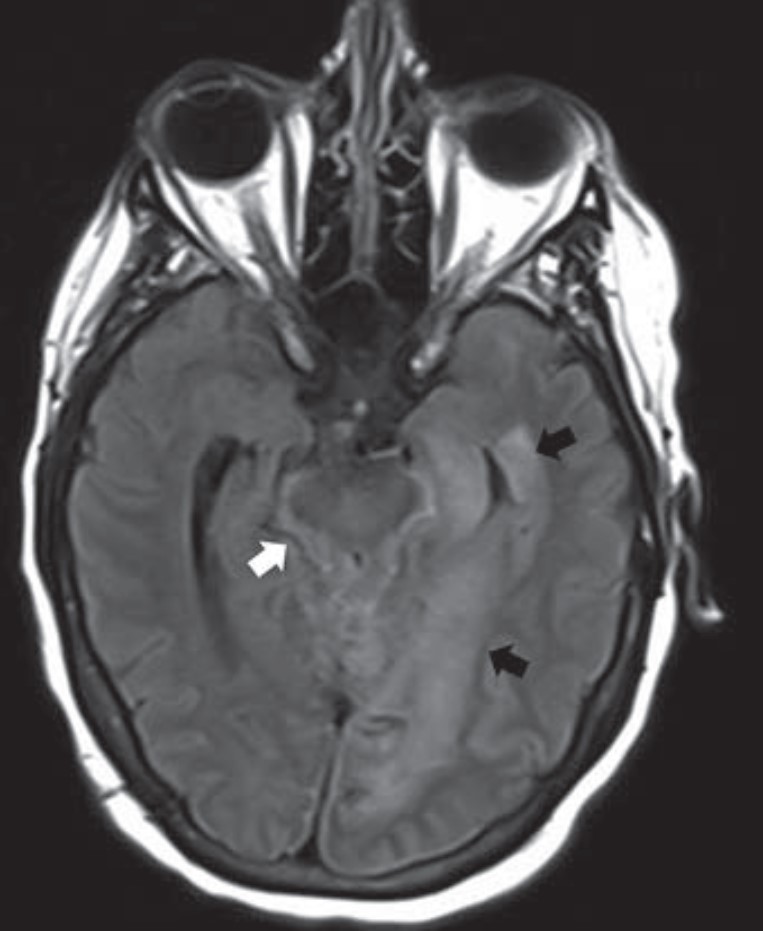
The manifestations of CNS infections are myriad, including diffuse (meningitis, encephalitis, ventriculitis), focal (abscess, empyema), and other special presentations, especially in immunocompromised patients. Diagnosing CNS infections is decidedly multidisciplinary, and as “Overview of Imaging Intracranial Infections” points out, radiology’s contribution to the clinical workup of a patient is paramount. The Roentgen Ray Review’s first entry in its Categorical Course Highlight series, this CME credit article focused on key confounders details how CT and MRI with FLAIR sequencing and DWI can focus your differential diagnosis—ultimately streamlining additional testing and management decisions by the clinical team (Fig. 1).
Fig. 1—64-year-old patient with acute myeloid leukemia and prosthetic hip infection that led to basilar meningitis due to unspeciated fungal infection. Axial FLAIR MR image shows parenchymal edema (black arrows) in left medial temporal lobe and CSF FLAIR nonsuppression (white arrow) in basal cisterns.
Approach to the Lung Cyst With Nodule
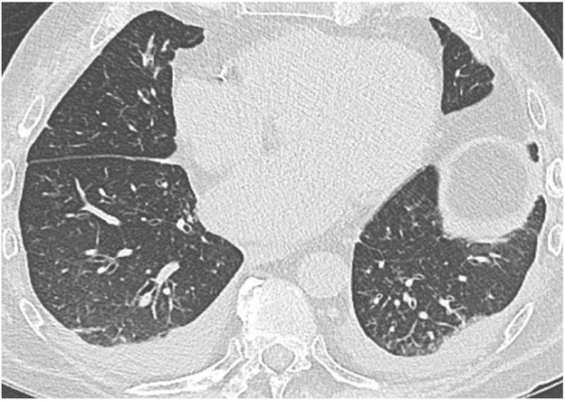
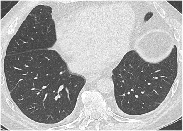
Because their imaging features can overlap with benign entities (bullae, fibrosis, bronchiectasis, etc.), lung cancers associated with cystic spaces are often diagnosed at later stages than cancers presenting as solid nodules. Compared to Lung-RADS version 1.1, Lung-RADS v2022’s updated classification and management criteria offer the potential for earlier detection and treatment of cystic cancers. This first Roentgen Ray Review Clinical Practice Challenge with CME tests readers on appropriate management of an atypical pulmonary cyst in a 66-year-old man with a 40-pack-year smoking history who had undergone right upper lobectomy for stage I lung cancer a decade earlier (Fig. 2).
Fig. 2—66-year-old man undergoing baseline lung cancer screening. Axial CT shows 29-mm pulmonary cyst in right lower lobe (arrow).
A Downward Spiral of Knee Pain and Swelling: Use of MRI to Distinguish Lyme Arthritis From Other Pediatric Arthritides
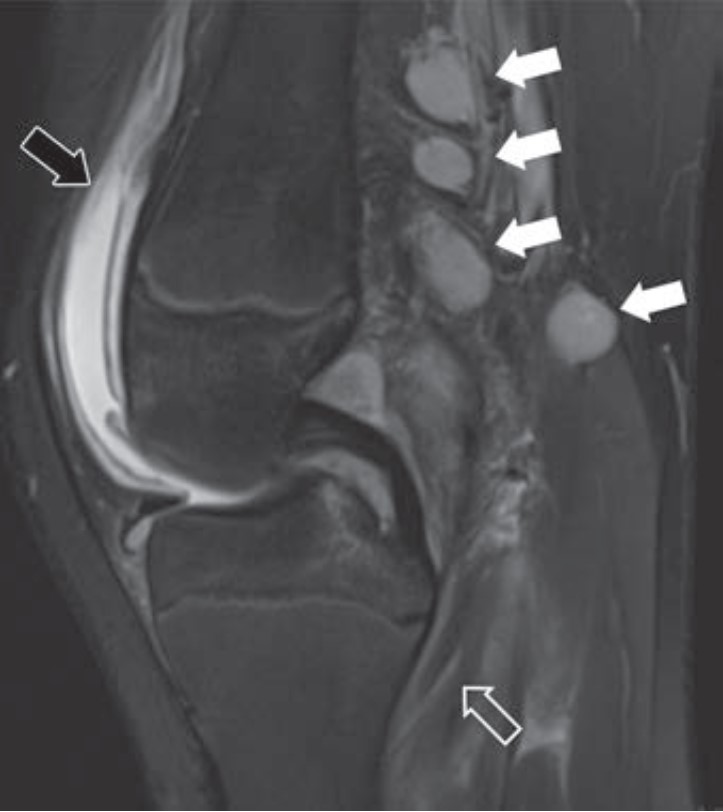
Clinical features of Lyme arthritis overlap with those of both septic arthritis and juvenile idiopathic arthritis, making the diagnosis of Lyme arthritis challenging. As “A Downward Spiral of Knee Pain and Swelling: Use of MRI to Distinguish Lyme Arthritis From Other Pediatric Arthritides” shows, MRI can help differentiate Lyme arthritis of the knee from other entities when certain imaging features, such as popliteal adenopathy, myositis, and lack of subcutaneous edema, are present. This first Roentgen Ray Review Case Report concerns a 13-year-old boy with a 1-month history of knee pain and swelling (Fig. 3).
Fig. 3—Sagittal T2-weighted fat-saturated MRI shows suprapatellar synovitis and effusion (solid black arrow), multiple enlarged popliteal lymph nodes (solid white arrows), and feathery increased popliteus muscle signal consistent with myositis (open arrow).
Crucial but Often Unrecognized Radiologic Signs in Thoracic Imaging
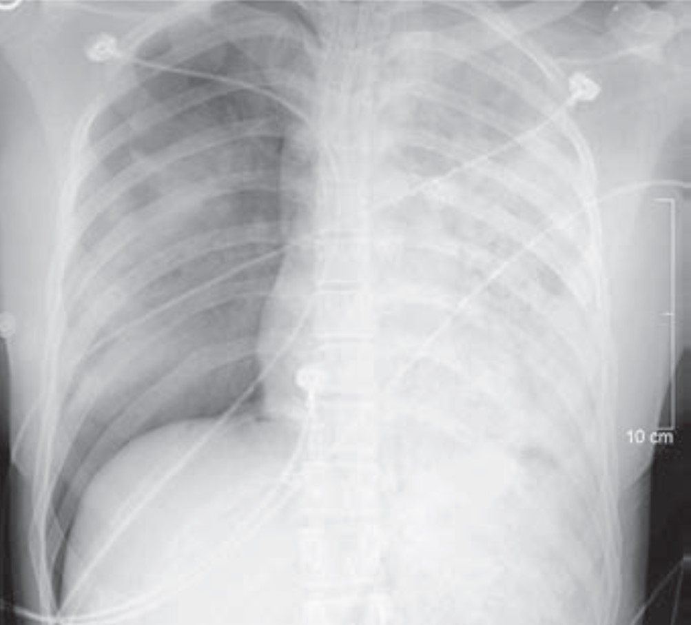
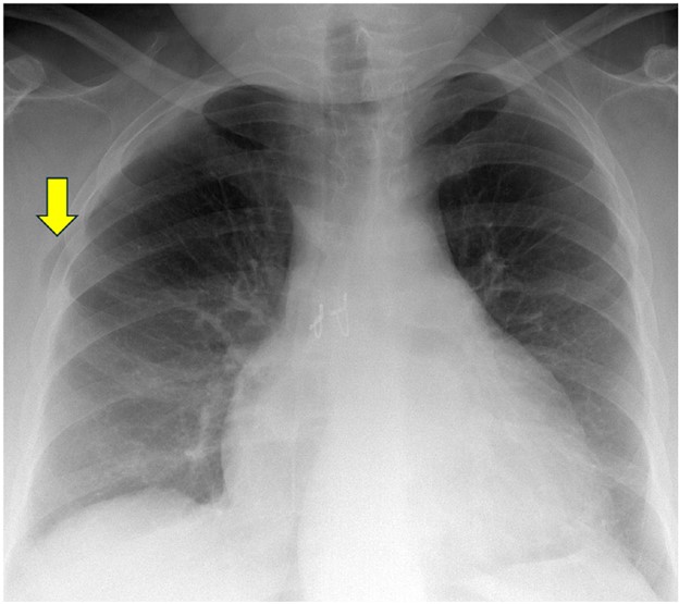
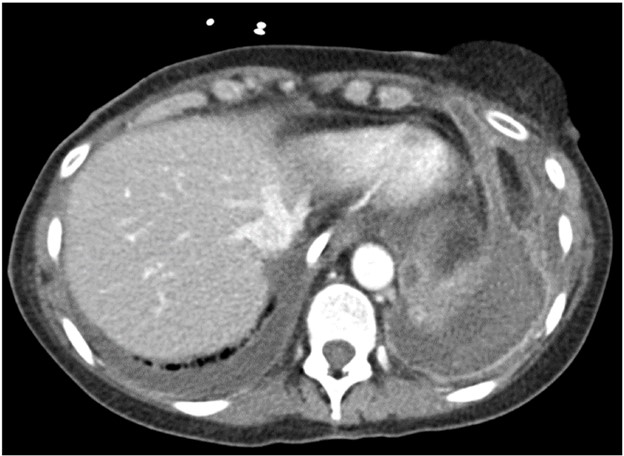
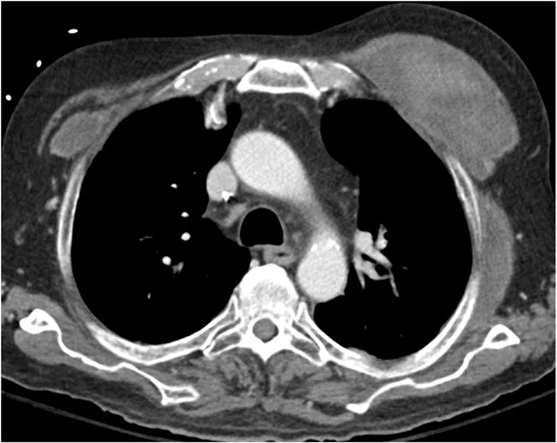
Despite its limitations, the chest radiograph is the world’s most frequently ordered and performed diagnostic imaging examination. However, accurately interpretating the chest radiograph remains challenging. “Crucial but Often Unrecognized Radiologic Signs in Thoracic Imaging” is the first of a two-part R3 Case-Based Review with credit highlighting key chest signs radiologists should recognize. Mark S. Parker, MD, presents six unknown cases of underlying abnormalities affecting the lungs, pleura, and mediastinum to help clinically steer your differential diagnosis (Fig. 4).
Fig. 4—Anteroposterior portable chest radiograph of 28-year-old man involved in high-speed motor vehicle collision.
First-Trimester OB Imaging in the Emergency Setting
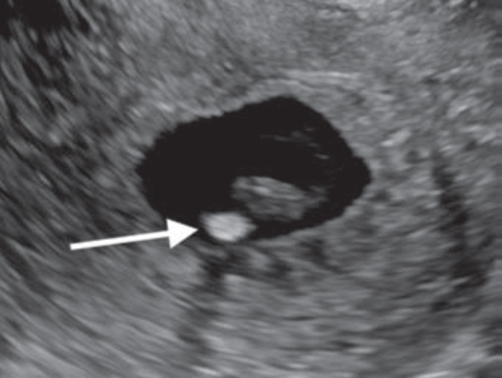
Radiologists play a key role in detecting early pregnancy, assessing cardiac activity and gestation location, as well as evaluating for complications in the acute setting. Knowledge of potential technical errors in image acquisition and an understanding of possible ectopic implantation locations are also necessary to optimize care of the pregnant patient in the first trimester. “First-Trimester OB Imaging in the Emergency Setting” reviews the pearls and pitfalls of first-trimester ultrasound—early pregnancy development, early pregnancy loss, intrauterine pregnancy of unknown location, ectopic pregnancy—applying the 2024 Society of Radiologists in Ultrasound Consensus Conference’s recommended lexicon throughout (Fig. 5).
Fig. 5—Calcified yolk sac in 23-yearold woman with vaginal bleeding and HCG level of 9300 mIU/mL. Transverse transvaginal ultrasound image shows calcified yolk sac (arrow).
Lateral Trochlear Osteochondritis Dissecans of the Elbow: A Rare and Subtle Finding With Important Implications for Management
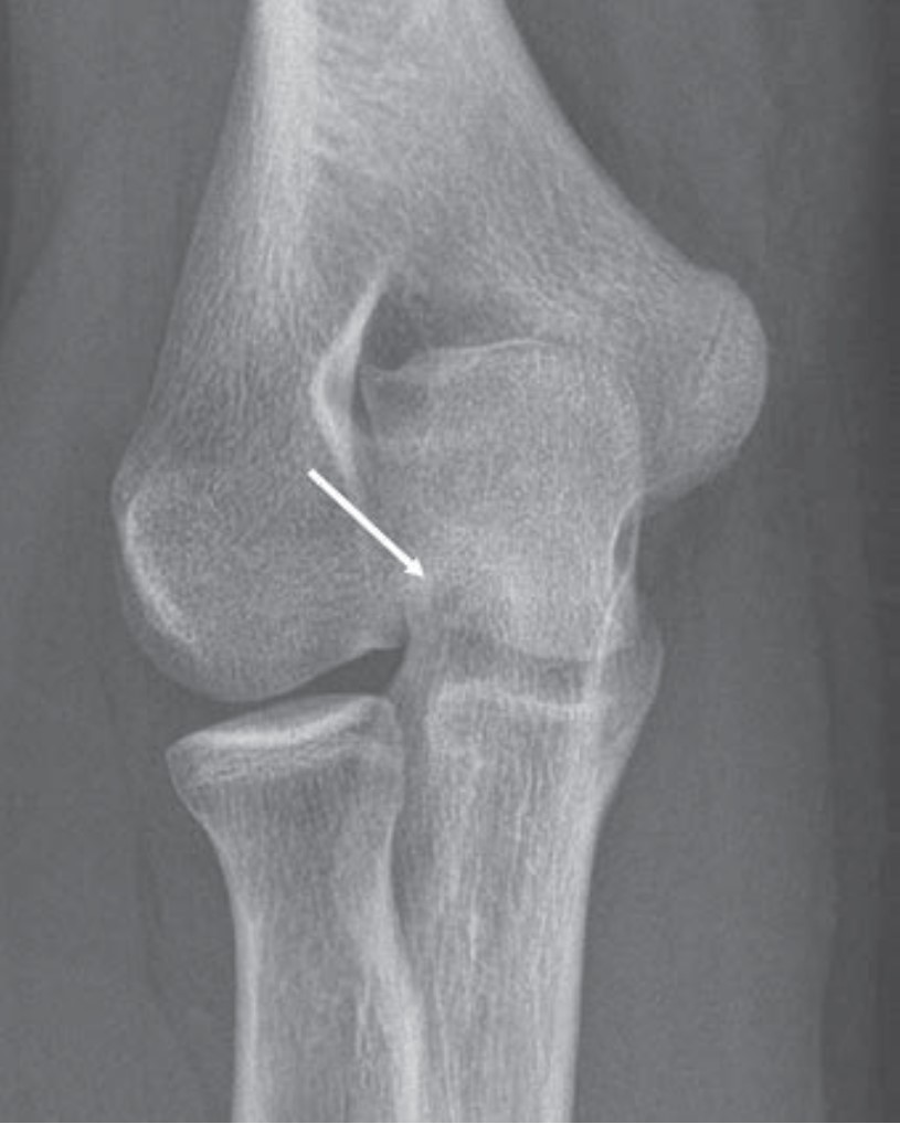
Fig. 6—Frontal radiograph of 14-year-old boy with right elbow pain shows rounded lucency at lateral trochlea (arrow).
Given the overlapping ulnar anatomy and subtlety of its radiologic features, even experienced radiologists can miss lateral trochlear osteochondritis dissecans (OCD) of the elbow. As the first Don’t Miss This article in R3 notes, early identification of lateral trochlear OCD allows for appropriate activity modification, as well as timely intervention to mitigate lasting disability (Fig. 6).
Coronary Artery Displacement and Narrowing on CT in the Setting of Giant Sinus of Valsalva Aneurysms
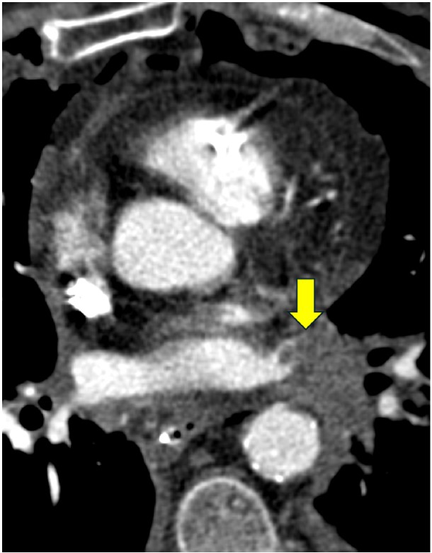
Whereas aortic valve regurgitation and rupture are known complications of sinus of Valsalva aneurysm, this Roentgen Ray Review Case Report illustrates that cardiac ischemia can be a manifestation, too. Presenting a 54-year-old man with large aneurysms involving all three aortic sinuses, “Coronary Artery Displacement and Narrowing on CT in the Setting of Giant Sinus of Valsalva Aneurysms” also illustrates the modality’s value in complex anatomical depictions for proper surgical planning (Fig. 7).
Fig. 7—Left ventricular outflow tract view from contrastenhanced ECG-gated cardiac CT scan shows large aneurysms of right (white arrow) and noncoronary (black arrow) sinuses of Valsalva.
The American Roentgen Ray Society (ARRS) first traveled to San Diego, CA, for the 1996 ARRS Annual Meeting. Almost three decades ago, Kay H. Vydareny of Emory University Hospital in Atlanta, GA, was named this society’s first female president on Sunday, May 5. (Apropos, of the four newly elected 2025–2026 ARRS officers installed on Sunday, April 27, 2025 at the Marriott Marquis Marina, half are women.)
The latest convening of North America’s first radiological society delivered the same clinically relevant experience for which ARRS has long been heralded: radiologists of each practice type and every training level relishing world-class instruction from trusted experts spanning every subspecialty. Pioneers in asynchronous continuing education, ARRS continues to offer both inperson and virtual registrants the most flexible meeting experience in radiology. All attendees retain on-demand access to the complete program for an entire calendar year, learning and earning CME well into 2026.
And speaking of 2026, ARRS looks forward to delivering yet another singular experience in Pittsburgh, PA. This city of steel and bridges has reinvented itself as a hub of health care innovation, making David L. Lawrence Convention Center the perfect host for #ARRS26. Like the city itself, ARRS is constantly evolving— offering new education, fresh perspectives, and invaluable opportunities to connect with peers in meaningful ways. Join us to be part of a dynamic meeting where you’ll gain knowledge, build connections, and experience the energy of a city that’s shaping the future of patient care.
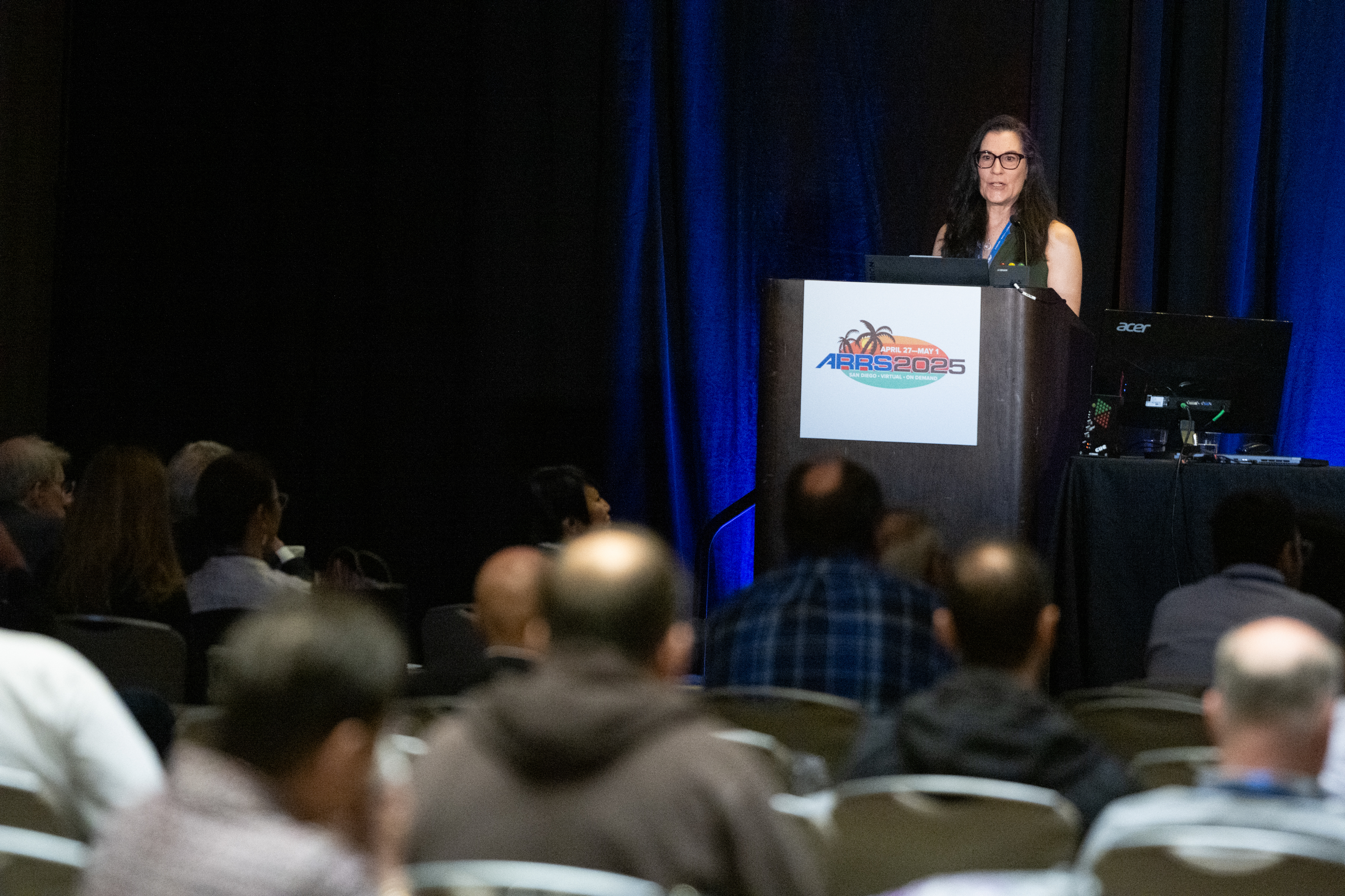
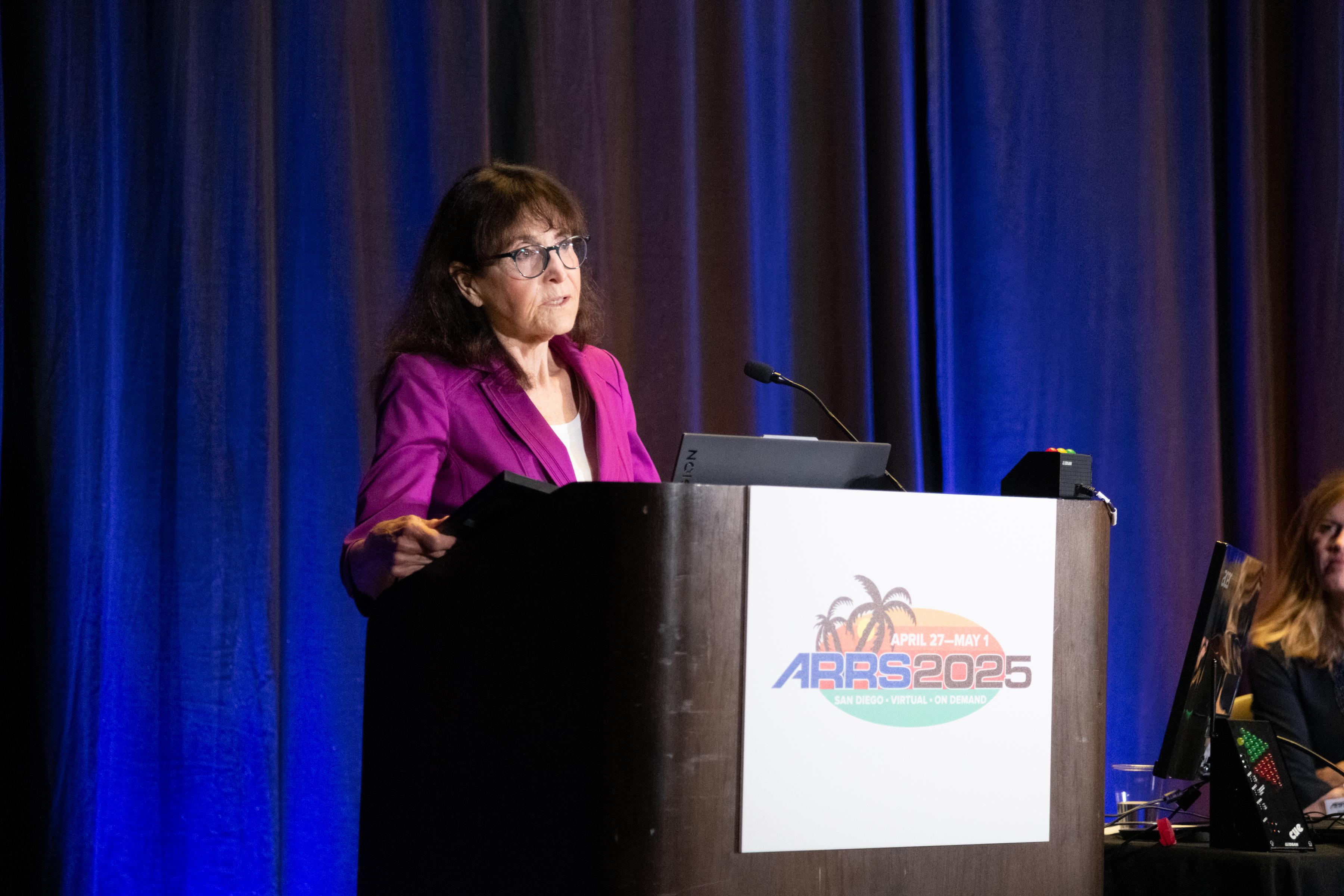
Dr. Deborah Baumgarten presiding over the 2025 ARRS Member Business Meeting
After ratifying an amendment to Article X of the bylaws to clarify that ARRS now publishes two radiology journals—AJR and R3—the ARRS membership officially installed Deborah A. Baumgarten, MD, MPH, of the Mayo Clinic in Jacksonville, FL, as the 125th president of ARRS. An internationally recognized leader in radiology and a steadfast advocate for education, Dr. Baumgarten assumes the presidency after years of distinguished service on the ARRS Executive Council.
Her appointment marks a new chapter in this society’s ongoing mission to advance medical imaging and patient care through expert education, cuttingedge research, and fruitful collaboration. Widely published and highly respected for her clinical expertise, instructional insights, and leadership within the imaging community at large, Dr. Baumgarten succeeds Angelisa M. Paladin, MD, who presented the ARRS presidential gavel to her successor.
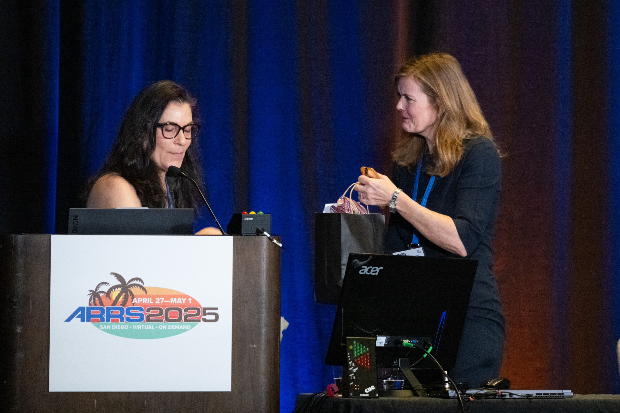
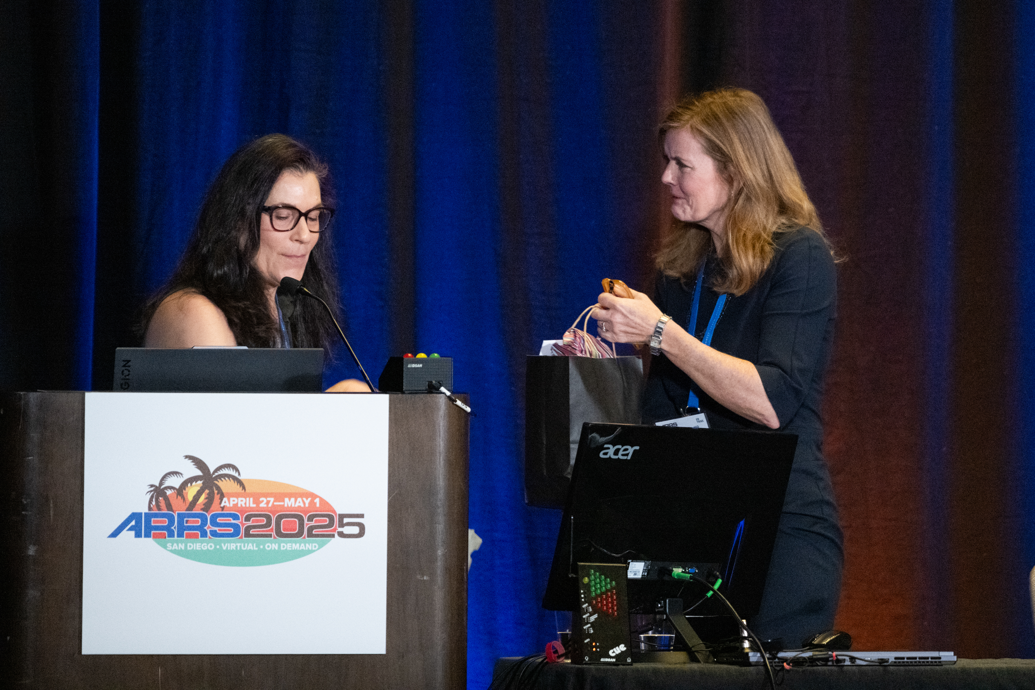
Dr. Angelisa Paladin passing the gavel, as well as a gift, to Dr. Deb Baumgarten.
First, Tanya W. Moseley, MD, of the University of Mississippi Medical Center was honored as the 2025 ARRS Distinguished Educator. For more than 20 years, Dr. Moseley’s contributions to ARRS have transformed radiological education. Through her service on key educational committees and roles as casebased breast imaging chair and AJR SA-CME Consultant Editor, she has fundamentally shaped the organization’s educational direction. Her pioneering vision led to the creation of the first multi-vendor tomosynthesis certification course, demonstrating her exceptional ability to build collaborative educational programs. As architect and director of the ARRS Longitudinal Course Series, she continues to advance innovative approaches to radiology education.
Drs. Baumgarten, 2025 ARRS Distinguished Educator Tanya Moseley, and Paladin.
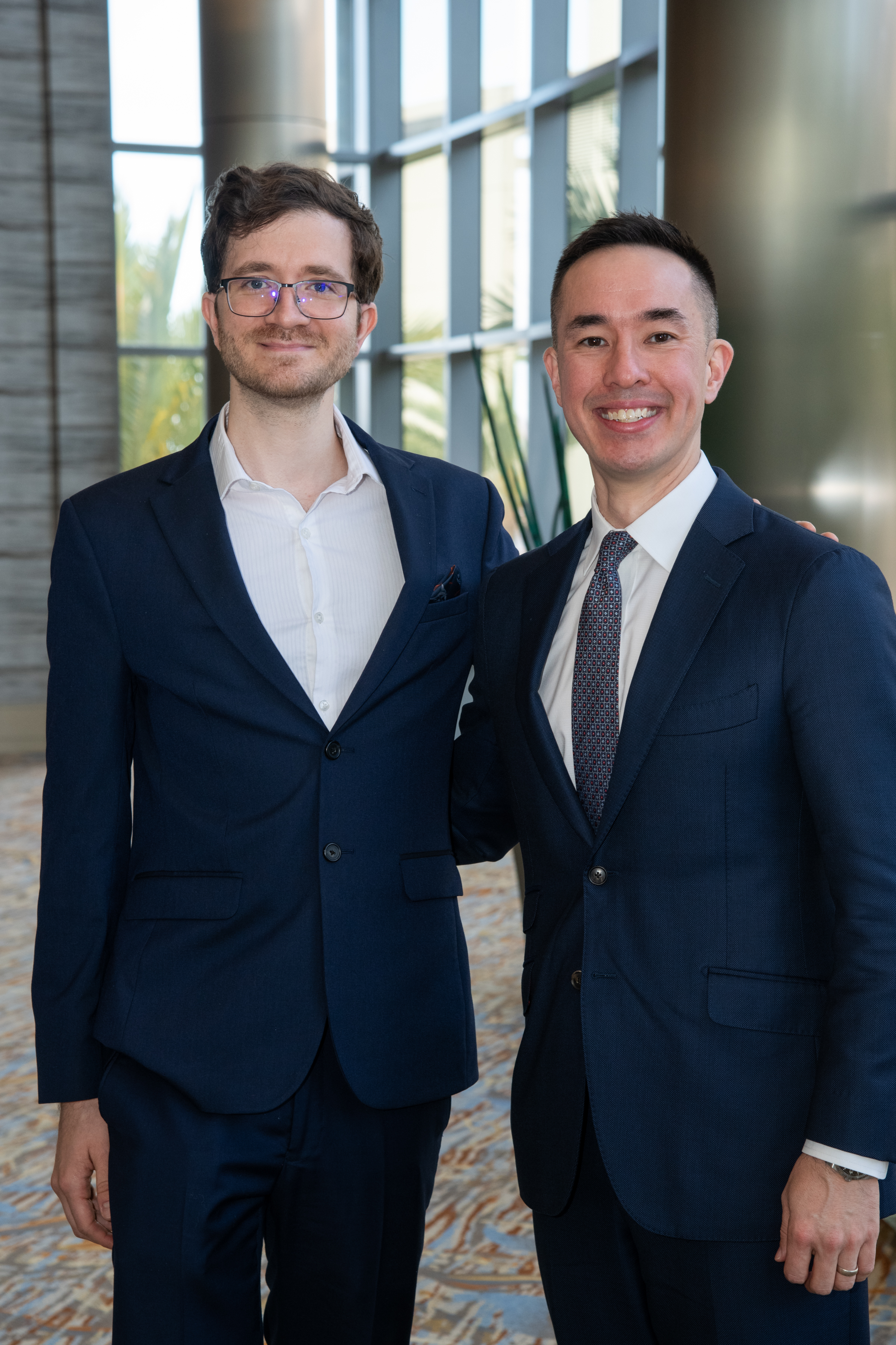
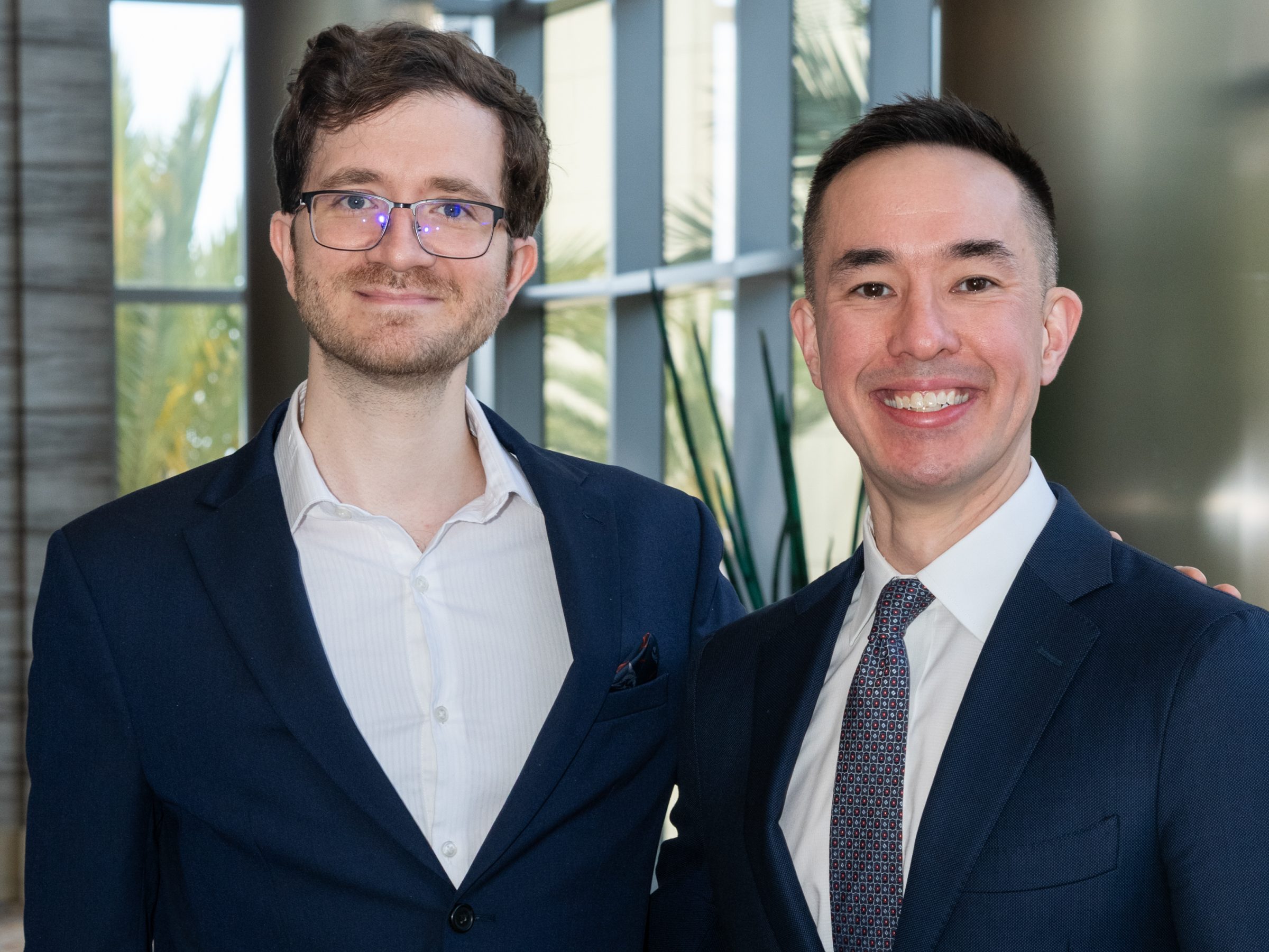
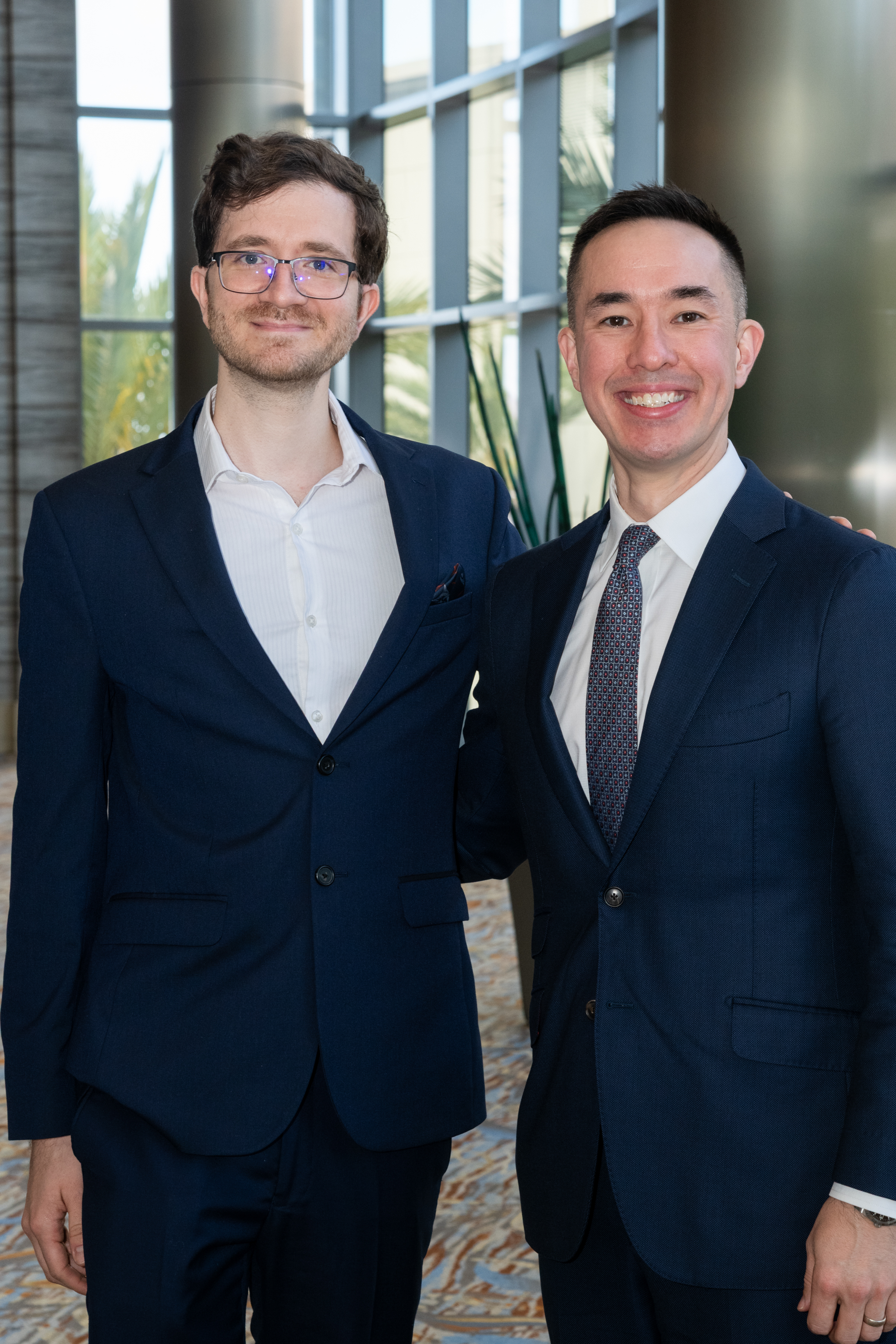
This society was then doubly proud to recognize two recipients of 2025 ARRS Scholarships: Matthew Lee, MD, at the University of Wisconsin School of Medicine and Public Health and Yale School of Medicine’s Luca Pasquini, MD, PhD. Provided by The Roentgen Fund, ARRS Scholarships support early-career faculty members pursuing radiological research that promises to change how medical imaging is practiced. A two-year grant totaling $180,000, the ARRS Scholarship aims to advance emerging scholars, as well as prepare them for positions of leadership.
Drs. Luca Pasquini and Matthew Lee, 2025 ARRS Scholars.
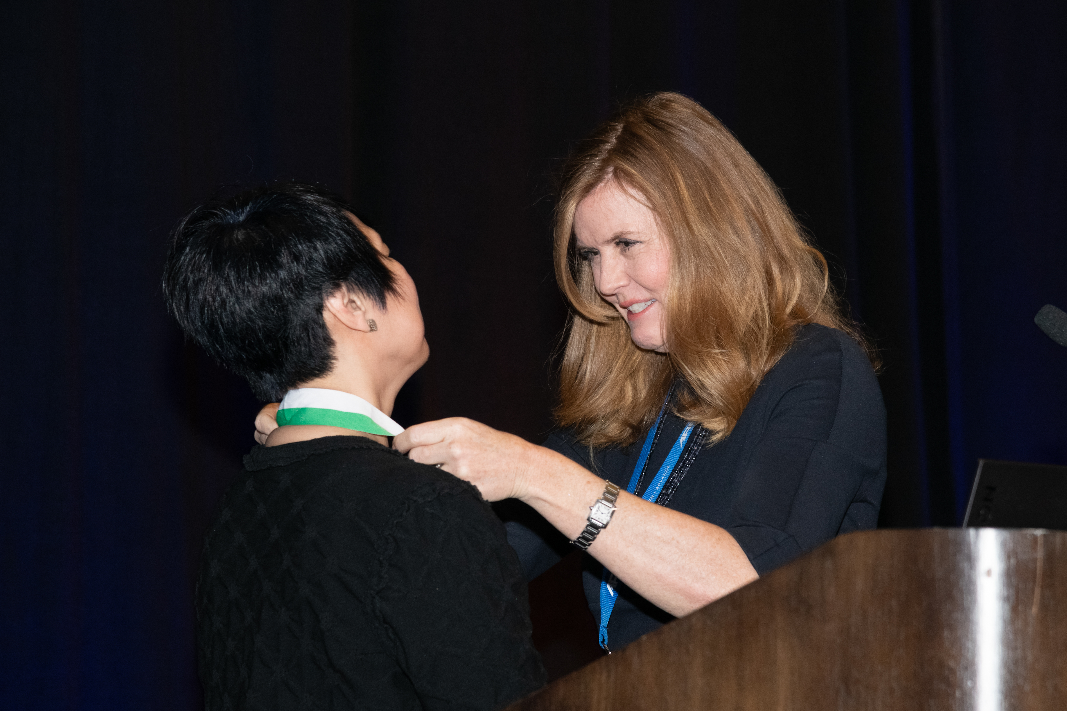
The biggest laurel of the morning, the ARRS Gold Medal, went to Ruth C. Carlos, MD, MS, FACR. Installed as president of ARRS during the 2019 Annual Meeting in Honolulu, HI, presently, Dr. Carlos is a professor at Columbia University Irving Medical Center and associate chair of research faculty development for the radiology department. And as fellow past ARRS president and longtime Michigan colleague N. Reed Dunnick introduced her: “Ruth Carlos isn’t just a superstar. I’d argue that she’s an entire galaxy!” Indeed, having served 11 years on our Executive Committee, as well as 11 ARRS committees prior, Dr. Carlos remains a fixture in our universe. There’s no one more deserving of the highest merit bestowed by this society, which has been honoring distinguished service to radiology for more than four decades.
Dr. Ruth Carlos receives her 2025 ARRS Gold Medal from Paladin.
Service Over Self: Longtime ARRS ED Receives New Lifetime Award
The entirety of the ARRS was eager to give the first-ever ARRS Lifetime Service Award to its former executive director Susan B. Cappitelli, MBA, CAE. “For many of us who have been attending the ARRS Annual Meeting for many years,” Dr. Paladin remarked, “you’ve seen a huge growth in our programming and our services.” Well, that growth started with Susan way back in 1991, when ARRS recruited her to bring every aspect of producing the world’s longest published general radiology journal, AJR, in house. Needing a home for all the talented and dedicated professionals Susan herself was recruiting, six years later, she helped ARRS secure its own office building in Leesburg, VA. The society would go on to use every inch of space, of course, because during Susan’s historic tenure as executive director, ARRS membership increased more than 155%. Meanwhile, the society’s net assets ballooned from just $5 million to over $30 million. Having steered the membership and organization through everything from RVUs to COVID and beyond, “Susan always led with grace,” Dr. Paladin added, “and she will never, ever be forgotten.”
“Innovation is bound to flourish, and innovating is fun. I believe that the society’s commitment to continually learning and improving is why ARRS has grown and produced far beyond its size.”—Susan Cappitelli | ARRS Executive Director, 1999-2024
Welcome to 2025 Honorary ARRS Member, Salvador Amézquita Pérez
Dr. Salvador Amézquita Pérez, president of Sociedad Mexicana de Radiología e Imagen (Mexican Society of Radiology and Imaging), received his honorary ARRS membership on day one, and he and his colleagues were welcomed to the 2025 ARRS Annual Meeting in San Diego as part of the Global Exchange Featuring Mexico. The ARRS-SMRI Sunday Featured Course, “Advances in Cardiac Imaging,” focused on techniques and considerations for evaluating structural heart conditions and coronary artery anomalies. This session addressed non-atherosclerotic coronary artery narrowing, CT’s role in transcatheter mitral valve replacement and adult congenital heart disease, CT perfusion and FFR-CT for myocardial ischemia, as well as CT in TAVR before and after surgery.
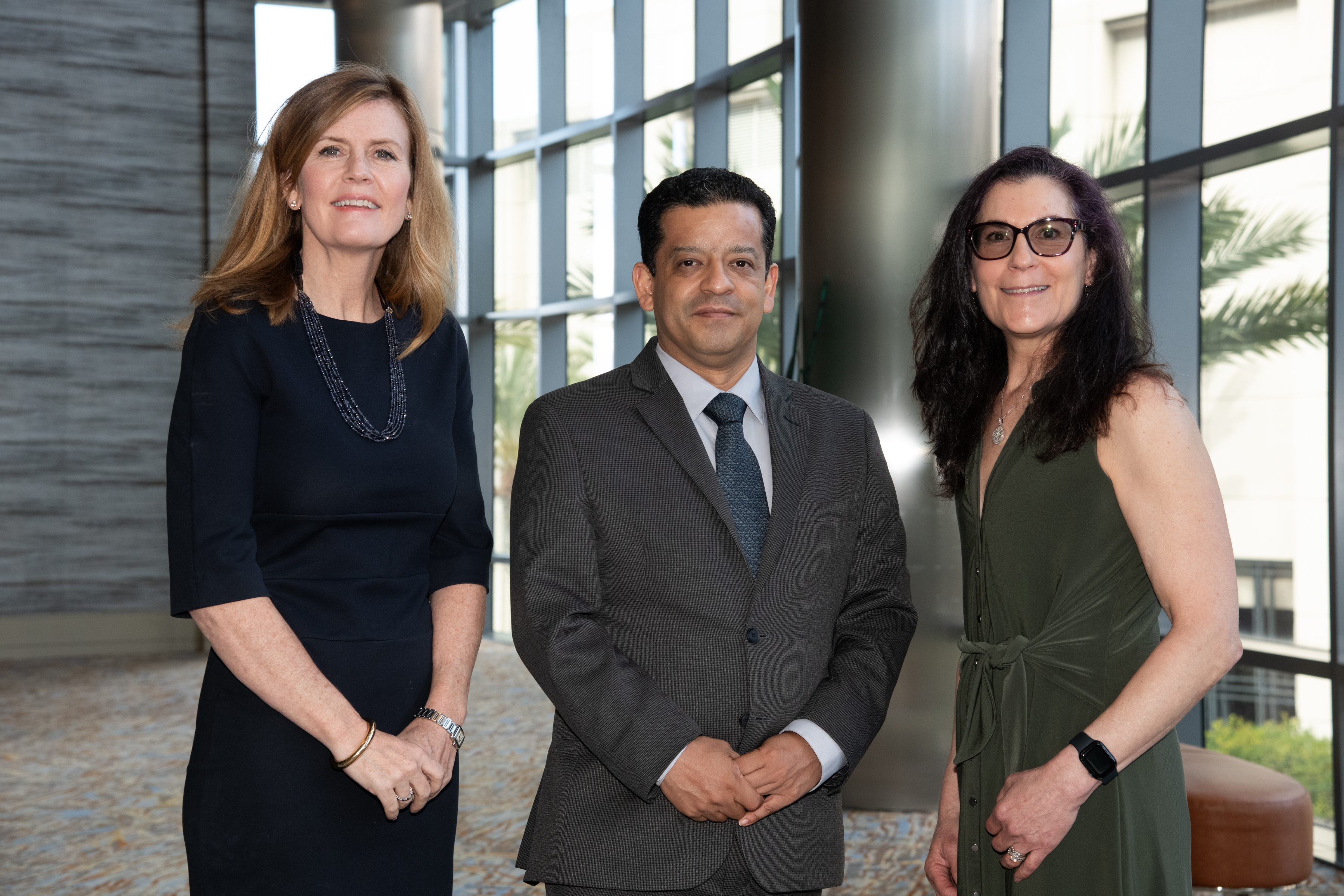
Drs. Paladin, Salvador Amézquita Pérez, and Baumgarten.
The mission of the ARRS Global Partner Society Program is to build long-standing relationships with key leaders and organizations in the worldwide imaging community—increasing awareness of our society’s services in specific nations, while raising the stature of Global Partner Societies among ARRS members. Every year, the ARRS Annual Meeting Global Exchange incorporates one partner society into the educational and social fabric of our meeting. ARRS members then reciprocate at the partner society’s meeting that same year. Our 2026 Annual Meeting Global Exchange will welcome a delegation from the Royal Australian New Zealand College of Radiologists to Pittsburgh, PA.
AJR Luncheon Recognizes 2025 Figley and Rogers Journalism Fellows
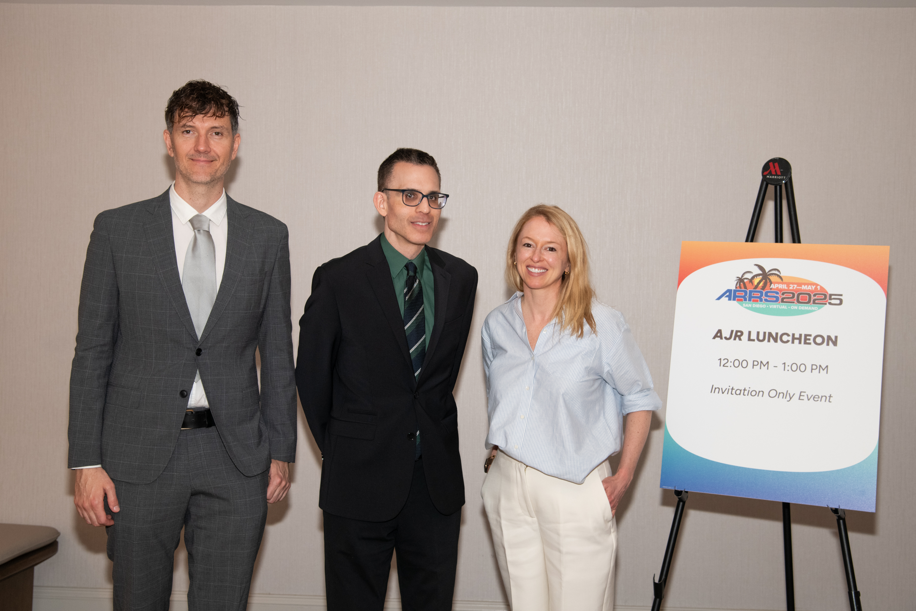
During the American Journal of Roentgenology (AJR) Luncheon on Monday afternoon, Erin Alaia, MD, of NYU Langone Health in New York City was honored as the 2025 Melvin M. Figley Fellow in Radiology Journalism. Domen Plut, MD, PhD, from Slovenia’s University Medical Centre Ljubljana was recognized as the 2025 Lee F. Rogers International Fellow in Radiology Journalism.
Drs. Domen Plut, AJR Editor in Chief Andrew Rosenkrantz, and Erin Alaia.
Named for two distinguished Editors Emeriti of AJR, the Melvin Figley and Lee Rogers Fellowships offer practicing radiologists an unparalleled opportunity to learn the tenets of medical publishing via “the yellow journal”—the world’s longest continuously published radiology journal. Through hands-on experience with ARRS staff and AJR personnel—as well as personal apprenticeship with AJR’s 13th Editor of Chief, Andrew B. Rosenkrantz— Drs. Alaia and Plut will receive expert instruction in scientific writing and communication, manuscript preparation and editing, peer review processes, journalism ethics, and digital production and publication.
With Distinction: Award-Winning Scientific Research Presented in San Diego
The 2025 ARRS Annual Meeting hosted hundreds of electronic exhibits and abstracts presenting leading-edge research. Below are but a few highlights from our award-winning Scientific Program posters.
Mitigating Disparities in MASLD—Advancing Early Image Detection and Management
According to the Summa Cum Laude Award-Winning Online Poster presented during the 2025 ARRS Annual Meeting, patients with metabolic risk factors and an image diagnosis of steatosis— but without a recognized diagnosis of metabolic-associated steatotic liver disease (MASLD)—were more likely to experience significant complications of chronic liver disease, including cirrhosis and hepatocellular carcinoma (HCC), when compared to patients with a formal MASLD diagnosis. A public health crisis with significant morbidity and mortality implications, “MASLD affects approximately one-fourth of the global population, making it the most chronic liver disease worldwide,” noted lead investigator Emmanuel Mgboji of the University of Michigan Medical School. Using data from his institution’s EMR, Mgboji and fellow Michigan researcher Jessica Fried, MD, identified a cohort of 10,280 subjects with a formal diagnosis of MASLD and 5,103 subjects without a formal MASLD diagnosis but with MRI evidence of hepatic steatosis and metabolic risk factors. After demographic extraction and comparison, Mgboji and Fried monitored each cohort for progression of disease-related outcomes from 2018 to 2023, including HCC, cirrhosis, myocardial infarction (MI), chronic kidney disease (CKD), coronary artery disease (CAD), and type 2 diabetes mellitus (T2DM). Then, after comparing the incident rate and relative risk for each cohort, subjects were stratified by race (n = MASLD Dx; n = Image Dx cohort), including Caucasian Americans (CAs) (n = 8,548; n = 4,227), African Americans (AAs) (n = 580; n = 399), Asian American (ASAs) (n = 599; n = 229), and Hispanic Americans (HA) (n = 433; n = 169). “Comparing incidence rates between the MASLD Dx and Image Dx cohorts (no MASLD Dx cohort),” Mgboji said, “we found a significant relative risk of 2.185 (95% CI: 1.6822–2.8395, p < 0.0001) for a diagnosis of HCC in the Image Dx cohort. Additionally, the relative risk of developing cirrhosis in the Image Dx cohort was 1.459 (95% CI: 1.2934–1.6461, p < 0.0001).” Mgboji and Fried also assessed the risk of being diagnosed with MI, CKD, CAD, and T2DM in the Image Dx cohort, with a relative risk of 1.236 (p = 0.0496), 1.240 (p = 0.0002), 1.346 (p < 0.0001), and 1.259 (p < 0.0001), respectively. “Further significant differences were observed when patients were stratified by racial groups,” added Mgboji. For developing HCC, the relative risk was 2.465 (p < 0.0001) in CAs, 3.488 (p = 0.0180) in AAs, 1.962 (p = 0.375) in ASAs, and 4.270 (p = 0.0451) in HAs. For cirrhosis, relative risk values were 1.590 (p < 0.0001) for CAs, 1.817 (p = 0.0248) for AAs, 1.933 (p = 0.0336) for ASAs, and 2.795 (p = 0.0003) for HAs.
BI-RADS 3 “Report Card” Decreases the Rate of Usage
In the Magna Cum Laude Award- Winning Online Poster at this year’s Annual Meeting, anonymous, peer comparison BI-RADS 3 “report cards” proved to be an effective method of rate reduction, particularly at community hospitals where preintervention rates were higher than at academic sites. Reserved for “probably benign” breast imaging abnormalities that have a low (< 2%) risk of being malignant, in practice, the actual use of ACR’s BI-RADS category 3 assessment varies among radiologists— often overutilized to equivocate a finding. Giving radiologists a recommended target rate of less than 12% as a benchmark, head presenter Bonmyong “Bora” Lee, MD, and her team of researchers from UPenn’s Perelman School of Medicine sent quarterly BI-RADS 3 report cards to each breast imaging radiologist via automated emails. This report card included personal BIRADS category 3 rates for each modality, as well as cumulative rates for the radiologist’s covering site and hospital. Each radiologist was blinded to others’ individual rates, participation was voluntary, and Lee et al. offered neither rewards nor punitive measures for performance. Noting that radiologists were not monitored for review compliance either, “after 4 cycles, we reviewed the data to determine if there were changes in the rate of BI-RADS 3 assessment among radiologists and across the institution using paired t-tests,” Lee said. Over Lee et al.’s 17-month assessment period, 38 radiologists issued BI-RADS 3 in 4,289 total patients: 1,171 diagnostic mammograms, 1,281 screening mammograms, 658 MRI, and 1,179 ultrasound examinations. After Lee and colleagues’ intervention, the average BI-RADS 3 rate decreased (all sites: p < 0.01; community sites: p < 0.01; academic sites: p = 0.07). Radiologists with preintervention BI-RADS 3 rates that were greater than the group median had larger reductions in BI-RADS 3 rates post-intervention (p < 0.05).
Significance of Nonspecific 18F-DCFPyL Rib Uptake on PET/CT in Prostate Cancer Patients
The Cum Laude Award-Winning Online Poster concluded that evaluating increased rib uptake on 18F-DCFPyL PET/CT may be challenging, even resulting in false-positive findings. “In patients without osseous metastasis, the uptake is often low (less than the mean liver SUV), stable, and likely represents benign etiologies, such as fibrous dysplasia, fibrous cortical defect, traumatic fractures, and hemangiomas. Thus, further evaluation is usually not required,” said presenter Aisha Alam, DO, from the Icahn School of Medicine at Mount Sinai in New York, NY. Dr. Alam and her all-Icahn School team performed a retrospective review of patients with prostate cancer who underwent PET/CT scans with radiotracer fluorine-18 2-(3-{1-carboxy-5-[(6-18F]fluoro-pyridine- 3-carbonyl)-amino]-pentyl}-ureido)-pentanedioic acid (18F-DCFPyL) at a single academic center from June 2021 to April 2024. Alam and colleagues’ EHR search identified patients with increased radiotracer rib uptake. The team excluded patients with imaged evidence of osseous metastasis at other sites or with CT suggestive of alternate diagnoses, then analyzed Gleason scores, most recent serum PSA levels, maximum standardized uptake values (SUVmax) in the rib foci, and mean liver SUV. Finally, for confirmation of benignity, follow-up evaluation included stability on 18F-DCFPyL PET/CT and/or imaging modalities such as CT, bone scan, or MRI. With 204 total 18F-DCFPyL PET/CT scans showing solitary or multiple foci of increased rib uptake without CT correlation and imaging evidence of osseous metastatic disease, the mean age was 68 years old, mean Gleason score was 7, mean PSA was 7.9 ng/mL (range: undetectable–50.2 ng/mL), mean SUVmax for rib uptake was 3.8 (range: 1.4–9.8), and mean liver SUV was 5.7 (range: 2.7–10.9). “Of the 204 scans,” Alam noted, “31 studies belonged to 13 patients who underwent follow-up 18F-DCFPyL PET/CT for restaging.” For patients with redemonstrated rib uptake, mean SUVmax was 3.5 and mean SUVmax percent change on subsequent scans was 15.1% (range: 0–46.4%). And for patients with multiple PET/CT scans, the SUVmax for each foci of rib uptake was less than the mean liver SUV (mean: 6.1, range: 4.0–8.8). Of the 23 single-scan PET/CT patients who had a rib uptake greater than the liver mean, 14 of those had a PSA less than 10 ng/mL. “Larger studies evaluating similar findings with histologic correlation and follow-up imaging are needed to improve diagnostic certainty,” added Alam et al.
Analysis of Breast Radiation Therapy and Breast Arterial Calcifications on Screening Mammography
Findings from a Certificate of Merit Online Poster presented during ARRS 2025 suggest that breast radiation therapy exposure does not impact the prevalence of mammographic breast arterial calcification— therefore, not impacting its utility as an imaging biomarker of cardiovascular disease risk. “Our study is the first retrospective analysis of the association between breast cancer radiation therapy exposure and the presence of breast arterial calcification on screening mammography,” noted presenter Jessica Rubino, MD, from Dartmouth Hitchcock Medical Center in Lebanon, NH. Rubino et al. performed an electronic health database query to identify women ages 40–75 years who had a screening mammogram between January 1, 2011 and December 31, 2012. After a chart review to extract data regarding breast cancer radiation therapy history, two breast imaging radiologists then reviewed mammograms for the presence of breast arterial calcification. The researchers used multivariate logistic regression to examine the association between breast radiation therapy exposure and breast arterial calcification, adjusting for age, BMI, smoking status, hypertension, type 2 diabetes, as well as use of statin and antihypertensive medication. Of the 1,155 women included in this analysis, 222 (19.2%) had mammographic evidence of breast arterial calcification, 122 (10.6%) had a history of radiation therapy exposure, and 39 (32%) women with radiation therapy exposure had breast arterial calcification on the index mammogram obtained at least 2 years after completing radiation therapy. Compared to women without radiotherapy, women with a history of breast radiation therapy exposure had higher odds of breast arterial calcification (OR: 2.18, 95% CI: 1.43–3.28; p = 0.0008). After multivariable adjustment, however, this association became nonsignificant, with the maximally adjusted model demonstrating an OR of 1.52 (0.95–2.40; p = 0.07).
Ruben Rajah, MD Comedian and friendly neighborhood radiologist
It is a wet mid-November night in 2022, and I am trudging to an open mic in Manhattan’s lower east side. Apple maps told me the bar was around the corner. It wasn’t. Instead, I found four cop cars boxing in a suspect on a trash-strewn street. I had just moved from Seattle to Jersey City, partly to relaunch my stand-up comedy career. Becoming a comedian is a nocturnal, unglamorous process best pursued by those who are comfortable being ignored and living in the dark—perfect for a radiologist.
I signed up for four lessons in stand-up comedy in the winter of 2007 while working as a nuclear medicine resident in Seattle. The last lesson was a “bringer” gig, meaning we had to invite our friends to watch us perform a five-minute “set” at a comedy club. Radiologists are often understated, but I’m also a comedian…I rocked the house! I blame it on gamma rays altering my neurochemistry (but unlike Bruce Banner, I transformed into a comedian instead of The Hulk). I went on to perform in and around Seattle for two years before the demands of private practice ended my nascent comedy career.
During a vacation in New York City in 2022, I stepped onto a grimy stage at a Manhattan open mic and delivered an old five-minute set. As I walked off the stage, a disheveled figure emerged from the rear of the club. It was the club booker, who told me he liked the set and invited me to perform at Industry Night, as long as I brought five people. I’d been discovered! Reality check: Industry Night books 20 new comics every week. I was just another hopeful among the thousands stepping onto New York stages every night, chasing stardom.
Now, I live two lives—teleradiologist by day and comedian by night. At 3:00 pm, I swap the workstation for a stage. I compete for stage time with people young enough to be my kid. They joke about their sad dating lives, and I make them chuckle with my thoughts on married life and radiology. Three years after that fateful open mic, my wife and I produce Hi Brow Comedy, with shows in Manhattan and Jersey City.
Radiologists are not meant to be humorous. It’s hard to be funny when the public don’t know even know we exist. Just surgeons and ER docs appear in medical dramas. We only have ourselves to blame; we look at black-and-white pictures of organs and talk to ourselves in the dark, like a bunch of color-blind serial killers. I think it’s time the wider world knows that radiologists are real and can be funny. In a way, stand-up can be like radiology. One stares into a spotlight and delivers a monologue. The difference is that the comedian gets rewarded with laughter. As physicians, our profession is about helping and healing, and I believe that laughter is indeed the best medicine. So, maybe we should step out of the dark and into the spotlight.
I am an abdominal radiologist in the abdominal and ultrasound divisions at Mayo Clinic Rochester. Being a radiologist, I have found much of my source of unwellness and burnout tends to be psychological, rather than physical. There is a lot of mental burden, and it is no surprise that radiology is the most mentally demanding physician specialty with increasing workload contributing to burnout [1].
Music is something I have always enjoyed, so I’ve added my three tunes to the ARRS Wellness Playlist on YouTube—songs curated for those mentally tougher days in the reading room, including “Rise Up” by Andra Day, “Fight Song” by Rachel Platten, and “Hero” by Mariah Carey.
Music has been found to improve mood and decrease anxiety and cortisol levels [2], even improving depression [3]. Physiologically, music can decrease blood pressure, heart rate, and respiratory rate [2]. In particular, listening to peaceful and low tempo music has been found to decrease heart rate [4].
There are times in the reading room where I fi nd it useful to have relaxing music to help decrease anxiety and irritation, as well as calm down. However, there are days where I want and need to get pumped up to help tackle a rougher day, just like getting pumped up at the gym for a workout. In fact, motivational music has been shown to combat cognitive and physical performance decline caused by exercise fatigue in sports [5].
The other factor in this study by Bentouati et al. that showed to combat cognitive and physical decline was a 30-minute nap. This also showcases the importance and power of sleep, which we are all very aware of in mitigating burnout and unwellness. Since we are on the topic of sleep, listening to relaxing music has been found to be as effective as diazepam in reducing anxiety [6], and thus benefi cial for aiding sleep. Music can lower our blood pressure, heart rate, and respiratory rate to help us fall asleep and achieve quality sleep, further boosting our wellness.
Build a Community of Belonging During ARRS’ Wellness Symposium in San Diego
Helping us move forward in the wellbeing space during the ARRS Annual Meeting, our 2025 Radiology Wellness Symposium in San Diego, CA will lay out a lot of the hard work done by many imaging centers to shift the narrative in our working environments. The widespread shortage of radiologists, combined with higher volumes and the prevalence of burnout, has been challenging radiology practices of all types.
Focused on hard-won practical solutions for workforce belonging and overall positivity, multi-generational leaders in radiology education, operations, and informatics will tackle the differing approaches for schedule optimization, multiple strategies to help those trainees who are unwell, better incorporation of international medical graduates, and the many benefi ts of proper coaching and mentoring.
As with all ARRS Annual Meeting sessions, live and virtual audience interaction remains welcome, especially during our question-and-answer portions, so I hope to see you in San Diego or online for the 2025 ARRS Wellness Symposium!
References
Harry E, Sinsky C, Dyrbye, LN, et al. Physician task load and the risk of burnout among US physicians in a national survey. Joint Commission Journal on Quality and Patient Safety 2021; 47:76–85. doi.org/10.1016/j.jcjq.2020.09.011
Fancourt D, Ockelford A, Belai, A. The psychoneuroimmunological effects of music: a systematic review and a new model. Brain Behav Immun 2014; 36:15–26. doi.org/10.1016/j.bbi.2013.10.014
Daykin N, Mansfi eld L, Meads C, et al. What works for wellbeing? A systematic review of wellbeing outcomes for music and singing in adults. Perspect Public Health 2018; 138:39–46. doi.org/10.1177/1757913917740391
Sandstrom, GM, Russo, FA. Music hath charms: The effects of valence and arousal on recovery following an acute stressor. Sage Publications 2010. doi.org/10.1177/1943862110371486
Bentouati E, Romdhani M, Abid R, et al. The combined effects of napping and self-selected motivation music during warming up on cognitive and physical performance of karate athletes. Front Physiol 2023; 14:1214504. doi.org/10.3389/fphys.2023.1214504
Berbel P, Moix J, Quintana S. [Music versus diazepam to reduce preoperative anxiety: a randomized controlled clinical trial]. Rev Esp Anestesiol Reanim 2007; 54:355-358. (Estudio comparativo de la efi cacia de la música frente al diazepam para disminuir la ansiedad prequirúirgica: un ensayo clínico controlado y aleatorizado.) doi.org/10.3389/fphys.2023.1214504
Mentors in both private and academic settings are invaluable, providing guidance, support, and expertise that foster professional growth and a sense of belonging. However, based on my experience, mentorship programs can be challenging to implement effectively due to factors such as time constraints, mismatched expectations, and the difficulty of pairing the right mentors with mentees. Despite these challenges, when mentorship programs are well-structured and supported by departmental leadership, they can significantly enhance the development of both trainees and faculty while improving overall job satisfaction.
In our university-based program, all first-year residents are paired with both faculty and upper-level peer mentors. Faculty advisors assist with research projects, while peer mentors provide additional support and guidance. Additionally, junior faculty collaborate with senior faculty to navigate the promotion process successfully. In private practices, many groups focus on integrating new team members and conducting regular check-ins, which are vital for professional development. Team building—especially in the post-COVID era—has become even more critical, and leaders should prioritize creating strong, cohesive teams. Research shows that the most successful work environments thrive when they cultivate a supportive, family-like culture. For example, our department has experienced great success with a monthly departmental walk/run that fosters camaraderie. Additionally, we offer imaging services at an annual clinic for the underserved in Seattle, where faculty, residents, and fellows can volunteer. These events provide valuable opportunities for everyone to connect and engage with each other outside of work.
As leaders, we must focus on ensuring that our trainees and faculty feel valued and that their work matters. Some important questions to consider include: How do we engage our departments in decision-making? How do we ensure equity and transparency, foster a culture of gratitude, and align faculty and trainees with the department’s mission?
In a healthy work environment, the ability to give feedback is essential. It allows team members to voice concerns, suggest improvements, and contribute to the overall health of the department, ultimately improving patient care. I’m fortunate to have a section head who conducts daily rounds, and I can’t overstate how meaningful this is to me. Every day I’m on service, he stops by to check in: “How are you doing? Is there anything you need?” It’s a simple gesture, but it makes me feel valued to have someone actively seek my feedback.
Equity remains a cornerstone of happiness at work. For example, transparency around scheduling, such as publishing call schedules and vacation blocks, is vital to ensuring fairness. Interestingly, I’ve recently heard from more colleagues across the country that their departments are appointing chiefs of wellness. During a visit to Wisconsin, I learned that they have a director of communications and wellness. This trend reflects a growing recognition of the importance of wellness in the workplace. Many departments are now creating newsletters to keep colleagues informed about personal milestones and professional achievements. Like many of us, I enjoy knowing when my colleagues have a birthday, when they publish new research, or when they receive awards. These types of communications help to create a more supportive work environment.
Fig. 1—The five essentials for workplace mental health and wellbeing help organizations develop, institutionalize, and update policies, processes, and practices that best support the mental health and wellbeing of all workers.
In conclusion, the U.S. Surgeon General’s Framework for Workplace Mental Health and Well Being (Fig. 1) is an invaluable resource for launching a wellness initiative within your departments. Ultimately, I hope I’ve demonstrated that happiness is vital to the future of health care. We must show individuals that they matter, that their work holds meaning, and provide the necessary support for them to thrive. By doing so, we will cultivate a resilient community and build a more productive, successful workplace.
Matthew Lee and Luca Pasquini Receive 2025 ARRS Scholarships
The American Roentgen Ray Society (ARRS) is pleased to announce two 2025 ARRS Scholarships have been granted to Matthew Lee, MD, at the University of Wisconsin School of Medicine and Public Health and Yale School of Medicine’s Luca Pasquini, MD, PhD.
Provided by ARRS’ own The Roentgen Fund®, the ARRS Scholarship supports early-career faculty members pursuing radiological research that promises to change how medical imaging is practiced. A two-year grant totaling $180,000, the ARRS Scholarship aims to advance emerging scholars, as well as prepare them for positions of leadership.
Drs. Lee and Pasquini were formally recognized as recipients of the ARRS Scholarship during the opening ceremony of the 2025 ARRS Annual Meeting at Southern California’s Marriott Marquis San Diego Marina.
Matthew Lee, MD, is an assistant professor of radiology in the abdominal imaging and intervention section at the University of Wisconsin School of Medicine and Public Health. He completed his medical degree, surgical internship, and diagnostic radiology residency at the University of Wisconsin School of Medicine and Public Health. Following residency, Dr. Lee served as an active-duty diagnostic radiologist in the United States Navy. Returning to the University of Wisconsin for an abdominal imaging and intervention fellowship, in 2022, he joined the faculty. Dr. Lee’s research focuses on artificial intelligence (AI) applications in abdominal imaging, “opportunistic” imaging, body composition analysis, workflow optimization, and advancing radiology’s role in population health. Having published and presented extensively on these topics, Dr. Lee is particularly interested in applying novel AI tools to radiology datasets to gain insights into disease diagnosis, risk prediction, outcomes, and health disparities—while optimizing radiology resource utilization. Committed to expanding radiology’s impact on public and population health, he is passionate about leveraging large-scale imaging data to add value through enhanced prevention, improved patient outcomes, and reduced health care costs, particularly for preventable diseases.
Luca Pasquini, MD, PhD, earned his medical degree from the University of Florence in 2014, followed by a radiology residency and a PhD in neuroplasticity at La Sapienza University in Rome—completed in 2019 and 2023, respectively. During his early career in Italy, he held neuroradiology positions at La Sapienza University and Bambino Gesù Children’s Hospital in Rome. In the United States, Dr. Pasquini advanced his career at Memorial Sloan Kettering Cancer Center (MSKCC), where he served as a research associate in the fMRI laboratory. Subsequently, he completed a Fellowship in neuro-oncology imaging and a nuclear medicine residency at MSKCC from 2020-2024. Having published more than 50 papers in peer-reviewed journals, he is internationally recognized for his contributions to the understanding of brain plasticity, earning multiple prestigious awards from leading radiological societies. Currently, he serves as assistant professor in radiology and biomedical imaging at Yale School of Medicine and as a neuroradiologist at Yale New Haven Hospital in Connecticut.
Medical schools, affiliated hospitals, and clinical research institutions with training interests in diagnostic radiology, nuclear medicine, allied sciences, or other professions fundamental to imaging techniques are invited to submit one nomination for the 2026 ARRS Scholarships. For full details, please visit ARRS.org/Scholars.
ARRS Scholar Update: Steven Rothenberg
Steven Rothenberg, MD, is in the second year of his ARRS Scholarship program, investigating methods for mitigating the nocebo effect in diagnostic reporting of lumbar spine MRI. Currently, he is recruiting for his first prospective randomized controlled clinical trial: NCT06103474. Since receiving his ARRS Scholarship during the 2023 ARRS Annual Meeting in Honolulu, HI, Dr. Rothenberg has been awarded Most Prolific Inventor by the Herbert Institute for Innovation and Entrepreneurship and the Light Bulb Award from the University of Alabama at Birmingham’s radiology department. His submission of eight invention disclosures have led to three distinct patent applications and one notice of allowance (US20240257947A1) from the United States Patent and Trademark Office. Meanwhile, Dr. Rothenberg’s research findings have yielded five co-authored published articles, two related editorials, and two AJR Original Research manuscripts presently in press. As an ARRS Scholar, thus far, he has presented 19 scientific abstracts, earning a Certificate of Merit during the 2024 ARRS Annual Meeting in Boston, MA. Dr. Rothenberg continues to donate to The Roentgen Fund to give back in support of other early-stage faculty applying for future ARRS Scholarships.
30 Radiologists Receive Grants for Clinician Educator Development Program
ARRS proudly announces the latest recipients of our longrunning Clinician Educator Development Program (CEDP) for radiologists. The CEDP Class of 2025 includes the following 30 clinical educators of medical imaging, selected from a competitive field of applicants, who have collectively received more than $40,000 in grant funding from The Roentgen Fund:
Ellen Sun Kathleen Eddy Danielle Kruse Ryan Adams Rory Cochran Maria El Homsi Sarah Beier Christopher Newman Maham Jehangir Meng (Cathy) Hao Daniel O’Neal Pat Whitworth Preethi Raghu Hyung Won Choi Iris Chen
Christine Boatright Aurela Clark Samer Soussahn Alyson Stacks Carraway Lei Yu Natalie Cain-Wisdom Rekha Krishnasarma Nicholas Voutsinas Janardhana Ponnatapura Mark Hoegger Babina Gosangi Livia Maria Kruger Nai-Wen Chang Kye Jin Park Jose Felices Farias
Each year, our CEDP recipients are selected to receive a travel grant to attend a specialized workshop during the ARRS Annual Meeting. With a curriculum offering increased proficiency in teaching skills, as well as educational activity design, the ARRS Clinician Educator Development Program remains a highly interactive day of learning. Focusing on new and emerging pedagogical tools, while improving already acquired clinical acumen, over half of this expertly curated syllabus consists of hands-on learning.
This year’s CEDP intensive was held on Saturday, April 26–the day before the 2025 Annual Meeting.
Chairpersons and directors at medical schools, affiliated hospitals, and clinical research institutions are invited to nominate two candidates per institution for the ARRS Clinician Educator Development Program. For full details, please visit ARRS.org/CEDP.
Introducing SPARC—Sparking and Promoting Academic Radiology Careers
Sparking and Promoting Academic Radiology Careers, or SPARC, is a new three-day program from ARRS designed to introduce PGY2 and PGY3 radiology residents to the many opportunities in academic radiology. The inaugural SPARC program took place Monday, April 28 through Wednesday, April 30 during ARRS 2025 in San Diego. Participants received a $1,000 stipend payable to their institution to support their involvement.
Led by course directors Ania Kielar, MD, and John-Paul “JP” Yu, MD, PhD, SPARC attendees received invaluable insights from academic radiologists on how to chart a viable career path, seek institutional employment, and even explore hybrid public-academic roles. The “freshman class” participants will also received mentorship from experienced faculty, participated in career focus groups, and learned about critical topics in the field, including the economics of academic imaging and work-life transitions and harmonization. Additionally, SPARC covered how to build a strong CV, time management tips, promotion considerations, committee involvement, and more pertinent topics. Panel discussions and professional networking will addressed the true diversity of career arcs from teaching to research to leadership.
The mission of ARRS’ SPARC program is to help ensure the future of academic radiology by exposing junior trainees to a variety of practice and career perspectives—guiding them on their journey of discovery to decide if academic imaging is indeed right for them. SPARC’s goal is to provide insight, mentorship, and career direction, which might otherwise be absent in their training curricula. All SPARC participants receive access to the ARRS Annual Meeting, including recorded content available for up to a year, extending their education beyond this program.
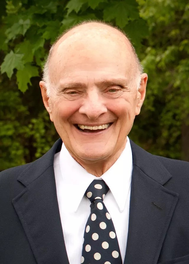
The American Roentgen Ray Society (ARRS) joins the radiology community in mourning the loss of Dr. Lee F. Rogers, a transformative leader, beloved educator, and devoted advocate for medical publishing. Named president of ARRS in 1988 and recipient of the 1993 Gold Medal, Dr. Rogers passed away on November 28, 2024, at the age of 90. He leaves an indelible mark on radiology and on ARRS, where his visionary leadership advanced the society’s mission of fostering excellence in radiologic education and research.
As just the 10th Editor in Chief of the American Journal of Roentgenology (AJR) from 1996 to 2004, Dr. Rogers ushered in a new era for “the yellow journal.” His editorial innovations, such as the medicolegal column by Dr. Leonard Berlin and his acclaimed Editor’s Notebook, enriched AJR as it transitioned into the digital age with advancements like AJR Online.
A look back at AJR Online in 2000—a pioneering step into the digital age, championed by Dr. Rogers during his tenure as Editor in Chief.
Reflecting on his impact, AJR’s current Editor in Chief, Andrew Rosenkrantz, MD, shared the following words:
“Lee Rogers had a tremendous influence on radiology. During his tenure with AJR, he greatly propelled the journal forward, introducing many changes that helped shape the journal into what it is today. He was an amazing educator, mentor, colleague, and friend to many throughout the field. He will be missed by those who had the chance to know him, with his legacy greatly remembered.”
Dr. Rogers’ influence extends beyond the pages of AJR through the Lee F. Rogers International Fellowship in Radiology Journalism. This Roentgen Fund fellowship invites radiologists from around the world to work closely with AJR’s Editor in Chief and publications staff, cultivating the next generation of medical journalists and enhancing the quality of radiology literature worldwide. Through this program, Dr. Rogers’ passion for academic excellence and mentorship continues to inspire radiologists to shape the future of the field. Writing Dr. Rogers’ memorial in the January issue of AJR, Ronald J. Zagoria, MD, duly noted: “Working under Lee’s guidance in the journal office, a generation of editors, authors, and reviewers now reflect his virtues” [1].
In his own Editor’s Notebook for AJR in November 2003 [2], Dr. Rogers shared his heartfelt belief in the value of participation:
“Meaningful participation is a broadening experience. You become informed, gain insights into the world about you, make new and often lasting friendships, and may have the opportunity to make significant contributions to things that are of interest and importance to you.”
ARRS honors Dr. Lee F. Rogers’ profound participation in radiology, his mentorship, and his enduring vision. His leadership and intellect will be deeply missed but remain a guiding light for all who follow in his footsteps.
ARRS is also saddened to note the passing of former president and Gold Medalist John P. Tampas of Colchester, VT. Having ably served on the ARRS Executive Council for many years, Dr. Tampas was gaveled in as president of this society during the opening ceremony of the 1982 ARRS Annual Meeting in New Orleans, LA.
ARRS is also saddened to note the passing of former president and Gold Medalist John P. Tampas of Colchester, VT. Having ably served on the ARRS Executive Council for many years, Dr. Tampas was gaveled in as president of this society during the opening ceremony of the 1982 ARRS Annual Meeting in New Orleans, LA.
Earning both undergraduate and medical degrees from his beloved University of Vermont (UVM), Dr. Tampas chaired the radiology department at his alma mater for a remarkable 26 years. In 2006, UVM endowed the Soule-Tampas Green & Gold Professorship in Radiology—a faculty-funded position named after him and predecessor, Dr. Bradley Soule.
Dr. Tampas’ medical career was marked by continuous service to his profession. In 1956-1957, he volunteered to serve as a U.S. Air Force flight surgeon in Korea and was awarded the rank of captain. A frequent contributor to AJR, he was bestowed lifetime membership in ARRS. And for his distinguished service to this society, in 1992 in Orlando, FL, Dr. Tampas received the ARRS Gold Medal.
Ruth Carlos Honored With American Roentgen Ray Society’s Gold Medal
The American Roentgen Ray Society (ARRS) proudly announces that former ARRS president Ruth C. Carlos, MD, MS, FACR, has been awarded the 2025 ARRS Gold Medal.
The highest accolade bestowed by North America’s first radiological society, the ARRS Gold Medal has been honoring illustrious service to radiology for more than four decades. The ARRS Gold Medal is awarded to physicians with a substantial record of service and who continue to render distinguished service to both the practice and science of medical imaging and its allied sciences.
Ruth Carlos was installed as the 119th president of ARRS during the 2019 Annual Meeting in Honolulu, Hawaii, succeeding Philip Costello, MD. She was honored as this year’s ARRS Gold Medalist on Sunday, April 27 during the opening ceremony of the 2025 ARRS Annual Meeting at California’s Marriott Marquis San Diego Marina.
Ruth Carlos, MD, MS, FACR, is a professor of radiology at Columbia University Irving Medical Center and associate chair of research faculty development for the department of radiology. She is a board-certified radiologist specializing in abdominal imaging.
Dr. Carlos also serves as the director of research in outcomes and care delivery for the Center for Imaging Biomarkers and Innovation in Integrated Diagnostics (CIMBID) at Columbia University Irving Medical Center.
A distinguished leader and pioneer in the field of radiology, Dr. Carlos has held several prestigious leadership positions that have shaped the direction of radiology research and practice. She is the first woman editor-in-chief of a major radiology journal, the Journal of the American College of Radiology (JACR), which guides the management and practice of radiology. She has served as president of ARRS, the Association of University Radiologists (AUR), and the Radiology Alliance for Health Services Research in Radiology. Currently, she chairs the GE AUR Research Radiology Academic Fellowship (GERRAF), a national program supporting early-stage investigators in health services research and care delivery.
Dr. Carlos’ influence extends to national policy, evidenced by her role as co-chair of the National Cancer Institute (NCI) Cancer Care Delivery Research (CCDR) Development Committee, and she lends her expertise as a thought leader to the National Academy of Medicine, the National Quality Forum, and the Joint Commission. Her wealth of expertise spans health services research and policy, health equity, and structural racism. Most recently, her work has focused on social genomics, the negative biological effects of social and economic marginalization and imaging biomarkers.
Dr. Carlos has received numerous awards and honors. She is an elected fellow of the American College of Radiology (ACR), as well as the Society of Computed Tomography and Magnetic Resonance (SCBT-MR). She has received the AUR Gold Medal, the Distinguished Educator Award from the Radiological Society of North America (RSNA), and the Stanford Medicine X Research Challenge Grand Prize. A funded investigator of the National Institutes of Health (NIH), she has been recognized for her research by induction into the Academy of Radiology Research Council of Distinguished Investigators, reflecting top 10% of all academic radiology faculty and awarding of the Paul C. Hodges Excellence Award.
Dr. Carlos received her medical degree from the University of Chicago, where she also completed her diagnostic radiology residency. She then completed an abdominal imaging fellowship at the University of Michigan in Ann Arbor. She also holds a master’s degree from the School of Public Health at the University of Michigan.
Tanya Moseley Receives American Roentgen Ray Society Distinguished Educator Award
ARRS is also proud to announce Tanya W. Moseley, MD, as the 2025 ARRS Distinguished Educator. The ARRS Distinguished Educator award recognizes outstanding individuals in the field of medical imaging who have a proven record of improving radiological education and remain committed to creating and implementing new and innovative educational activities.
ARRS’ Distinguished Educator award celebrates truly inspirational teachers—like Dr. Moseley— dedicated to education that results in improved participant performance, ultimately leading to enhanced patient outcomes. She was recognized as our 2025 Distinguished Educator during the opening ceremony of the ARRS Annual Meeting in San Diego, CA.
Newly installed ARRS president Deb Baumgarten with 2025 Distinguished Educator Tanya Moseley, and immediate past president Angelisa Paladin at the ARRS Annual Meeting at Marriott Marquis Marina in San Diego, CA.
Tanya Moseley exemplifies transformative leadership in radiology education, making her an exceptional choice for our Distinguished Educator. Following her distinguished tenure at MD Anderson Cancer Center as professor of breast imaging and breast surgical oncology, she now brings her visionary leadership to the University of Mississippi Medical Center, where she serves as vice chair of faculty development, division chief of breast imaging, and breast imaging fellowship director.
Her impact on radiology education has been both innovative and profound. As director of MD Anderson’s breast imaging fellowship program, she developed comprehensive curricula that have influenced generations of specialists. Her leadership of the Breast Imaging Technologist Continuing Education Committee demonstrated her commitment to technologist advancement, while her work as clinical image reviewer for multiple American College of Radiology accreditation programs underscores her dedication to maintaining the highest educational standards.
For more than two decades, Dr. Moseley’s contributions to the ARRS have transformed radiological education. Through her service on key educational committees and roles as case-based breast imaging chair and AJR SA-CME Consultant Editor, she has fundamentally shaped the organization’s educational direction. Her pioneering vision led to the creation of the first multi-vendor tomosynthesis certification course, demonstrating her exceptional ability to build collaborative educational programs. As architect and director of the ARRS Longitudinal Course Series, she continues to advance innovative approaches to radiology education.
Dr. Moseley’s leadership influence extends from institutional to national levels. In her role as chair of the FDA’s National Mammography Quality Assurance Advisory Committee, she helps establish national quality standards. Her leadership of both MD Anderson’s Diagnostic Imaging Council on Gender Inclusion Committee and the Society of Breast Imaging’s Inclusion, Diversity, and Equity Alliance reflects her commitment to creating a more inclusive field that elevates both trainees and faculty.
What truly sets Dr. Moseley apart is her comprehensive approach to education and leadership. Through strategic academic appointments across multiple institutions, she has created vital connections between breast imaging, surgical oncology, and clinical sciences. Her extensive committee service encompasses scientific programming, faculty achievement, quality improvement, and executive leadership, demonstrating her profound impact on medical education and practice.
Her teaching excellence has garnered prestigious recognition, including the University of Texas System Regents’ Outstanding Teaching Award and induction into the University of Texas Shine Academy of Health Science Education. Her influence reaches globally through international breast imaging observership programs and visiting professorships.
Dr. Moseley’s educational philosophy rests on seven core principles: clinical excellence in teaching, innovative program development, rigorous quality standards, interdisciplinary collaboration, dedicated mentorship, technological advancement, and an unwavering commitment to diversity and inclusion in medical education.
Her selection as ARRS Distinguished Educator recognizes a career dedicated to transforming radiology education and leadership. Dr. Moseley’s exceptional ability to combine clinical expertise with innovative teaching methods, while championing mentorship, quality, and inclusivity, makes her the ideal recipient of this honor. Her career exemplifies the highest standards of an educator-leader who shapes the future of radiology through both educational excellence and visionary leadership.
ARRS Names 2025 Honorary Member: Salvador Amézquita Pérez
Drs. Paladin, Pérez, and Baumgarten at the 2025 ARRS Annual Meeting in San Diego, CA.
And, finally, ARRS is happy to report that Dr. Salvador Amézquita Pérez, president of Sociedad Mexicana de Radiología e Imagen (Mexican Society of Radiology and Imaging), will receive honorary ARRS membership on day one of the society’s 2025 Annual Meeting in San Diego.
A distinguished army veteran and vascular and interventional radiologist with more than two decades experience in clinical practice and medical education, Dr. Pérez has chaired the radiology and imaging department at Hospital Central Militar (Central Military Hospital) in Mexico City since 2019, following his seven- year tenure as deputy department chair. He has been a prominent figure in the Mexican Society of Radiology and Imaging (SMRI), receiving the Award for Academic Excellence in 2018. Previously, Dr. Pérez served SMRI as treasurer (2018-2020) and general secretary (2020-2022). He will remain SMRI President until 2026.
ARRS looks forward to welcoming Dr. Pérez and his colleagues to the 2025 ARRS Annual Meeting in San Diego as part of the Global Exchange Featuring Mexico. The global exchange includes the ARRS-SMRI Sunday Featured Course, “Advances in Cardiac Imaging,” focused on techniques and considerations for evaluating structural heart conditions and coronary artery anomalies. This session will address non-atherosclerotic coronary artery narrowing, CT’s role in transcatheter mitral valve replacement and adult congenital heart disease, CT perfusion and FFR-CT for myocardial ischemia, as well as CT in TAVR before and after surgery.
Since its founding in 1946, SMRI has focused its mission on maintaining high levels of academic and scientific partners, while working with radiology groups across the country on annual courses and weekly, on-site ultrasound and general radiology meetings. In recent years, SMRI has organized international events with the International Society of Magnetic Resonance in Medicine and the European School of Radiology. SMRI also maintains a technical school for radiologists—the oldest and most prestigious in Mexico. Anales de Radiología México, founded in 2002, is the country’s only publication of its kind specializing in radiology.
The mission of the ARRS Global Partner Society Program is to build long-standing relationships with key leaders and organizations in the worldwide imaging community—increasing awareness of our society’s services in specific nations, while raising the stature of Global Partner Societies among ARRS members. Every year, the ARRS Annual Meeting Global Exchange incorporates one partner society into the educational and social fabric of our meeting. ARRS members then reciprocate at the partner society’s meeting that same year.
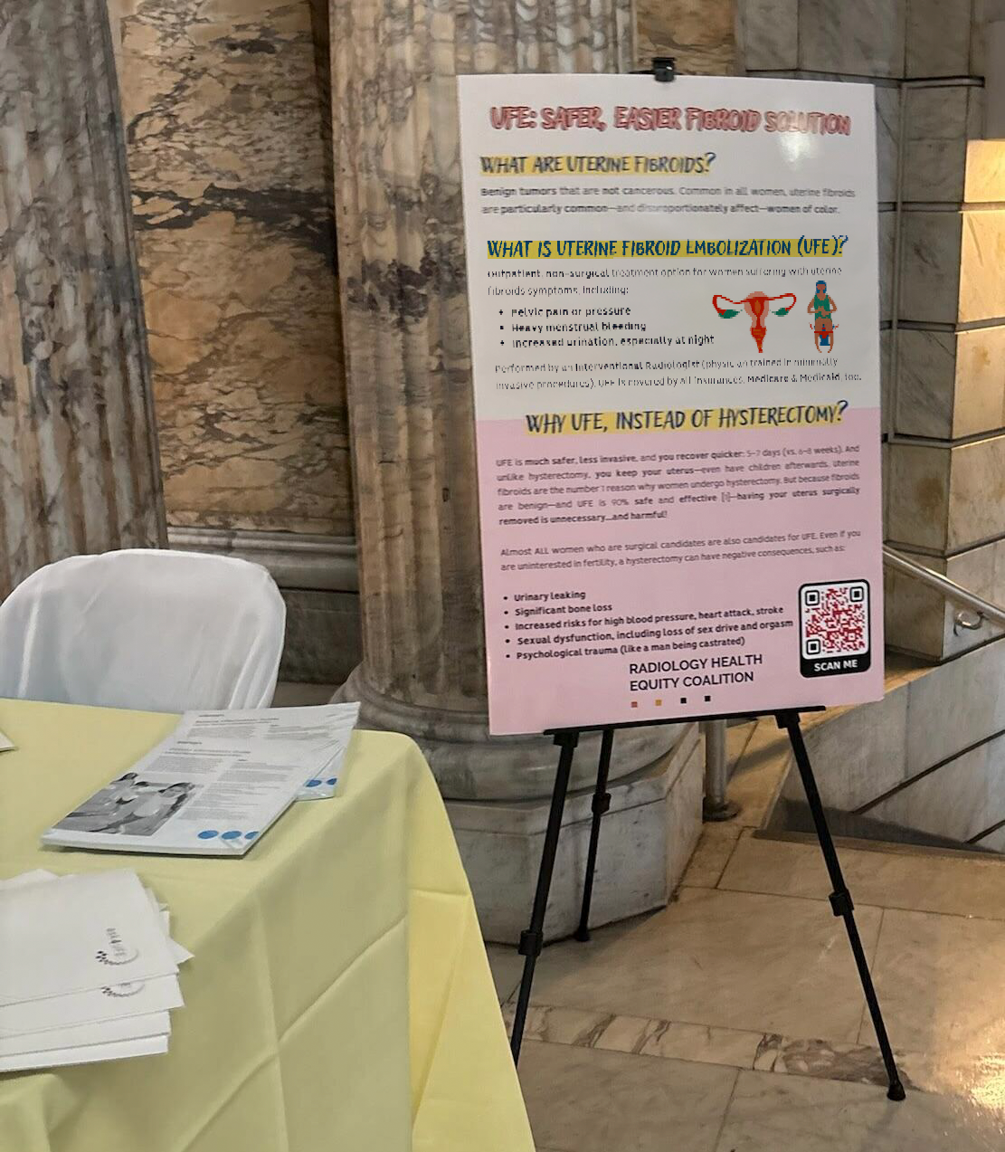
A member of the Radiology Health Equity Coalition (RHEC), ARRS continues to collaborate with RHEC on curating and disseminating trusted resources and best practices for improving access to and utilization of preventive and diagnostic imaging.
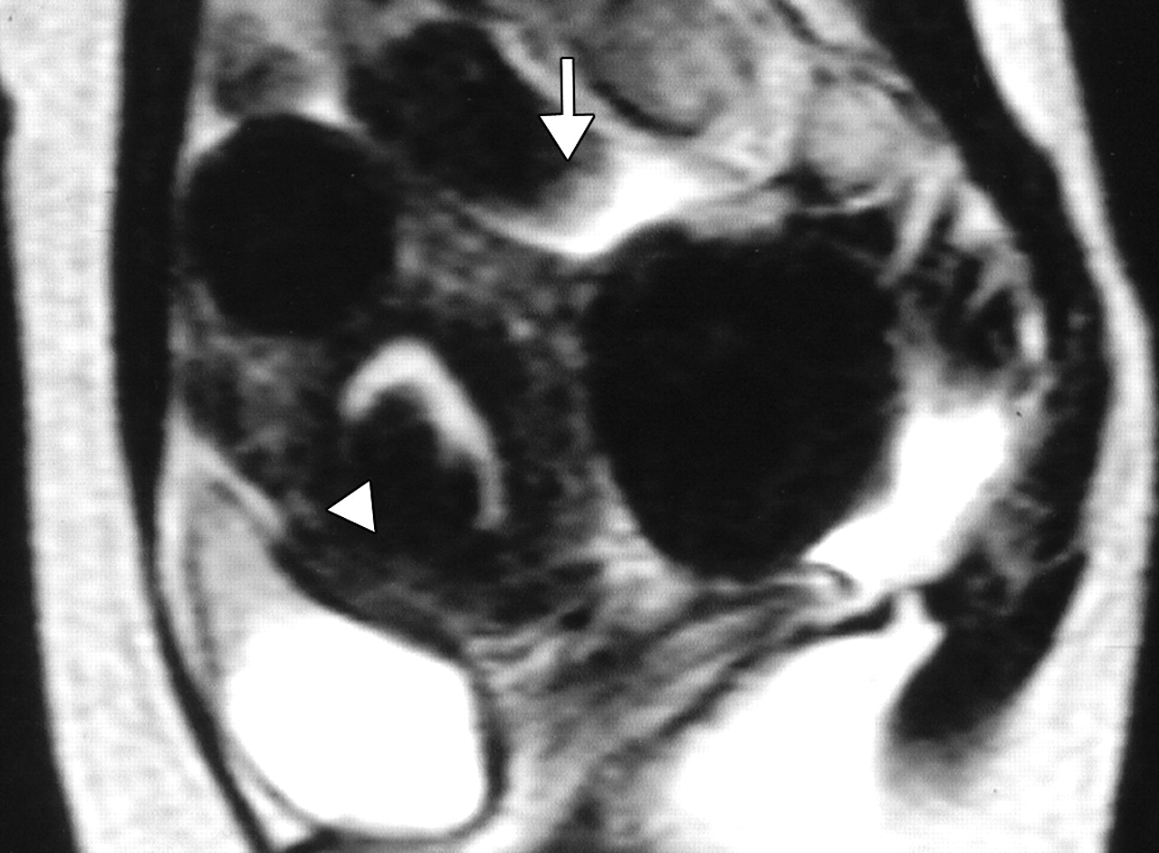
Uterine fibroids are benign tumors that develop in the uterus. While common among all women, fibroids disproportionately affect women of color. Fibroids are the leading cause of hysterectomy, with the average age of women undergoing this major surgery being just 39 years old (Fig. 1).
Fig. 1—44-year- old woman with menorrhagia and bulk-related symptoms caused by multiple uterine fibroids. T2-weighted MR image obtained before embolization reveals submucosal fibroid (arrowhead) and pedunculated subserosal fibroid 6 cm in maximal diameter. Diameter of stalk (arrow) is 2 cm.
Why Patients Should Consider Uterine Fibroid Embolization
UFE offers numerous benefits compared to hysterectomy or other surgical options:
Minimally Invasive
Performed by an interventional radiologist, UFE involves no surgical incisions
Outpatient Procedure
Patients typically go home the same day
Shorter Recovery Time
Recovery from UFE takes only 5–7 days compared to the 6–8 weeks often required after a hysterectomy
Uterus Preservation
Unlike a hysterectomy, UFE allows women to keep their uterus, maintaining their fertility and hormonal balance
Safe and Effective
UFE has a success rate of 90%, providing relief from fibroid symptoms like heavy menstrual bleeding, pelvic pain, and increased urinary frequency (Fig. 2)
Fig. 2—44-year- old woman with menorrhagia and bulk-related symptoms caused by multiple uterine fibroids. Contrast-enhanced T1-weighted MR image obtained 1 year after embolization reveals pedunculated subserosal fibroid, now 4.5 cm in maximal diameter, is not enhancing. Diameter of stalk (arrow) is now 1.1 cm. Rate of volume reduction in pedunculated subserosal fibroid is 61%. Menorrhagia and bulk-related symptoms markedly improved.
Why Keeping Your Patient’s Uterus Matters
Even for women not planning to have children, the uterus plays a vital role in overall health. Removing the uterus through hysterectomy can lead to serious complications, such as:
Psychological distress (often compared to the emotional impact of castration in men)
Sexual dysfunction, including reduced libido and loss of orgasm
Urinary incontinence
Significant bone loss
Increased risks of high blood pressure, heart attack, and stroke
Nearly all patients who qualify for surgery are also candidates for UFE, and UFE is covered by all major insurance providers, including Medicare and Medicaid.
Artificial intelligence (AI) now impacts radiologists at all stages of training and in all practice settings. Every day, radiologists hear of new opportunities and new challenges that AI will bring to their work and lives. Most practicing radiologists, and many radiologists in training, have not received the education necessary to navigate this new world, discern truth from hype, and recognize the dangers and responsibilities facing them as they integrate these systems into hospitals and clinics.
In order to safely, responsibly, and successfully develop, implement, and use AI systems to improve patient care and our work lives, radiologists must learn the basics in key areas. On Sunday, April 27 during the 2025 ARRS Annual Meeting at Marriott Marquis San Diego Marina, “Critical Education for Safe, Ethical, and Successful AI in Radiology” will provide expert guidance in:
Reliable AI: From Research to Production;
Practical AI: Implementation, Workflow and Monitoring;
Successful AI: Product Development, Validation, and Regulation;
Ethical AI: Bias, Diversity, Accessibility, and Privacy.
Most practicing radiologists have not received basic, trustworthy education that will allow them to understand, assess, implement, and successfully utilize AI products in their practices. Training programs now offer basic education about AI, but this often lacks teaching from those with broad experience and the ability to provide a comprehensive overview, including research, ethics, clinical implementation, and the business and economics of AI. Radiologists in practice and in training must be prepared to evaluate products, understand the important role of standards for AI integration into workflow, and be aware of pitfalls and dangers that could harm patients and the practice if not recognized. Led by course director Wende Gibbs, MD, attendees of “Critical Education for Safe, Ethical, and Successful AI in Radiology” will benefit from the unique group of speakers who are trailblazers in the areas of AI research, education, and business, and current and future leaders who are breaking down silos and facilitating multidisciplinary, multispecialty cooperation in the creation of safe, successful, and ethical AI for all.
Later in the afternoon on Sunday, April 27 in San Diego, CA, “Clinical Implementation of AI” will provide a timely review of best practices for selecting and a primer for deploying AI products to maximize return on investment. Course director Manisha Bahl, MD, has assembled four leading researchers in AI who also practice in different subspecialties. Their goal? Enhancing the overall value of any AI system by streamlining clinical workflows, while instilling effective methodology for monitoring said system’s practical impacts. Combining expert-led lectures with an interactive Q&A session, this ARRS Annual Meeting Sunday Session is open to all registrants.
Stacy J. Kim, MD Mallinckrodt Institute of Radiology Washington University in St. Louis
The first human lung transplant was performed in 1963. Since then, the number of lung transplant cases in the United States has steadily increased due to continued advancements in surgical technique and immunosuppressive medication. There were over 3000 lung transplants performed in the United States in 2023, and the number of lung transplants is likely to continue to increase [1]. The patients who undergo lung transplant are those with end-stage lung disease, which can result from a variety of pathologies including emphysema, fibrosing interstitial lung disease, cystic fibrosis, and pulmonary arterial hypertension. Postoperatively, these lung transplant recipients are vulnerable to complications for the remainder of their lives. The complications can be categorized by time course; that is, the time period after transplant during which the complications occur or most often occur.
The postoperative time periods can be organized as follows: immediate, within 24 hours of transplant; early, from 24 hours to 1 week after transplant; intermediate, from 1 week to 3 months after transplant; and late, more than 3 months after transplant [2]. Some complications can occur during more than one time period or span multiple time periods. This chapter will discuss the complications that occur or most often occur during the 1st month or so after lung transplant, which includes immediate, early, and some of the intermediate complications. Late complications of lung transplant will be discussed during the 2025 ARRS Annual Meeting Categorical Course, “Comprehensive Insights Into Transplant Imaging,” in San Diego, CA, and online April 27-May 1.
Imaging Techniques
Chest radiography is the most commonly used imaging study in the immediate and early postoperative setting. Chest radiographs are easy to acquire at the bedside and are useful in evaluating the positions of tubes and lines, which are ubiquitous immediately after transplant, such as endotracheal tubes, central venous catheters, and chest tubes. The lung parenchyma and the pleura can also be evaluated with chest radiographs for complications such as pneumonia and pleural effusion. Given the lower radiation dose of chest radiography when compared with CT, chest radiographs are useful for serial imaging; that is, image acquisition over multiple days to assess for change over time.
CT of the chest is performed if a more detailed assessment of the chest is required. Example scenarios in which a detailed assessment may be necessary include if there is concern for bronchopleural fistula in the setting of a persistent pneumothorax, if a pulmonary embolism (PE) is suspected due to new-onset tachycardia, and if a patient with decreasing hemoglobin values must be evaluated for hemorrhage. A noncontrast chest CT examination is sufficient for evaluation of the lung parenchyma, airways, and bones. A contrast-enhanced chest CT examination should be acquired (if the patient’s renal function permits and if the patient does not have a contrast media allergy) for evaluation of the vasculature and the pleura and assessment for active hemorrhage. The protocol or phase of contrast should be tailored to the diagnosis being evaluated; for example, a PE protocol should be used when evaluating for PE.
A noncontrast high-resolution chest CT examination is rarely necessary for the evaluation of early lung transplant complications. However, it is useful for the evaluation of late lung transplant complications as it can be used to detect air trapping and fibrosis (discussed in the next chapter). MRI, sonography, and nuclear medicine imaging are not typically used in the evaluation of early lung transplant complications.
Hyperacute and Acute Rejection
Hyperacute rejection occurs during the lung transplant surgery or within 24 hours of transplant when preformed recipient antibodies react to donor antigens in the allograft [3]. It is exceedingly rare because ABO blood group antigens and human leukocyte antigens are taken into account when lung donation is arranged, to ensure donor-recipient compatibility. Hyperacute rejection manifests as fulminant multiorgan system failure, and most patients with hyperacute rejection die within a few days to 2 weeks after lung transplant. The imaging findings of hyperacute rejection are nonspecific and resemble severe pulmonary edema, including consolidation, ground-glass opacities, and septal-line thickening. Acute rejection can occur anytime after lung transplant. It consists of two types, acute cellular rejection (ACR) and antibody-mediated rejection (AMR), which can coexist. ACR is the more common of the two types and occurs when recipient T lymphocytes attack donor antigens within the lung allograft. Approximately 35% of lung transplant recipients experience at least one episode of ACR during the 1st year after transplant [2]. During these episodes, patients may be asymptomatic or may present with nonspecific symptoms such as dyspnea and cough. The imaging findings of ACR are nonspecific and include consolidation, ground-glass opacities, and septal-line thickening; as with hyperacute rejection, ACR resembles pulmonary edema. Given its nonspecific clinical and imaging manifestations, ACR requires transbronchial biopsy and tissue analysis for diagnosis. Timely treatment, typically by increased immunosuppression with steroids, is important because ACR is the greatest risk factor for chronic lung allograft dysfunction [4]. Figure 1 shows a patient with biopsy-proven ACR.
Fig. 1—Patient with history of bilateral lung transplant who developed acute cellular rejection (ACR). Left: Chest CT at time of biopsy-proven ACR shows bronchial wall thickening and nodular ground-glass opacities in both lungs, as well as small pleural effusions. Right: Chest CT several months after treatment of ACR shows resolution of bronchial wall thickening, ground-glass opacities, and pleural effusions.
AMR, the less common of the two types of acute rejection, occurs when recipient B lymphocytes create donor-specific antibodies (DSA), donor-specific antigens and DSA form complexes, and the complexes trigger the immune system’s complement pathway. Like patients with ACR, patients with AMR can be asymptomatic; can have nonspecific symptoms such as dyspnea and cough; and can have normal chest imaging or nonspecific imaging findings resembling pulmonary edema such as consolidation, ground-glass opacities, and septal-line thickening. Transplant physicians diagnose patients with clinical versus subclinical AMR and definite versus probable versus possible AMR on the basis of the presence or absence of allograft dysfunction, histology results suggestive of AMR (such as neutrophil arteritis and capillaritis), immunostaining results (positive C4d staining of the capillary endothelium), and the presence or absence of DSA in peripheral blood [4]. Treatments include plasmapheresis and IV immunoglobulin to remove harmful antibodies and to suppress antibody production, respectively. Steroids are not typically used to treat AMR, unlike ACR. Figure 2 shows a patient with AMR.
Fig. 2—Patient with history of bilateral lung transplant who developed antibody-mediated rejection (AMR), which was diagnosed by transbronchial biopsy and donor-specific antibody blood testing. Chest CT at time of AMR diagnosis shows ground-glass opacities and reticulation in both lungs and small pleural effusions.
Primary Graft Dysfunction
Primary graft dysfunction (PGD) is a transient complication that occurs within 24–72 hours after lung transplant and tends to resolve by postoperative day 5–10. PGD is thought to be secondary to ischemic injury of the allograft before and during transplant and secondary to reperfusion injury after transplant. PGD occurs in approximately 10–30% of lung transplant recipients [5]. On imaging, it manifests as perihilar and lower lung–predominant airspace and interstitial opacities and is similar in appearance to pulmonary edema. Clinically, PGD is graded from 0 to 3 on the basis of the presence or absence of imaging abnormalities and the severity of hypoxemia [6]. In patients who have undergone a unilateral lung transplant, PGD and pulmonary edema can be differentiated by observing the distribution; PGD affects only the lung allograft, whereas pulmonary edema affects both the lung allograft and the native lung. Like acute rejection, PGD is considered a risk factor for chronic lung allograft dysfunction. It is treated with supportive care, such as mechanical ventilation and extracorporeal membrane oxygenation. Figure 3 shows a patient with PGD.
Fig. 3—Patient with right lung transplant who developed primary graft dysfunction (PGD). Left:Chest radiograph on postoperative day 0 shows clear right lung allograft and fibrosis of native left lung. Center: Chest radiograph on postoperative day 3 shows interval development of hazy airspace opacities throughout right lung allograft. Right:Chest radiograph on postoperative day 4 shows near resolution of right lung allograft airspace opacities, consistent with PGD.
Infection
Infection of the lung parenchyma and airways is exceedingly common after lung transplant due to immunosuppression and decreased mucociliary clearance in the airways after transplant. Infection can occur anytime after transplant, including the early postoperative period. Patients are vulnerable to bacterial pneumonia as well as viral and fungal pneumonias not commonly encountered in immunocompetent patients. Common pathogens include Pseudomonas organisms, Staphylococcus aureus, cytomegalovirus, Epstein-Barr virus, herpes simplex virus, Aspergillus organisms, and Candida organisms [2]. The clinical and imaging manifestations of pneumonia in transplant recipients are similar to those of nontransplant patients. Lung transplant recipients who present with dyspnea, cough, or fever are evaluated for pneumonia. Imaging findings of pneumonia include consolidation, ground-glass opacities, septal-line thickening, and pulmonary nodules. Pulmonary nodules can be single or multiple; they may be solid or ground-glass in attenuation. Cavitary nodules and nodules with ground-glass halos can occur, especially in patients with fungal pneumonia. Imaging studies should be scrutinized for complications of infection such as pulmonary abscess and bronchopleural fistula. Patients may also have reactive pleural effusions or reactive mediastinal or hilar lymphadenopathy. Treatment is the same as in nontransplant patients and consists of antibiotics, antivirals, or antifungals depending on the causative pathogen. Figure 4 shows three different lung transplant recipients with pneumonia.
Fig. 4—Three different patients with history of bilateral lung transplant who presented with pneumonia. Left:Chest CT image shows consolidation and numerous small nodules in lungs due to bacterial pneumonia, in addition to left lower lobe cavitation and left empyema with gas in pleural space. Center: Chest CT image shows ground-glass opacities and septal-line thickening throughout both lungs caused by viral pneumonia (SARS-CoV-2). Right:Chest CT image shows cavitary lesion and multiple small nodules in right lung in patient with fungal (Aspergillus) pneumonia.
Pleural Complications
Simple pleural effusions and small pneumothoraces are frequently encountered in the immediate and early posttransplant setting and typically resolve within a few days to 1 week or so after lung transplant. Pleural fluid collections and pneumothoraces that are large, increasing in volume over time, or persist over 1 week may indicate a potentially serious complication such as hemothorax, empyema, bronchial anastomotic dehiscence, or bronchopleural fistula.
Hemothorax, the presence of blood products in the pleural space, should be suspected if there is rapid increase in the volume of pleural fluid over serial imaging or if pleural fluid is hyperattenuating relative to simple fluid on CT. Hemothorax can be heterogeneous in attenuation on CT due to mixing or layering of new blood products with old blood products; a fluid-fluid level may be present. Figure 5 (left) shows a patient with hemothorax.
Fig. 5—Two different patients with history of bilateral lung transplant who presented with hemothorax and empyema. Left: Chest CT image shows large heterogeneous-attenuation collection throughout right pleural space, consistent with hemothorax. Right: Chest CT image shows small- to moderate-volume collection in left pleural space with associated visceral and parietal pleural thickening and enhancement extending into prior left chest tube track, consistent with empyema. Patient also has small right pleural effusion with mild pleural thickening.
Empyema, the presence of infected material (i.e., pus) in the pleural space, should be suspected if there is persistence of pleural fluid over serial imaging for more than 1 week or so after transplant and if visceral and parietal pleural thickening or loculated pleural fluid is present on chest CT. On contrast-enhanced CT of the chest, patients with empyema may have abnormal thickening and enhancement of the visceral pleura and parietal pleura with fluid between the two pleural layers, which is known as the split pleura sign. Figure 5 (right) shows a patient with empyema.
Pneumothorax, the presence of gas in the pleural space, may be indicative of a bronchial anastomotic dehiscence or a bronchopleural fistula if it persists more than 1 week after transplant or increases in volume over time. A tension pneumothorax with cardiomediastinal shift away from the affected hemithorax should be immediately communicated to the transplant medicine team, as an untreated tension pneumothorax can cause cardiovascular and respiratory collapse. Small pneumothoraces on immediate postoperative chest imaging that resolve over the next few days, at which point any chest tubes present would be removed, are considered to be expected postoperative findings.
Treatment of pleural collections typically involves drainage of the pleural space material via pleural catheters or thoracostomy tubes. Surgical intervention may be required if drainage via catheters and tubes is unsuccessful or if the pleural collections are caused by complications such as bronchial anastomotic dehiscence or a bronchopleural fistula.
Vascular Complications
PE can occur after lung transplant. Hemorrhage most commonly occurs during the immediate and early postoperative periods and can manifest as hemothorax, other forms of thoracic hemorrhage such as mediastinal hematoma and chest-wall hematoma, and nonthoracic hemorrhage such as retroperitoneal hematoma. Approximately 4.5% of lung transplant recipients experience posttransplant hemorrhage severe enough to require surgical intervention [7]. The causes of hemorrhage include inadequate coagulation, vascular anastomotic dehiscence (which is rare but can be catastrophic when it occurs), and injury of other vessels. CT of the body part of concern (for example, CT of the chest if there is concern for mediastinal hemorrhage) should be performed, ideally with IV contrast material. If active hemorrhage is suspected, CT can be performed before and after the administration of IV contrast material in the arterial and venous phases to detect contrast material extravasation.
PE also most commonly occurs during the immediate and early postoperative periods. Patients are typically bedbound for at least the first few days after lung transplant, and some patients require mechanical ventilation during that time. Some patients may have been bedbound and/or may have been receiving mechanical ventilation while awaiting the transplant surgery. Immobility increases these patients’ risk for developing deep venous thrombosis (DVT) and subsequently developing PE. It is important to understand that lung transplant recipients are at increased risk of pulmonary infarction secondary to PE because the bronchial circulation is not reestablished during the transplant, and until collateral vessels form in a few weeks, these patients are relying on blood supply from the pulmonary circulation. If PE is suspected, CT of the chest with IV contrast material should be performed per the PE protocol.
The imaging findings of hemorrhage and PE are the same in lung transplant recipients as in nontransplant patients. Hemorrhage manifests as hyperattenuating fluid (higher attenuation than that of simple fluid) or a hyperattenuating mass (if it is a hematoma) that often has a heterogeneous appearance. PE manifests as hypoattenuating and well-defined filling defects in the contrast material–opacified pulmonary artery branches. These filling defects can be occlusive or nonocclusive; nonocclusive acute PE is centrally located in the vessel lumen, rather than eccentric. Patients with coagulopathy and patients who are receiving anticoagulation therapy for DVT or PE may have both hemorrhage and PE on imaging. Figure 6 shows a patient with bilateral chest-wall hematomas.
Fig. 6—Patient with history of bilateral lung transplant who developed deep venous thrombosis in early postoperative setting was prescribed anticoagulation therapy and then spontaneously developed chest-wall hematomas, left greater than right. CT image shows heterogeneous-attenuation collections in right and left chest walls deep to pectoralis major muscles, which represent hematomas.
Pulmonary vein thrombosis and pulmonary venous anastomotic stenosis occur during the immediate and early postoperative periods, usually within 48 hours of lung transplant [8]. In pulmonary vein thrombosis, CT of the chest with IV contrast material shows a filling defect within a pulmonary vein, which may or may not be accompanied by consolidation, ground-glass opacities, and septal-line thickening in the lung parenchyma drained by the thrombosed pulmonary vein; the airspace opacities and septal-line thickening represent edema and hemorrhage due to venous ischemia and infarction. Endovascular intervention can be attempted, but cases of severe pulmonary vein thrombosis require surgery. Figure 7 (left) shows a patient with pulmonary vein thrombosis.
Fig. 7—Patient with history of bilateral lung transplant who presented with thrombosis of left inferior pulmonary vein. Left: Chest CT image acquired day after lung transplant shows occlusion of left inferior pulmonary vein (arrow). Right: Chest CT image shows consolidation and ground-glass opacities in left lower lobe indicative of edema and hemorrhage due to venous ischemia.
The pulmonary venous anastomosis is created adjacent to the left atrium. Stenosis of the pulmonary venous anastomosis is rare compared with stenosis of the pulmonary arterial anastomosis. On imaging, stenosis manifests as a focal narrowing of the pulmonary vein anastomosis that may or may not be accompanied by findings of venous ischemia and infarction in the lung parenchyma drained by the affected pulmonary vein, such as consolidation, ground-glass opacities, and septal-line thickening. As with pulmonary vein thrombosis, treatment options include endovascular intervention and, in severe cases, surgical repair. Figure 7 (right) shows a patient with pulmonary venous anastomotic stenosis.
Mechanical Complications
Mechanical complications include pulmonary torsion and lung herniation. Pulmonary torsion is a very rare complication of lung transplant and occurs during the immediate and early postoperative periods. A risk factor for lung torsion is when the donor lung is small relative to the recipient thoracic cavity, which means that the allograft is more mobile and likely to twist around its vascular pedicle after the transplant [9]. Careful size matching between the donor lung and the recipient chest cavity before the transplant surgery has greatly reduced the risk of lung torsion; however, given the potentially catastrophic consequences of torsion and the need for emergent surgical intervention, it remains an important diagnosis to be aware of. As previously stated, lung transplant recipients are particularly vulnerable to allograft ischemia and infarction because the bronchial circulation is not reestablished during transplant and the lung allograft must rely on pulmonary circulation until collaterals can form. Vascular compromise of the allograft due to torsion can result in severe allograft damage, allograft failure, or even death.
Imaging findings of pulmonary torsion can involve a lobe (in lobar torsion) or the entire lung (if the entire lung has twisted around its vascular pedicle). Pulmonary torsion can manifest as volume loss or collapse of the affected lobe or lung; it can also manifest as rapid expansion or opacification of the affected lobe or lung. Because of the twisting that occurs in torsion, patients with torsion have abnormal orientations and positions of anatomic structures such as lobes, hila, fissures, vessels, and airways. There may be abrupt cutoff of vessels and bronchi at the site of twisting. If pulmonary torsion is confirmed or suspected, the transplant physicians should be notified immediately to salvage as much of the allograft as possible. Pulmonary torsion requires emergent surgery to prevent allograft infarction and patient death.
Lung herniation can occur anytime after lung transplant. A major risk factor for lung herniation is increased intrathoracic pressure, as can be seen in patients with persistent cough due to pneumonia or aspiration after transplant. On imaging, herniated lung has an abnormal contour with a portion of lung bulging into the chest wall; this can occur at surgical sites (such as thoracotomy incisions) or at intercostal spaces. Mild herniation involving a small portion of the lung allograft with normal-appearing parenchyma is not worrisome. However, herniation involving a large portion of the allograft or deep herniation into the chest wall places the patient at risk for atelectasis, ischemia, infarction, and gangrene of the herniated lung, all of which can manifest as opacities within the herniated portion of lung. Surgical repair of the chest wall may be necessary in cases of pulmonary infarction or gangrene. Figure 8 shows a patient with lung herniation.
Fig. 8—Patient with history of bilateral lung transplant who presented with lung herniation. Frontal chest radiograph shows mild herniation of small portion of right upper lung into chest wall at thoracotomy incision site (arrow).
Conclusion
Lung transplant is increasingly becoming a cure for many patients with end-stage lung disease. In addition to academic chest radiologists, private practice radiologists and general radiologists are likely to encounter lung transplant recipients at some point in their careers. It is therefore essential for all radiologists to have a basic understanding of lung transplant complications—both common complications such as pneumonia and rare but life-threatening complications such as torsion. Although great progress has been made since the 1960s, the mean life expectancy of lung transplant recipients lags behind that of other organ recipients at only 6–7 years after transplant [10]. Early complications account for much of the morbidity and mortality in lung transplant recipients. These complications must be accurately detected and described when interpreting imaging studies, and they should be taken into account when protocoling imaging studies for lung transplant recipients. By doing so, radiologists can contribute to the postoperative care of lung transplant patients and can help optimize the quality and the duration of their posttransplant lives.
ERRATA:The winter issue featured this article with production errors, including inadvertent unauthorized changes to the title, introduction, and figures 3 and 4. We regret these errors and have republished the complete, corrected article for clarity.
References
Organ Procurement and Transplantation Network, U.S. Department of Health & Human Services. National Data: Transplants in the U.S. by Region. optn.transplant.hrsa.gov/data/view-data-reports/national-data. Published 2024. Accessed August 25, 2024
Kim SJ, Azour L, Hutchinson BD, et al. Imaging course of lung transplantation: from patient selection to postoperative complications. Radiographics 2021;41:1043-63
Masson E, Stern M, Chabod J, et al. Hyperacute rejection after lung transplantation caused by undetected low-titer anti-HLA antibodies. J Heart Lung Transplant 2007;26:642-45
Shah RJ, Diamond JM. Primary graft dysfunction (PGD) following lung transplantation. Semin Respir Crit Care Med2018;39:148-54
Snell GI, Yusen RD, Weill D, et al. Report of the ISHLT Working Group on Primary Lung Graft Dysfunction, part I: definition and grading – a 2016 consensus group statement of the International Society for Heart and Lung Transplantation. J Heart Lung Transplant 2017;36:1097-1103
Adelmann D, Koch S, Menger J, et al. Risk factors for early bleeding complications after lung transplantation – a retrospective cohort study. Transpl Int 2019;32:1313-21
Kim SJ, Short RG, Beal MA, et al. Imaging of lung transplantation. Clin Chest Med 2024;45:445-460
Amadi CC, Galizia MS, Mortani Barbosa EJ Jr. Imaging evaluation of lung transplantation patients: a time and etiology-based approach to high-resolution computed tomography interpretation. J Thorac Imaging 2019;34:299-312
Verleden GM, Glanville AR, Lease ED, et al. Chronic lung allograft dysfunction: definition, diagnostic criteria, and approaches to treatment – a consensus report from the pulmonary council of the ISHLT. J Heart Lung Transplant 2019;38:493-503
Going into 2025—the 125th anniversary of the American Roentgen Ray Society (ARRS)—I will be using my remaining InPractice columns as your president to provide more context regarding working happier as a radiologist. Centered in worker voice and equity, the happy radiologist is connected most to a safe and secure community—where everything from mentorship to scheduling works together to help foster a true sense of belonging. Opportunities for learning and accomplishment afford radiologists a dignity and meaning beyond our workstations, too.But let us start in 2022. I think we can all remember then. In the wake of the COVID pandemic, the United States Surgeon General, Dr. Vivek Murthy, released an updated framework for mental health and wellbeing in the workplace, outlining the foundational roles that workplaces must play in promoting health and wellbeing alike in our communities [1]. The U.S. Department of Health and Human Services was formally recognizing that our health care industries had to start focusing on working well. I want to share an accompanying figure from this federal framework with InPractice readers because it provides a great platform to think about ground-up, actionable initiatives that could work for our own specific radiological workplaces (Fig. 1).
Fig. 1—The five essentials for workplace mental health and wellbeing help organizations develop, institutionalize, and update policies, processes, and practices that best support the mental health and wellbeing of all workers.
Let’s turn our attention to the right side of this graphic: specifically, making connections and belonging to a community. The first step here is developing a culture. What does your chair or section chief value? What does your CEO treasure? We always talk about mission and vision, and answering these big questions are critically important to those conversations. Personally, I would like to know what my chair and my section value. And what are their goals for my own department? Additionally, what resources are our leaders able to provide to ground us in the institution’s values?
Lately, I’ve been looking at how the social supports of community are connected to a crucial distinction that the Surgeon General’s chart makes here. At the end of the workday, it’s not really a “balance” between that work and our lives at home. It is much more about work-life “harmony,” which Dr. Murthy’s office also notes is based in autonomy and flexibility.
When I was in training, we worked from seven to six o’clock, and then, we’d call from six o’clock to the next morning. Our schedule was rigid. It didn’t allow for a family, much less proper family time. During residency, I didn’t even go to the dentist; I simply didn’t have the time! And as a fellow, I remember tearing up when I got home from a RiteAid, of all places. I was crying because I’d never taken enough care of myself to go and buy products. I was just working so hard.
Given all the types of scheduling we have now, there are such wonderful opportunities with shift work. In January, our group moved to shift-type areas, where some people arrive at noon, some people come in at 4pm. This flexibility has provided a lot of satisfaction for our group, as directors have empowered us to choose the shifts that best fit our lives at that moment.
Moving clockwise, you’ll notice another essential element of working happier. Everyone reading this piece right now is a leader, or a valued member of a group with a leader at the helm. So, how do good leaders accommodate the group’s varied interests, while making individuals feel that they matter? In an academic setting, we can further foster flexibility with research and education, especially subspecialty experience. As you know, data have come out noting that if only 20% of your workday is spent doing something you like, you will be a much happier worker.
For me, my happiness at work is temporal bone. When I see a temporal bone case, I just start smiling because it brings me such great joy. Of course, our colleagues are happier when they’re reading cases that spark joy, so until next time, let’s really think about related worklist opportunities. I’ve seen some creative solutions in which worklists are done not by modality, but by image or imaging interests. Overall, reported happiness is much higher.