Large language models (LLMs) are reshaping radiology, but their integration into the reading room is far from straightforward. 2026 ARRS Annual Meeting Categorical Course Director Yee Seng Ng, MD, outlines the most significant barriers to adoption and why the solutions are more complicated than they appear.
Security Slows Adoption
Most widely available LLMs live on proprietary cloud platforms. Sending protected health information (PHI) outside a hospital network creates immediate compliance issues, and current guidelines from major organizations explicitly prohibit using public LLMs for protected patient information. Even if models claim not to store or reuse data, rads cannot verify how patient information is handled, refined, or monetized.
Private Solutions, But Not for All
Institutions can build private LLM instances behind their firewall, but this requires substantial infrastructure, IT support, and vendor partnerships—resources generally limited to large academic centers. Local installations using open-source models (via interfaces such as webUI) avoid cloud exposure but introduce new challenges: maintenance, computing requirements, and accessibility across the department.
Where Is NLP Already Helping?
Even though LLMs aren’t widely used at the point of care, rads are already benefitting from improved natural language processing (NLP) embedded in commercial reporting tools, including
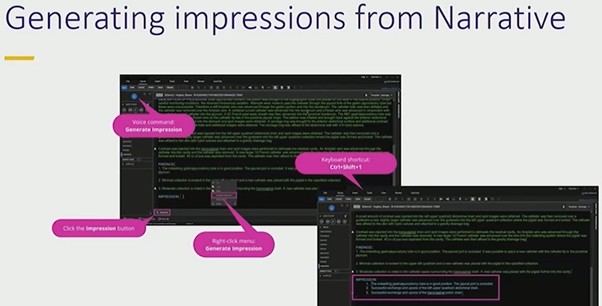
Automated impression generation from narrative text
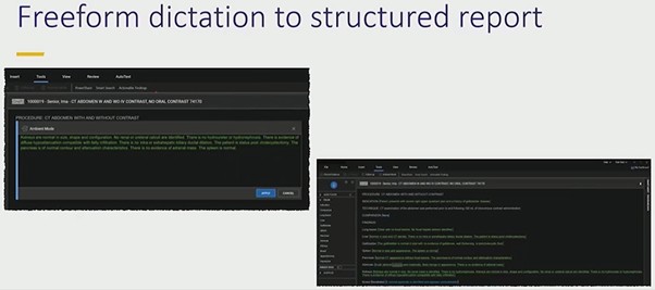
Converting freeform dictation into structured reports
Organizing sentences under correct headings
These features accelerate reporting and reduce cognitive load without exposing PHI externally.
Error Prevention Still Matters
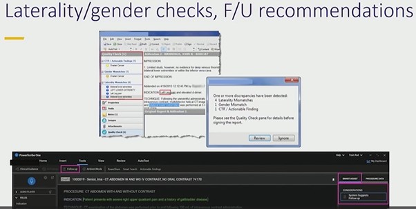
Simple NLP tools remain some of the most valuable. PowerScribe’s laterality and gender checks prevent avoidable mistakes that can undermine confidence in a report. Tools that flag mismatched anatomy—such as referencing a prostate in a female patient—provide immediate, low-friction safety nets that rads consistently appreciate.
Bottom Line
Security and workflow realities remain the biggest obstacles to adopting LLMs for radiology reporting. Until private, institution-controlled LLMs become practical and widely available, rads will continue to rely on integrated NLP tools that improve.
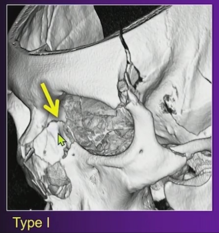
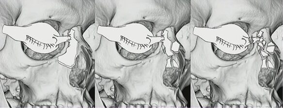
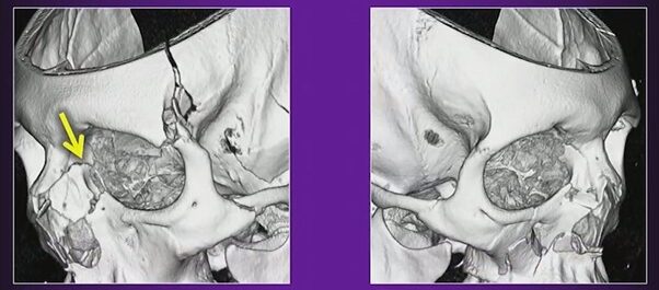
When evaluating orbital trauma, one detail rads should address is the Markowitz and Manson (M&M) classification. As Blair A. Winegar, MD, explains, this system focuses on the degree of comminution in the region of the lacrimal fossa and helps predict whether the medial canthal tendon is likely to be injured.
Left:Type I, Intact medial canthal tendon connected to single large fracture fragment; Center:Type II, Intact medial canthal tendon connected to single comminuted fracture fragment; Right:Type III, Disrupted medial canthal tendon with severe comminution about the lacrimal fossa
What M&M Describes
The key question is whether the lacrimal fossa remains intact or is significantly fragmented.
Intact lacrimal fossa: Low likelihood of medial canthal tendon injury.
Heavy comminution in the lacrimal fossa: Higher suspicion for medial canthal tendon disruption, which may require surgical repair.
Why Does It Matter?
The medial canthal tendon anchors at the anterior lacrimal crest. When that region is fractured extensively, the surgical team needs to prepare for possible tendon repair. Including this observation in your rad report sets appropriate expectations and guides planning for reconstruction.
Left:Type I; Right:Type II
Practical Approach
On CT, evaluate the anterior lacrimal crest and adjacent lacrimal sac fossa.
Describe whether this region is intact, minimally displaced, or extensively comminuted. Explicitly link substantial comminution to the potential for medial canthal tendon involvement.
Bottom Line
In orbital trauma, reporting the Markowitz and Manson classification provides actionable information. Identifying comminution in the lacrimal fossa helps surgeons anticipate medial canthal tendon repair and improves communication between rads and the operative team.
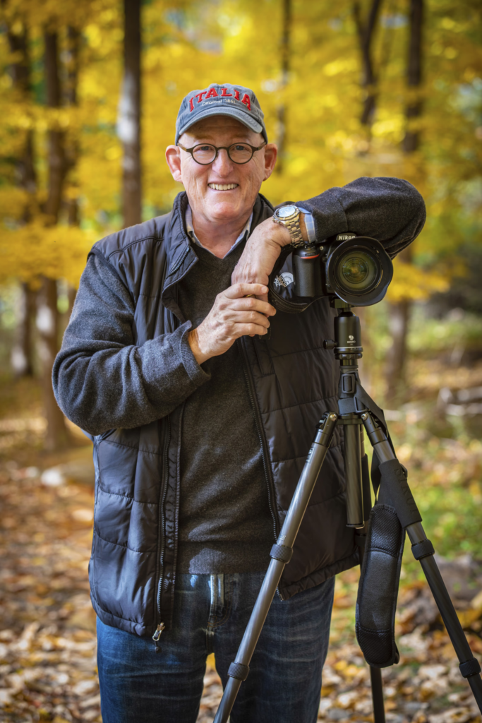
Retirement is one of the most unique, fulfilling, and exhilarating opportunities we will ever experience. Yes, I know, some of you may find that hard to believe. So did I, as I saw this major life transition sneaking up over the horizon and then, certainly, as I started to actually live it 10 years ago.
Why wouldn’t this be a major concern, a challenge, a mystery. We’ve been working at medicine and radiology for decades—at least since college or before (most of us finished our education/training in 22nd grade). Only a few vocations require this degree of involvement, so seeing it change or end can be a major shock. But have faith—there is more than hope down the line.
The first big question: “Is it time for me to retire?” There are many factors that determine when we decide, as they say, to put down the stethoscope (that is a piece of instrumentation used before an MRI or PET/CT is ordered). It is deeply personal, involving the interplay of fulfillment, health, finance, stress, and one’s specific life circumstances. Many ask, “What am I going to do in this vast new space?,” “What will give me a sense of purpose?,” and “Will I be able to create new personal connections?”
The good news is that, as radiologists, we are curious, self-motivated, goal-oriented, and like to learn. Whether you are simply contemplating retirement or are well into it (but need a fresh look at who you are and where you wish to be), one approach to retirement (which I prefer to call “rewirement”) is to ask a few questions, such as:
“What gives me satisfaction?”—for me; learning, teaching, being creative and productive
“What am I willing to try (new or reconnecting)?” e.g., writing, volunteering, acting classes, and improving my photography
Unlike prior generations, our next phase will be a more dynamic and fluid process as it may last a quarter of a lifetime. Therefore, self-awareness becomes extremely important in order to have a truly fulfilling and joyful experience,
Several things can make our retirement easier. The first is our “Experienced Brain.” We have learned a great deal about medicine, radiology, and life. It is a wonderful thing to pass this knowledge on to residents or medical students. I have loved voluntarily sharing what I have learned with radiology residents because it makes me stay informed and allows me to be engaged with a younger generation. You may want to teach in a totally different field, bringing to mind one physician who became a grade school teacher.
Another big advantage: If you don’t want to give up imaging entirely, there is always teleradiology, especially if you can set your own level of commitment. As this is still isolating, I would recommend balancing it with other non-medical interests, so that you keep developing and can become part of new communities.
Suffice it to say that when you are absolved of the responsibilities of work, the things you can explore and accomplish are endless. Plus, there are several other advantages that come with retirement, such as freedom from failure and freedom from comparing ourselves to others. The idea is to dive into new interests just for the sake of trying something different. If it is rewarding, keep going; if it is not, just move onto the next thing. No judgment involved. You may want to get back in touch with passions from your past or think about “What would I have done, had I had not become a physician?”
You never know where these new forays may take you. My photography ultimately led to something that gave me great satisfaction – donating my prints to hospitals to create a more welcoming and calming environment for patients, their families, and the staff who care for them. (When you are donating, competition goes out the window.)
So, time to see yourself in a different light and be open to a new, invigorating, and adventure-filled world. Enjoy the journey!
Dr. Agress retired 10 years ago, following a 36-year practice of diagnostic radiology and nuclear medicine. He continues to voluntarily teach at Columbia Presbyterian and Weill Cornell Medical Centers. You can learn more about Next Years Best Years, his resource for personal and emotional well-being, at NextYearsBestYears.com.
With therapies evolving and technology updating, radiologists the world over are ready to rise to the challenge of delivering more precise staging and follow-up for rectal cancer.
On Sunday, April 12 in Pittsburgh, PA, abdominal imaging experts from both hemispheres will convene at the David L. Lawrence Convention Center and online for Rectal MRI, the 2026 ARRS Annual Meeting Global Exchange course featuring faculty from the Royal Australian and New Zealand College of Radiologists (RANZCR).
Codirectors Aliya Qayyum (ARRS) and Kirsten Gormly (RANZCR) are bringing together the field’s finest to share evidence-based insight and technical pearls designed for immediate clinical application. For radiologists seeking to refine staging, elevate posttreatment evaluations, and stay ahead of emerging imaging biomarkers, this expertly curated course offers globally relevant guidance.
Rectal MRI: A Cornerstone of Modern Care
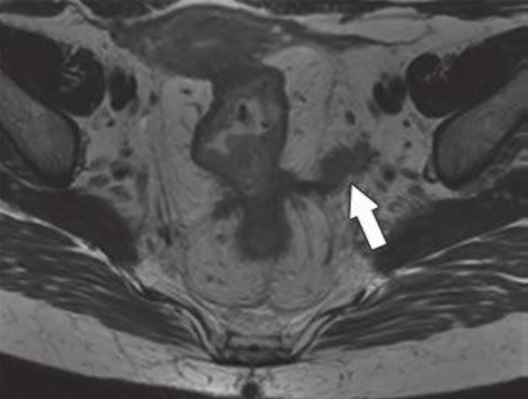
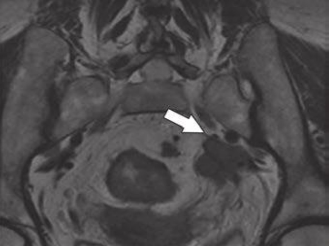
Once an emerging tool, rectal MRI is now the gold standard for staging, enabling assessment of the T stage and detecting key prognostic features such as mesorectal fascia involvement (MRF) and extramural venous invasion (EMVI) (Fig. 1).
Fig. 1—59-year-old patient with rectal cancer with extramesorectal vessel involvement, consistent with T category of T4b. Axial (top) and axial oblique (bottom) T2-weighted images show rectal tumor that extends through extramesorectal vein (arrow), which according to expert opinion warrants classification as T4b.
Dr. James Costello (ARRS) will lead a session on foundational principles of T staging, MRF, and EMVI, emphasizing actionable strategies to refine reports and guide multidisciplinary teams. Building upon Dr. Costello’s foundation, Dr. Verity Wood (RANZCR) addresses the myriad nuances of assessing lymph nodes and how to identify poor prognostic tumor deposits. Pointing out pitfalls left and right, her lecture during the 2026 ARRS Annual Meeting Global Exchange with RANZCR will provide the latest imaging criteria to help radiologists render more confident interpretations.
From Early Detection to Posttreatment Decision-Making
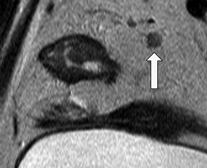
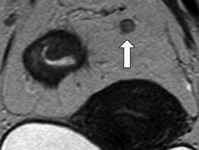
As screening programs detect more early-stage tumors, MRI also has the ability to evaluate these lesions. Dr. Gormly will discuss how to assess early cancers and why acquisition technique for high-resolution T2-weighted images directly impacts interpretive accuracy for all rectal MRI parameters (Fig. 2).
Fig. 2—Morphologic features of metastatic mesorectal nodes on MRI and potential pitfalls in assessment. 58-year-old woman with rectal adenocarcinoma. Oblique axial T2-weighted MRI (left) shows apparently spiculated node (arrow). Graininess of image is related to poor signal-tonoise ratio (SNR). Coronal T2-weighted MRI (right) shows that same node is homogeneously T2 hyperintense with dark capsule (arrow), which is typical of reactive mesorectal node. This image has superior SNR. Suboptimal images can lead to erroneous assessment of nodal morphologic features. This patient proceeded directly to surgery. Total of 38 lymph nodes (0.3–1.3 cm) were harvested. Eleven of larger lymph nodes were serially sectioned before submission for histologic processing. Final pathology revealed T3N0 disease.
Of course, staging is only part of the story. With the emergence of total neoadjuvant therapy (TNT) and “watch and wait” (W&W) protocols, radiologists play an increasingly vital role in posttreatment imaging. Dr. Raj Mohan Paspulati (ARRS) will outline W&W strategies in the United States, comparing tumor regression grading (TRG) systems and illustrating how MRI supports individualized care plans that may spare patients surgery.
The ability of MRI to predict patient outcomes can be considered more important than direct pathological correlation. Dr. Gormly will close the 2026 ARRS Annual Meeting Global Exchange course with her experiences in assessing emerging imaging biomarkers such as the “split scar sign,” aiming to improve patient selection for W&W, following a detailed explanation of how to assess residual tumor on high-resolution T2-weighted images. This is a key part of any posttreatment assessment system and is the cornerstone of the mrTRG system—the most validated MRI-based grading method globally.
Alliances Advancing Imaging
The mission of the ARRS Global Partner Society Program is to build long-standing relationships with key leaders and organizations in the worldwide imaging community—increasing awareness of our society’s services in specific nations, while raising the stature of Global Partner Societies among ARRS members. Every year, the ARRS Annual Meeting Global Exchange incorporates one partner society into the educational and social fabric of our meeting. ARRS members then reciprocate at the partner society’s meeting that same year.
Founded in 1949, RANZCR promotes and continuously improves the standards of training and practice in radiology and radiation oncology for the betterment of the people of Australia and New Zealand.
Rectal MRI not only reflects the robust collaboration between ARRS and RANZCR but also celebrates the global standardization of rectal MRI protocols.
And as Dr. Gormly told InPractice, “Australia and New Zealand’s early and ongoing partnerships with global leaders like Professor Gina Brown have positioned our region at the forefront of rectal MRI innovation. And this course is about sharing those insights with the world.”
Deborah A. Baumgarten, MD, MPH 2025-2026 ARRS President
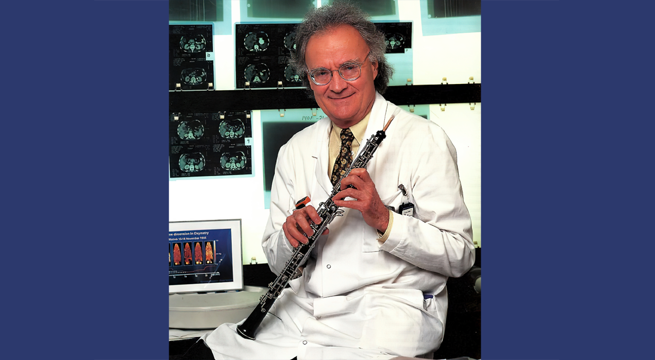
For our next installment of serendipity here in InPractice, we’re jumping ahead pretty far in time, as you’ll note by the color photograph of Professor Torsten Almén. As a young radiologist from Malmo, Sweden (not far from Copenhagen, Denmark), he was concerned about the pain of injected iodinated contrast, especially when it was administered arterially.
The father of non-ionic iodine contrast media: oboist Torsten Almén (1931-2016)
Dr. Almen’s serendipitous contribution is that he was struck by how little his eyes stung when swimming in the relatively isotonic Baltic Sea, as compared to the more hypertonic North Sea. He then wondered if a patient’s pain had anything to do with the tonicity of the contrast material that was administered. In 1967, Almen went to Temple University in Philadelphia, PA as a postdoctorate fellow and was given the choice on working on a steerable catheter that he’d helped design or working on this notion of toxicity and tonicity of contrast. Fortunately, he chose the latter. Almen was able to show in a bat model that his theory was correct, but he had to go about designing something that could be utilized in humans—a more ideal contrast agent. He wasn’t a chemist, so he bought some textbooks, taught himself the basics of organic chemistry, and tried to get someone interested in producing this theoretical compound. He finally persuaded Nygard, which later became Nycomed, to produce his low osmolar contrast material metrizamide in 1969. It was released several years later, quickly followed by other contributions to low osmolar contrast: Isovue by Bracco in 1981, Omnipaque by GE in 1982, and Optiray by Mallinckrodt in 1989.
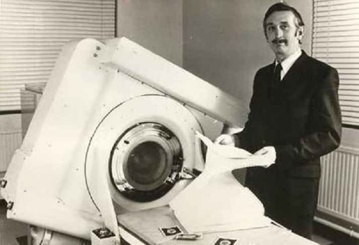
I dare say this gentleman is at least as recognizable as our society’s namesake, Wilhelm Conrad Roentgen. Sir Godfrey Newbold Hounsfield has had such a profound effect on our field. It was during an outing in the country when the idea came that he could determine what was inside a box by taking x-rays from all angles around the object, then somehow combine the different shadows the x-rays would produce to form an image of that object.
Sir Godfrey Newbold Hounsfield (1919-2004)
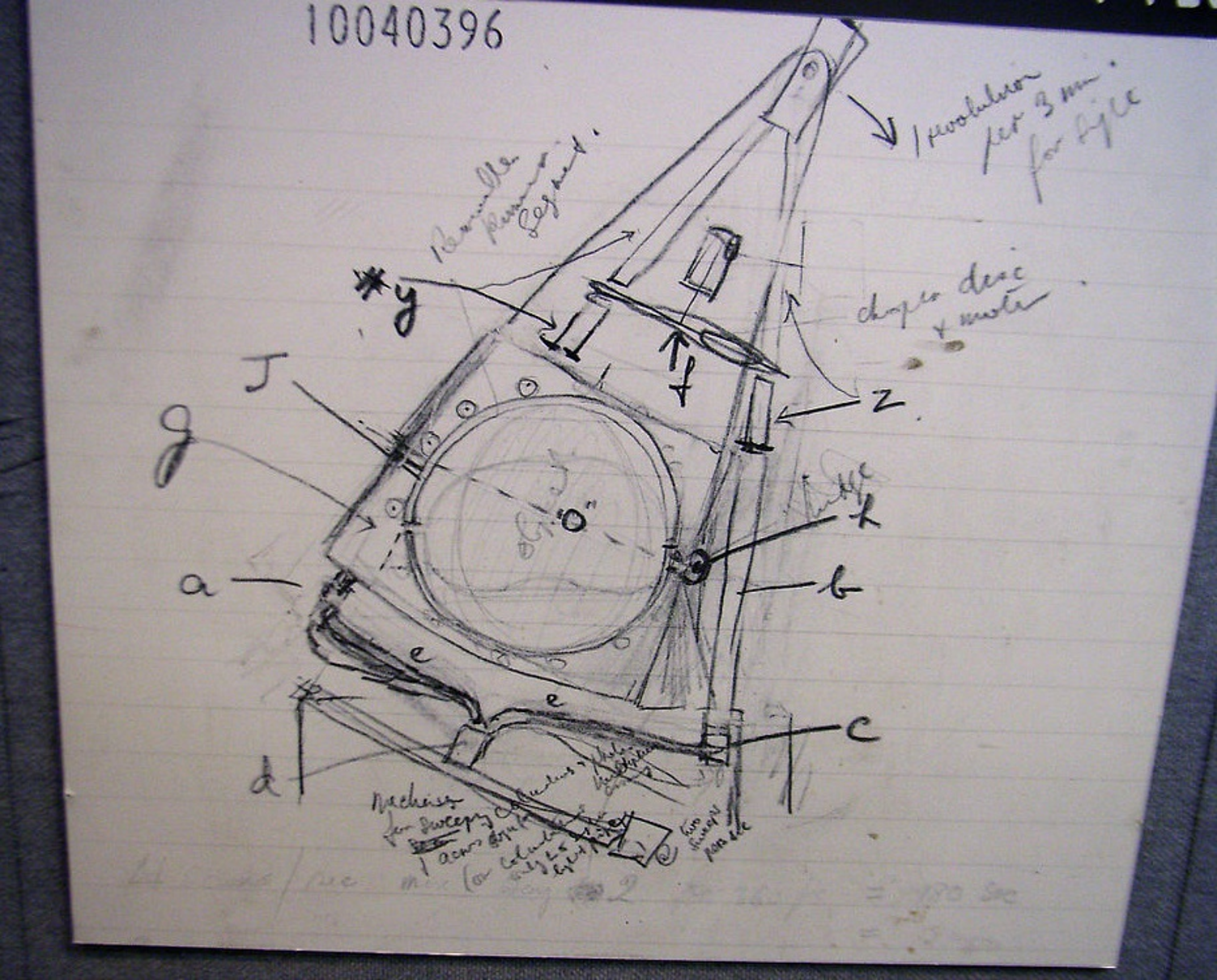
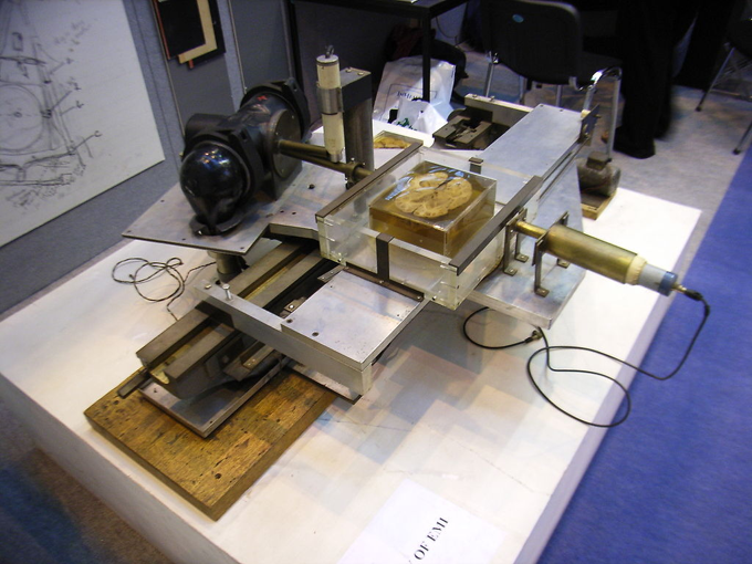
This is a sketch of his first idea of the CT scanner and the prototype. It’s one thing to formulate the idea of using x-ray readings, of course. But it was Dr. Hounsfield’s open mind that allowed him to realize not only the potential, but the necessity of using computers to analyze the data generated by taking all of the x-ray angles needed to create a CT image.
Hounsfield’s sketch of his CT scanner…
. . . compared to his prototype!
The following quotation by Dr. Louis Pasteur is particularly appropriate in this particular case: “In the field of observation, chance favors only the prepared mind.” Doctors Allan Cormack and Hounsfield shared the 1979 Nobel Prize in Physiology or Medicine for their work in CT.
Moving away from radiological discoveries for a moment, I love this quotation: “Eureka, I found what I wasn’t looking for!” This is associated with a book called Happy Accidents by a renowned abdominal radiologist named Dr. Mort Meyers, who spent his career at the State University of New York at Stony Brook. Again, emphasizing that the open mind is primed to take advantage of serendipity, how many of you know that the invention of the microwave oven had its origins when an engineer working with radar sets had a candy bar melt in his pocket, realizing it must’ve been from emitted radio waves? Or that Viagra was discovered by Pfizer when looking for a drug that increased blood flow to the heart, only to find that it increased blood flow a little lower? Or the discovery of artificial sweeteners by Constantin Fahlberg, a chemist working on coal tar derivatives, came from his inadvertent tasting of saccharine? Aspartame’s discovery came later, thanks to James Shaler, a chemist working on anti-ulcer drugs. Sure, their tasting of unfamiliar lab compounds is completely not up to today’s standards, but we can excuse them for that.
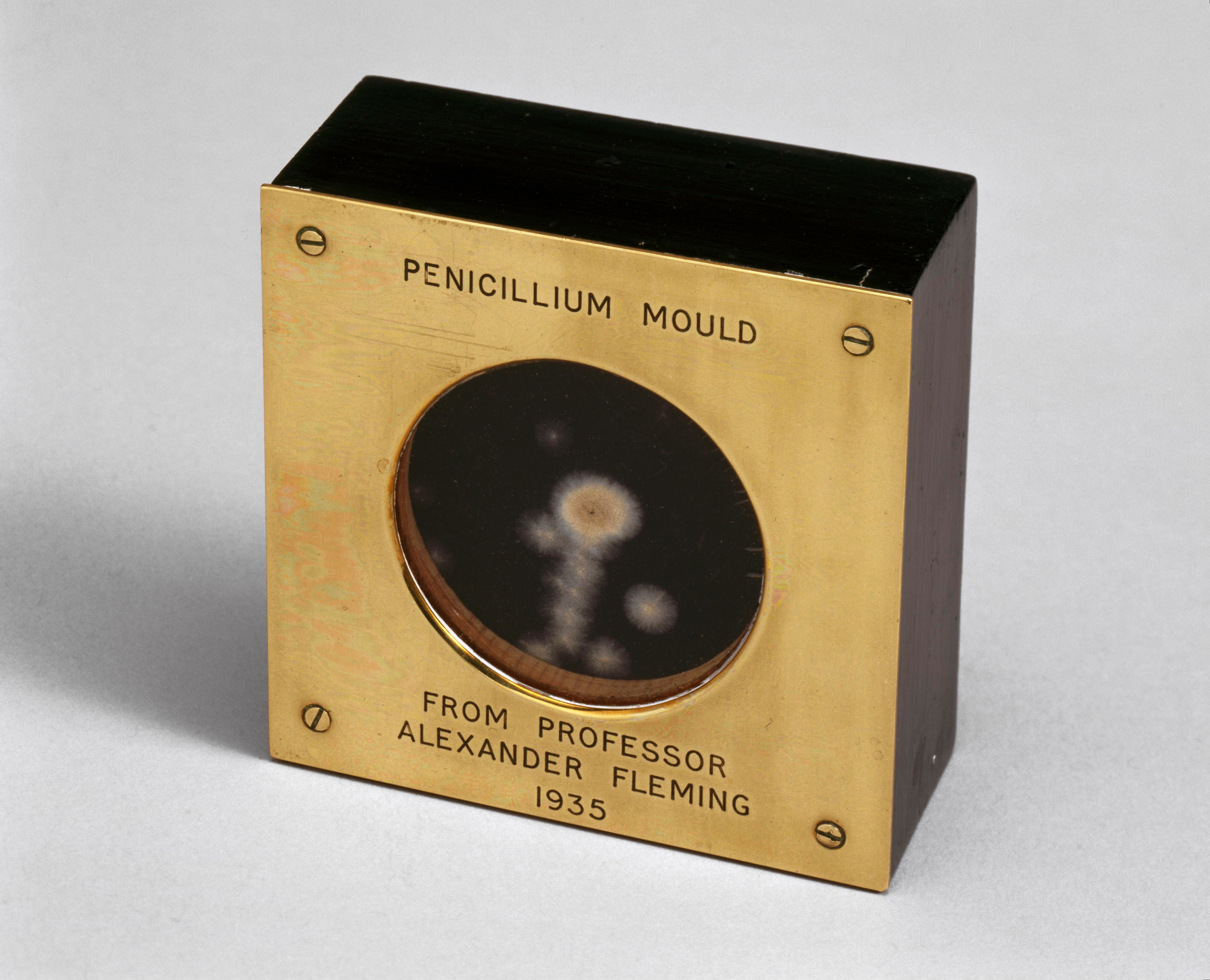
There are so many more examples of serendipity in science. Sir Alexander Fleming‘s discovery of penicillin came while working on compounds to inhibit bacterial growth, serendipitously noting one plate that had some mold on it seemed to be doing better than the other ones. Or veterinary pathologist Frank Schofield‘s discovery that spoiled sweet clover hay led to a hemorrhagic illness in cows, eventually leading to the isolation of dicoumarol, or Warfarin. Or the serendipity of the population in the United Kingdom erroneously getting a half-dose of the Oxford AstraZeneca COVID-19 vaccine before a second full dose that gave a 90% effectiveness rate against COVID-19, as compared to the 62% when two full doses were tested in Brazil and South Africa.
British biologist and pharmacologist Dr. Alexander Fleming gave this sample of penicillium notatum to a colleague at St. Mary’s Hospital, London, in 1935.
Physiologist Menko Victor “Pek” van Andel at the University of Groningen in the Netherlands studies serendipity patterns in knowledge and discovery, and he’s categorized three main types of serendipity. Positive serendipity is when a surprising fact is seen and followed by a correct interpretation (e.g., x-rays). Pseudo-serendipity is to discover something you were looking for, but in a surprising way. An example that van Andel uses is penicillin. Meanwhile, negative serendipity is when a surprising fact is seen, but not optimally investigated. And I’d like to think Dr. Donald Cameron never pursuing sodium iodine salts would be that kind of negative serendipity, although it was found positively later by Dr. Earl Osbourne (see Part I here).
Pek van Andel was one of the first researchers to extract serendipity patterns for accidental unsought knowledge discovery.
Others have built on van Andel’s work, including Darbellay et al., who published a paper titled “Interdisciplinary Research Boosted by Serendipity.” Their contention is that getting rid of the silos that house most of our disciplines and opening one’s mind to the unexpected are fundamental to the work of researchers who position themselves between and beyond disciplines. That serendipity itself is an important part of interdisciplinary research. As a creative process, serendipity is foundational in interdisciplinary collaboration, boosting the exchange of ideas to exploit the unexpected.
Examples of this interdisciplinary research aided by serendipity mentioned by Darbellay and colleagues include a team at Dow Chemical tasked with inventing a chemical compound for protecting the windscreens of airplanes. Upon application of substance 401, the researchers realized they could no longer remove the measurement device that they were utilizing on the windscreen. The team was worried because this was very costly equipment, so they called their boss, Harry Coover, who had a doctorate in chemistry. When faced with this unexpected effect of the substance 401, he realized the researchers had unknowingly and unintentionally discovered superglue, which was a substance that could bond metal and glass. Under Coover’s initiative, Dow abandoned trying to find something better for windscreens, channeled its efforts into developing superglue, and began marketing the product so many of us use today. Other accounts dispute Darbellay et al.’s retelling of this discovery, instead noting that Coover discovered superglue (cyanoacrylate) at the Eastman Kodak company, where he realized clear plastic gun sights stuck to everything. He first rejected the substance but later recognized its potential as an adhesive, so serendipitous no matter the origin!
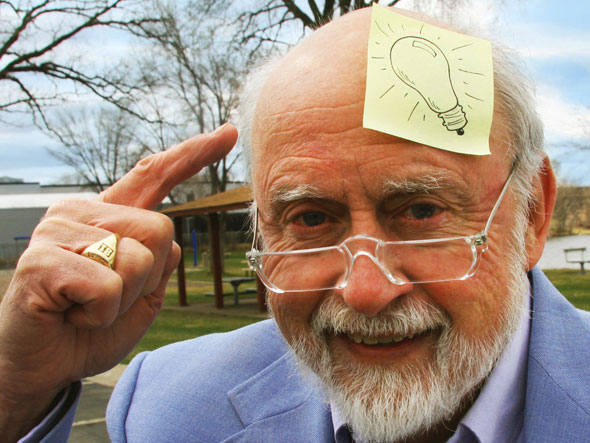
Ironically, the near complete opposite of superglue was also a serendipitous discovery. Spencer Silver and colleague Arthur Fry stumbled upon the idea for Post-it® notes while working at 3M. Silver discovered an adhesive that sticks permanently without a permanent bond and only does so in one direction, allowing an object to be repositioned. Fry’s contribution was finding use for the adhesive while singing in his church choir and lamenting that his bookmarks kept falling out of hymnals—his own eureka moment when he remembered Silver’s invention. The original yellow? Also, an accident; it was the color of a scrap of paper in an adjoining lab.
Art Fry, 93, in Saint Paul, Minnesota
Switching gears for just a moment, how about looking at serendipity on a more personal level? I’ve always been fascinated by the role of luck in my life and in the lives of others. When I was a child, whenever anything good in my life happened, I would tell my mom “I got lucky,” to which my mom would say, “we make our own luck.” This phrase has stuck with me for a very long time, and may be my own definition of serendipity: being open to possibilities when they happen and taking advantage of them. In one sense, I could describe my ascent to becoming president of the American Roentgen Ray Society as luck. Again, lucky in the sense that I repeatedly said “yes” to volunteer opportunities, even if that opportunity wasn’t glamorous or meant a lot of work.
So, here is what I conclude about serendipity. For those of you already well involved in research, getting your feet wet in research, or wanting to become involved in research, know that chance, luck, serendipity—whatever you want to call it—will play a role in your career. Keep an open mind, expect the unexpected, then turn it on its head. Seek out ways to collaborate with people who are in unrelated fields. Take advantage of the opportunity to network and build your community at meetings such as ARRS with those outside of your chosen field or discipline and those inside your chosen field or discipline. There will be so many more serendipitous discoveries in our lifetime and many lifetimes to come. It’s what keeps our field and so many others so very interesting. I understand everybody’s time is limited and valuable, but truly consider an opportunity before saying “no.” You never know which opportunities will lead to something bigger, so keep an open mind about saying, “yes.” You never know who will be touched by your words and actions, who will change your life with a chance encounter, or whose lives will be changed by a chance encounter with you.
References:
Nyman U, Ekberg O, Aspelin P. Torsten Almén (1931-2016): the father of non-ionic iodine contrast media. Acta Radiol 2016. 57:1072–1078
Recognizing this hallmark pattern can spare unnecessary confusion, guiding the right surgical call.
The Big Picture
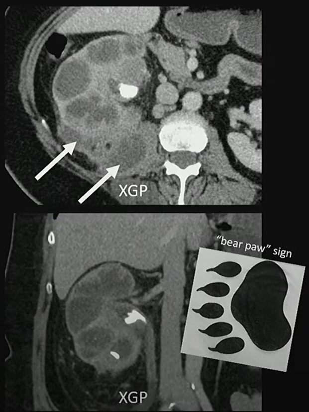
Xanthogranulomatous pyelonephritis (XGP) is a rare, destructive renal infection, most often in women with staghorn calculi. It replaces functioning parenchyma with lipid-laden macrophages and inflammatory tissue, often extending beyond the kidney.
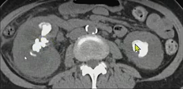
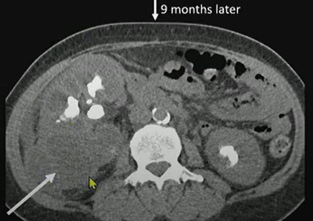
Nine months later, we see much more exuberant inflammation—soft-tissue thickening extending out from the kidney into the retroperitoneum.
Patient underwent renal scintigraphy prior to getting a nephrectomy, and we can see that that right kidney is not contributing to the renal function.
Key Takeaways
Classic appearance: Enlarged kidney, staghorn calculi, and a contracted pelvis—the “bear paw.”
Functional loss: Renal scintigraphy often shows little or no contribution from the affected kidney . . .
Parameter
Left Kidney
Right Kidney
Peak Time
6.7 minutes
2.3 minutes
Relative Function (Integral, 1–3 min)
85%
15%
Extrarenal extension: Look for inflammation tracking into the retroperitoneum, psoas, or paraspinal muscles.
Definitive treatment: Because the kidney is typically nonfunctional, nephrectomy is the standard.
Challenges Ahead
Distinguishing XGP from renal cell carcinoma or pyonephrosis can be difficult without correlating imaging and functional data.
Awareness of extrarenal spread is crucial to surgical planning.
Early recognition can prevent unnecessary biopsy or delay in definitive management.
Presented by Anup Shetty, MD, during ‘Radiology Case Review: Genitourinary Imaging,’ part of the ARRS 2025 Annual Meeting. Watch the full session now: arrs.org/am25
Bottom Line
When you see that “bear paw” (i.e., staghorn calculus, perinephric inflammation, and an enlarged, poorly functioning kidney), think XGP!
For radiologists especially, artificial intelligence (AI) is no longer just over the horizon; it’s in the reading room, right now. This practical immediacy is precisely the premise behind the 2026 ARRS Annual Meeting Categorical Course, Clinical Artificial Intelligence in Radiology. Presented live and virtually from the David L. Lawrence Convention Center in Pittsburgh, PA, this two-day ARRS Categorical Course continues our 125-year-old legacy of forward-looking education by arming radiologists with a robust understanding of how AI is reshaping the specialty.
Dr. Shandong Wu | Cat Course Codirector
Clinical Artificial Intelligence in Radiology brings together more than 20 distinguished faculty from leading institutions across the globe, all led by Shandong Wu, PhD, founding director of the University of Pittsburgh’s Center for AI Innovation in Medical Imaging, a cross-campus initiative including more than 130 researcher and clinician members. Also a professor of radiology, biomedical informatics, bioengineering, intelligent systems, clinical and translational science, one of Dr. Wu’s ARRS Cat Course codirectors is an abdominal radiologist and director of diagnostic AI at the University of Washington, Yee Seng Ng, MD.
Dr. Shandong Wu | Cat Course Codirector
Alongside codirector and 2024 AJR Lee F. Rogers International Fellow in Radiology Journalism Hyun Soo Ko, MD (Peter MacCallum Cancer Centre, Australia), the trio is curating a curriculum of more than two dozen lectures and panels across seven thematic sections, giving registrants a comprehensive view of AI’s current and future roles in everyday practice.
SUN, APRIL 12—From Concept to Clinic: Building AI Literacy
Day one of Clinical Artificial Intelligence in Radiology kicks off with “Getting to Know AI,” a primer tailored for all levels of experience. Tessa Cook, MD, PhD (University of Pennsylvania), provides an overview of radiological progress in AI, while Dr. Ko demystifies essential concepts, such as machine learning, deep learning, radiomics, as well as generative and agentic AI.
Dr. Linda Moy | Vice Chair of AI, NYU Radiology
Up next, inaugural vice chair of AI at New York University’s radiology department, Linda Moy, MD, will provide an invaluable look into leveraging AI to improve workflow efficacy and effectiveness alike. Dr. Wu himself closes the Cat Course’s first session. The leader of Pittsburgh’s Intelligent Computing for Clinical Imaging lab will explore and explain how AI is enhancing imaging interpretation for computational insights—from screening and triage to diagnosis and prediction.
Clinical Implementation: From Regulation to Real-World Deployment
Section two of Clinical Artificial Intelligence in Radiology, “AI Clinical Implementation,” addresses legal, regulatory, and operational frameworks essential for radiologists seeking to implement or evaluate AI tools in practice. Didactic highlights will include guidance on U.S. Food and Drug Administration (FDA) regulations and performance monitoring by Melissa Davis, MD, MBA (Yale), as well as insights into distinguishing high-quality AI models from market hype.
In a uniquely insightful presentation, Julian Rivera, JD (University of Pittsburgh), will tackle the legion of legal considerations accompanying AI adoption: liabilities, ethical perspectives on signing contracts, collaborative business modes with AI companies, etc. Dr. Cook returns to share her expertise on evaluating local versus commercial solutions when measuring ROI, while a panel moderated by Dr. Moy will outline best practices and common pitfalls.
Beyond the Pixel: Multimodality and Multidimensional AI
The promise of any good AI expands significantly when paired with non-image data. The “Going Beyond Images to Multimodality” session explores emerging applications that leverage large language models, vision-language models, and foundation models. Presenters Heather Whitney, PhD (University of Chicago), and Lifeng Yu, PhD (Mayo Clinic), will delve into data curation, federated learning, and the physics of AI model performance. With Christian Bluethgen, MD (University Hospital of Zurich), having assessed multimodal data methodologies in his presentation, a panel discussion on tackling technical challenges to find opportunities rounds out day one of this ARRS Cat Course.
MON, APRIL 13—Practical Impact Across Subspecialties
AI’s reach across subspecialties is the focus on Monday. Presenters including Constance Lehman, MD, PhD (Harvard), Ali Guermazi, MD, PhD (Boston University), and 2022 ARRS Gold Medalist Edward Y. Lee, MD, MPH (Harvard) will detail AI tools in breast, musculoskeletal, pediatric imaging, respectively. Dr. Ng’s highly anticipated survey of AI and abdominal imaging will be followed by a lecture from neuroradiologist Paulo De Aguiar Kuriki, MD (UT Southwestern).
Dr. Edward Y. Lee | ARRS Gold Medalist
That’s not all either. Real-world cardiothoracic, interventional, and nuclear medicine cases will further demonstrate how AI is already reflowing imaging workloads, improving diagnostic accuracy, and personalizing care across organ systems and patient populations.
Shaping Tomorrow: Research, Education, and Ethical Engagement
Day two of Clinical Artificial Intelligence in Radiology continues with “AI Research and Education,” including a model development demonstration by Dooman Arefan, PhD (University of Pittsburgh), and an exploration of MD–PhD collaboration opportunities from Dr. Wu. Justin Peacock, MD, PhD (Uniformed Services University), will discuss educational roadmaps and training resources, addressing a key concern for attendees seeking to build or deepen their AI competencies.
This 2026 ARRS Annual Meeting Categorial Course concludes with “Humanity and AI,” a thought-provoking session covering radiologist–AI collaboration, fairness and bias, and imaging’s ever-evolving role in AI-powered services. Florence Doo, MD (University of Maryland) will help us find a foothold in our present human–AI ecosystem, followed by a warning for all the disparities AI run amok could actually exacerbate care of Judy Gichoya, MD, MS (Emory). Eduardo Mortani-Barbosa, MD, MBA (University of Pennsylvania), will then detail specific skill sets that AI-forward radiologists will need to hone in their practices and in their communities. Finally, ARRS Scholar and Gold Medalist and editor of Radiology: Artificial Intelligence Charles E. Kahn, MD (University of Pennsylvania), joins to facilitate a panel discussion on action items and what to do next.
Dr. Charles E. Kahn | Editor, Radiology: Artificial Intelligence
With each live lecture accompanied by an e-book chapter, Clinical Artificial Intelligence in Radiology will provide strategic context and tactical guidance for imagers of each practice type and at every level of training.
And as Dr. Wu tells InPractice, “AI in radiology is not just a technical shift—it’s a cultural one. This ARRS Categorical Course is about empowering radiologists to shape that future, not just react to it.”
With content spanning conceptual foundations to the most practical of pearls, the curriculum curated by Wu, Ng, Ko and colleagues this April is poised to be an essential learning experience for working radiologists looking to engage with AI at the frontlines of medical imaging care.
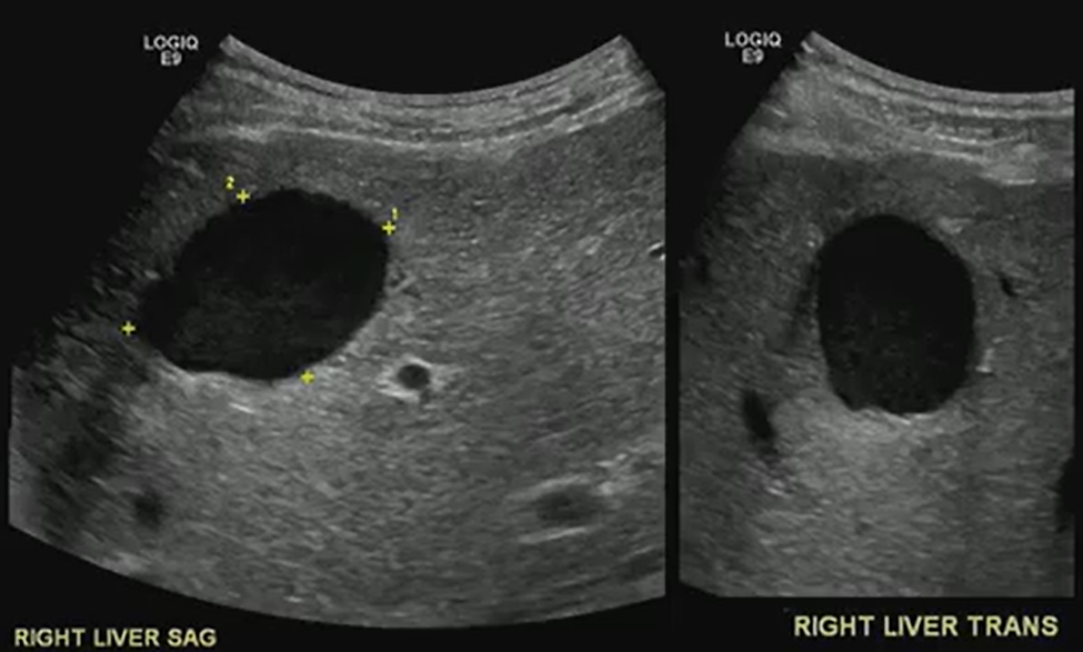
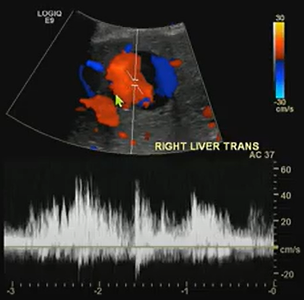
A benign-looking liver lesion turned out to be a hepatic artery pseudoaneurysm—all thanks to color Doppler.
The Big Picture
What looks like a simple hypoechoic cyst on ultrasound may hide a critical vascular pathology. In this ARRS Annual Meeting case from Kristin Rebik, DO, color Doppler proved essential for distinguishing cystic lesions from vascular anomalies like pseudoaneurysms.
Key Takeaways
Always Doppler: Even cyst-like structures require Doppler evaluation to rule out vascular causes.
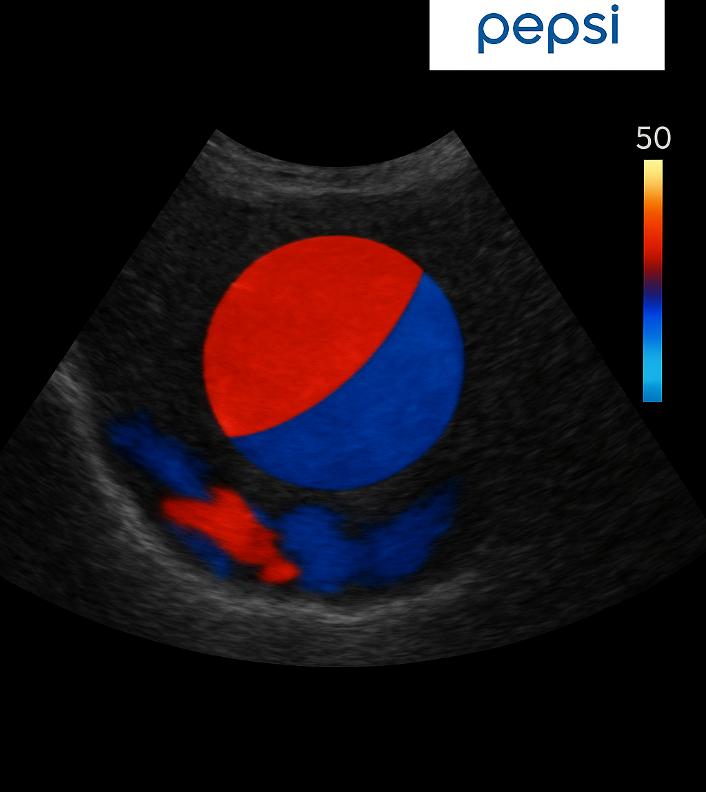
Pepsi Sign: Swirling vascular flow within a lesion may signal a pseudoaneurysm.
High stakes: Hepatic artery pseudoaneurysms can mimic benign lesions but require urgent recognition and intervention.
Next steps: Interventional radiology embolization can be lifesaving.
Challenges Ahead
Differentiating pseudoaneurysms from other vascular or cystic lesions remains tricky.
Missing Doppler evaluation risks misdiagnosis and delayed treatment.
Awareness of teaching signs like the “Pepsi sign” is uneven among trainees.
Bottom Line
Never skip Doppler. The “Pepsi sign” may be the clue that transforms a benign-looking lesion into a critical vascular diagnosis.
Deborah A. Baumgarten, MD, MPH 2025-2026 ARRS President
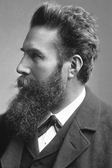
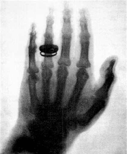
There’s no discussion about serendipity without mentioning the namesake of our society, Wilhelm Conrad Roentgen. I’m sure we all also recognize the world’s first radiograph on the right, taken on November 8, 1895. It’s an image of his wife Anna Bertha’s hand only a couple of weeks after Roentgen discovered what he termed “x-rays”—x for something unknown. Roentgen was studying cathode rays in his lab in a dark room, and he noted a glow several feet away from where he was standing. Although he was not the first to note that cathode rays could permeate thin metal sheets or light up a fluorescent screen near them, he was the first to put those pieces together. And for that, he won the first Nobel Prize in physics in 1901.
Wilhelm Roentgen (1845-1923) and the left hand of his wife, Anna Bertha Ludwig Roentgen, in 1895
I love this quotation attributed to Albert Szent-Györgyi, a Hungarian biochemist, who, among other things, aptly won a Nobel Prize in 1937 for the isolation of vitamin C: “Discovery consists of seeing what everyone else has seen and thinking what no one else has thought.” Many have called the discovery of x-rays serendipity—defined by the Oxford Dictionary as the occurrence in development of events by chance, in a happy or beneficial way, and by Merriam-Webster as a faculty or phenomenon of finding valuable or agreeable things not sought for. The term was first used in 1754 by Horace Walpole, the fourth Earl of Orford, who is remembered for a silly fairytale about three princes from the country of Serendip. Due to a series of accidents and keen mental judgment, these princes were able to discern the nature of a lost camel, of all things. But there’s another factor that lets serendipity thrive: keeping an open mind. There are many other points in the history of science and radiology where serendipity and open-mindedness played a role, and we’ll look at a few of those stories here in InPractice.
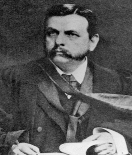
John McIntyre (1857-1928)
This is Dr. John McIntyre of Glasgow, Scotland. Although not a radiologist, he was actually the first person to in the world to set up an x-ray department in March of 1896, after lecturing on “The New Light: X-rays” a mere month after their discovery. The serendipity in his story is the chance intersection of electricity and medicine. An apprentice electrician before studying medicine, Dr. McIntyre was fascinated by the potential uses of electricity in medicine. He was named consulting medical electrician at the Glasgow Royal Infirmary in 1895, and although mainly interested in diseases of the ears, nose, and throat, Dr. McIntyre was the first to image a renal stone in vivo in April of 1896. He was also the first to show movement of a frog’s leg using cineradiography, later a beating heart, and then a bismuth meal in the stomach.
But even before the word serendipity made it into the English vernacular, the notion of serendipity existed. “The greatest part of the invention being, but a luckey bitt of chance [sic]” is attributed to English polymath Robert Hook in 1679. A man of many talents, he is credited for being the first to describe cells under a microscope. In fact, Hook designed his own compound microscope and coined the term cell. He also described the force applied to a spring and its deformation, what’s known as Hook’s law of elasticity.
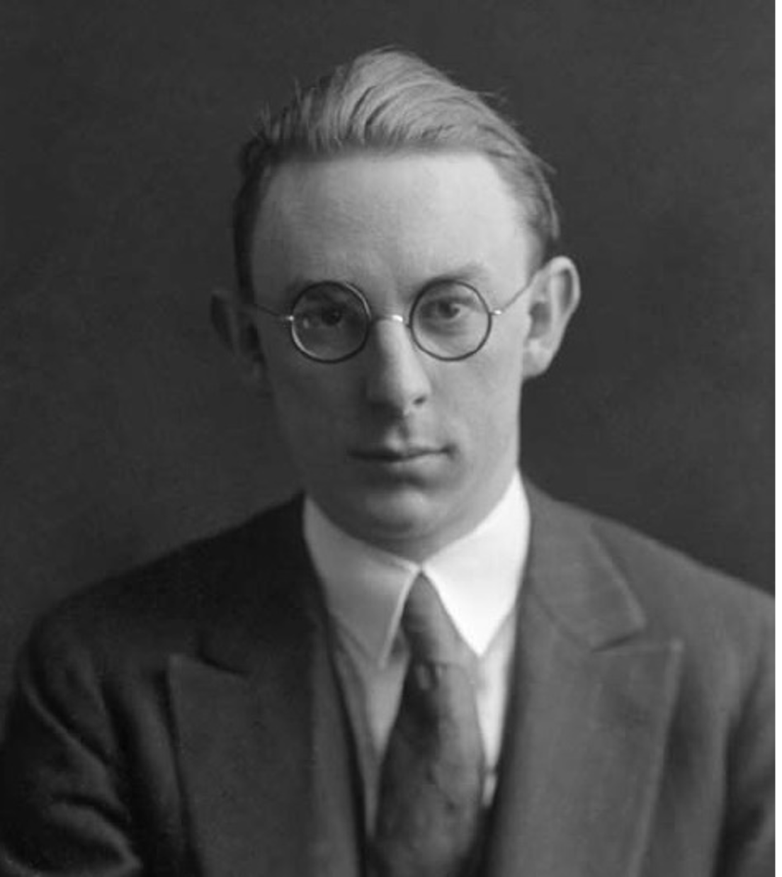
Earl Osborne (1895-1960)
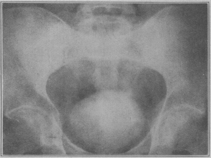
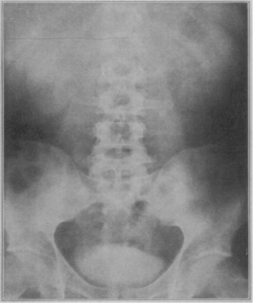
Our next story nicely illustrates Robert Hook’s thoughts about invention and chance. This is Dr. Earl Osborne, who was a dermatology resident at the Mayo Clinic. Dr. Osborne, along with physician and pharmacologist Leonard Rowntree, is credited with discovering that intravenous administration of sodium iodine salts for treating syphilis faintly opacified the urinary tract. Dr. Osborne is said to have serendipitously noted the opacification of the bladder in one of his patients who had an abdominal roentgenogram during treatment. When asked about this discovery, he stated, “it occurred to one of us that if in roentgenology of the urinary tract advantage could be taken of the fact that sodium iodine, after its introduction into the body is normally excreted in the urine, roentgenograms of the kidneys, ureters, and bladder might be secured without the need for catheterization.” Anybody recognize the term KUB in this statement?
Initial investigations of sodium iodide also included a radiologist named Charles Sutherland and a urologist named Albert Scholl and were published in JAMA in February of 1923. The image on the left is from that paper, taken two hours after the administration of 200 cc of a 20% solution of sodium iodine showing opacification of the bladder. The roentgenogram on the right was taken about an hour after the administration of 100 cc of a 10% solution. You can see that the bladder is opacified, and there’s faint opacification of the kidneys, spleen, and liver. Ironically, neither Dr. Osborne nor Dr. Rowntree pursued this any further, but Dr. Osborne did go on to help found the American Board of Dermatology.
Abdominal roentgenograms with bladder—as well as kidney, spleen, and liver—opacities
Dr. Donald Cameron, a young Minnesota surgeon, is seemingly forgotten in all of the fuss about Drs. Rowntree and Osborne. He published a preliminary report about the oral and intravenous administration of sodium iodine as an opaque medium in roentgenology in JAMA in 1918, five years before Sutherland’s paper came out. Cameron published this preliminary report in case he didn’t return from World War I. He published one more article a few months later, but then never pursued this line of research any further. But he did return from WWI and established a hospital in the Midwest now called the Cameron Memorial Community Hospital that still exists today.
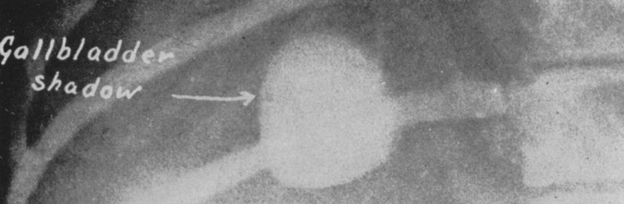
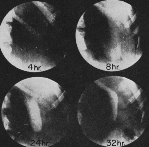
Like the discovery that sodium iodine opacifies the urinary tract, the origins of oral cholecystography are serendipitous, too. As the story goes, Dr. Warren Cole was working in a lab experimenting with compounds that were known to be excreted by the liver into bile. Four and a half months went by before he even ever saw a gallbladder image, until one day this image was obtained in one of the lab dogs.
Oral cholecystogram in canineOral cholecystogram in human
A new Harvey L. Neiman Health Policy Institute study, published in the American Journal of Roentgenology (AJR), investigated whether office-based providers are more likely to refer imaging to a radiologist, or to self-refer and interpret the ordered imaging studies themselves. “Self-Interpretation of Imaging Studies by Ordering Providers: Frequency and Associated Provider and Practice Characteristics” found that 43.6% of office-based studies were interpreted by the ordering provider, while 58.5% were interpreted within the ordering provider’s practice. Additionally, rates of self-interpretation varied by the specialty of the ordering provider and the imaging modality.
Self-Read Rates Vary by Modality and Specialty
Based on more than 1.6 million office-based imaging claims for Medicare fee-for-service beneficiaries in 2022 that were ordered by non-radiologists, by modality and across all specialties, this AJR Original Research manuscript by Christensen et al. found that the self-interpretation rate for office-based imaging was as follows:
50.4% for radiography and fluoroscopy
52.0% for ultrasound
39.5% for nuclear medicine
5.3% for CT
6.1% for MR
Meanwhile, self-interpretation by specialty broke down accordingly:
19.9% for primary care physicians
75.7% for orthopedic/sports medicine physicians
30.5% for non-physician practitioners
Another key determinant of self-interpretation rates was the size of the practice. Rates of self-interpretation were substantially less in the largest practices (500 or more providers) than the smallest practices (only 1-9 providers): 24.2% versus 48.9%, respectively, according to the researchers’ findings.
Access to Radiologists Could Curb Self-Reading
Eric W. Christensen, PhD
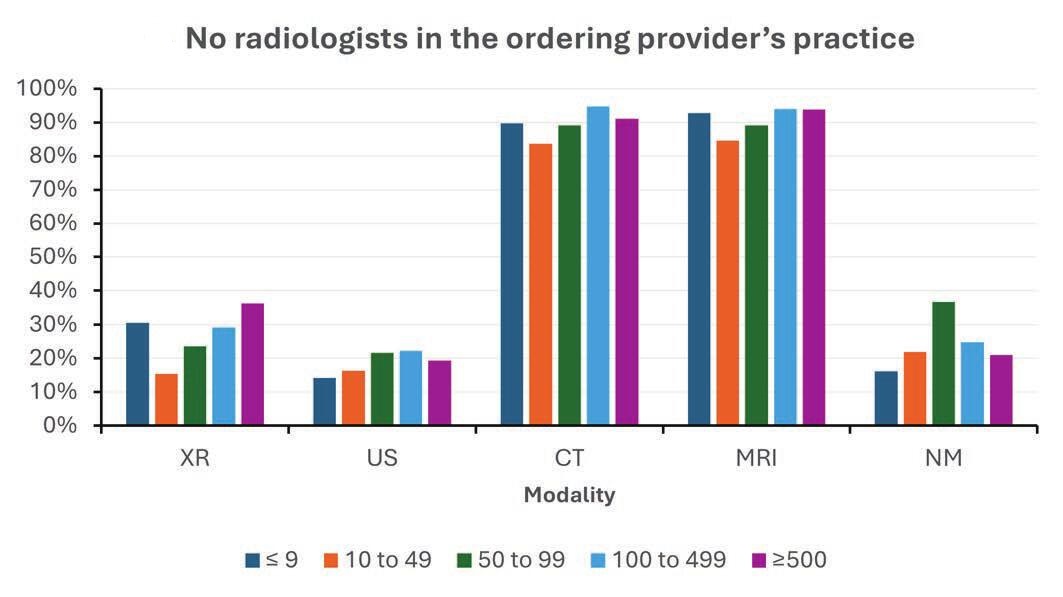
Whether or not there was a radiologist in the ordering provider’s practice was highly correlated with self-interpretation of imaging by the ordering provider (Fig. 1). “If there was a radiologist in the practice, the odds of self-interpretation were about one-half compared to practices without a radiologist on staff,” said Eric W. Christensen, PhD, research director at the Neiman Institute in Reston, VA. “Conversely, the odds of within-practice interpretation were generally 2 to 3 times higher if there was a radiologist in the practice.”
Continuing consolidation of health care—“which is creating larger, multispecialty practices,” Dr. Christensen added—could reduce imaging self-interpretation via increased access to radiologists within practice. “Such centralization of radiology services may encourage referral of imaging interpretation to the within practice radiologist, in lieu of potential self-interpretation.”
Fig. 1—Percentage of office-based diagnostic imaging studies interpreted by radiologist, stratified by modality, presence of in-practice radiologist, and ordering provider’s practice size. NM = nuclear medicine; US = ultrasound; XR = radiography and fluoroscopy.
Training Gaps, Policy Questions, and Equity Decisions Remain
Vijay M. Rao, MD
“Our results raise potential implications for quality of patient care. Nonradiologist specialties, aside from cardiology, lack the rigorous and comprehensive training in imaging interpretation that occurs during the 4 years of a radiology residency program,” said Vijay M. Rao, MD, senior vice president of enterprise radiology at Jefferson Health and professor of radiology at Thomas Jefferson University in Philadelphia, PA.
Pointing out that some specialties may provide targeted training in imaging interpretation that is narrow in scope, such as in ultrasound for emergency medicine or obstetrics/gynecology, “the large differences between radiologists and non-radiologists in interpretation training could lead to differences in diagnostic accuracy,” Dr. Rao said.
Kerry L. Thomas, MD
And as Kerry L. Thomas, MD, noted in her AJR Editorial Comment, while defined personnel qualifications—including for the interpreting physician—are clearly delineated when accrediting outpatient imaging centers, “such quality standards are lacking for the interpreting provider of in-office examinations.”
Importantly, to avoid secondary disparities related to health care quality, “images should be interpreted by providers with appropriate training for the specific examination being reviewed, agnostic of the care setting,” said Dr. Thomas from H. Lee Moffit Cancer Center and the department of diagnostic imaging and interventional radiology at University of South Florida’s Morsani College of Medicine in Tampa.
A “Stark” Surprise
Concerns of financial conflicts of interest related to self-referral contributed to the passage of the federal Stark Law in 1989. Named after U.S. House Representative Pete Stark of California, the Stark Law prohibits physician self-referral for designated health services, including radiology, under Medicare. Over time, certain exceptions (à la the in-office ancillary services rule) were added to allow necessary in-practice imaging referrals within guidelines.
Intended to curb the practice of self-referral, in reality, there remain too few restrictions on imaging interpretation by non-radiologists. And as the authors of this AJR article found, the Stark Law’s in-office ancillary exception greatly weakens its ability to restrict imaging self-referral. “Hence, our results highlight a need to revisit the in-office ancillary exception policy to impact the potential financial incentives that lead to self-referral,” Dr. Rao concluded.
Ralph Drosten, MD Professor Department of Medical Imaging University of Arizona Creighton University Medical School
The term “burnout” dates as far back as 1974. Coined by psychologist Herbert J. Freudenberger in a Journal of Social Issues article entitled “Staff Burn-out,” he described the phenomenon based upon his own experiences working with volunteers at a free clinic in New York City. Freudenberger observed that dedicated workers in this profession were becoming emotionally exhausted, cynical, and less effective—a state he dubbed “burn-out.” This publication marked one of the earliest formal discussions of burnout as a psychological syndrome, particularly among health care and social service professionals.
On May 28, 2019, burnout first appeared in the World Health Organization’s (WHO) 11th Revision of the International Classification of Diseases (ICD-11)—as an occupational phenomenon, however. Highlighting its relevance to health care workers, the ICD-11 filed burnout under “factors influencing health status or contact with health services,” but stopped short of classifying it as a disease. Although burnout is still not classified as a medical condition, in its definition of burnout as a syndrome, the ICD-11 identified three key components that contribute to chronic stress associated with work:
Specific contributors of said burnout include the following: time demands, lack of control, work planning and organization, as well as inherently difficult job situations and interpersonal relationships.
Fortunately, there are several workplace interventions for mentors to mitigate burnout with in-training physicians, such as wellness workshops, workload modifications (e.g., increased diversity of work duties), and better stress management education or appropriate emotional intelligence training.
As individuals, we have our own behavioral interventions to make: meditation, counseling, etc. Social interventions matter, too, especially when promoting our professional relationships. We can’t forget the importance of exercise and other physical activity either.
If not addressed, the risks of burnout are myriad. In addition to increased cardiovascular disease and inflammatory biomarkers, burnout elevates rates of depression and suicidal ideation. Thankfully, plans and attempts in burnout states do tend to decline with recovery.
Importantly, clinician depersonalization is associated with lower patient satisfaction and longer post discharge patient recovery time, so we must be able to identify key physical/psychological symptoms and associated elements of burnout—in ourselves and in others:
The American Roentgen Ray Society (ARRS) proudly recognizes the following four radiologists and their research projects with the 2025 ARRS Resident/Fellow in Radiology Awards:
Jordan H. Chamberlin, MD
Resident/Fellow in Radiology President’s Award “Machine Learning Based Optimization of Root Factors Associated with Image Quality of Myocardial Late Iodine Enhancement”
Shadi Ebrahimian, MD
Resident/Fellow in Radiology Executive Council Award “Evaluating AI Performance in Large Vessel Occlusion Detection: Impact on Time-to-Thrombectomy and Clinical Workflow Efficiency”
Ariel S. Kniss, MD, PhD
Resident/Fellow in Radiology Executive Council Award “Mammographic Breast Density and Its Impact on Screening Performance Metrics: An Analysis of 489,439 Screening Mammograms”
Capt. Tej Mehta, MD
Resident/Fellow in Radiology Melissa Rosado de Christenson Award “Multiparametric, Multimodal Delta-Radiomics for the Prediction of Transarterial Chemoembolization Outcomes in Hepatocellular Carcinoma”
Supported by The Roentgen Fund®, the ARRS Resident/ Fellow in Radiology Awards are available to all ARRS In-Training Members in imaging and allied sciences research to acknowledge their work and present their results during the ARRS Annual Meeting. ARRS Resident/Fellow in Radiology Awards are based on the competence and promise of the candidate in radiological research, education, or administration and the scientific merit and potential impact of the candidate’s research.
Since 1990, The Roentgen Fund has granted millions of dollars to hundreds of imaging professionals for both research pursuits and professional development. Today, through six vital scholarship and fellowship programs, the generosity of The Roentgen Fund’s donors is channeled to every corner of the globe—establishing dual foundations in innovation and leadership for a true diversity of radiology’s next generation.
Scholar Update: Derek Nguyen on Imaging-Focused Management of Architectural Distortions
According to research published in the American Journal of Roentgenology (AJR), imaging surveillance may be a safe alternative to surgical excision for architectural distortions (ADs) detected by digital breast tomosynthesis (DBT) alone with no sonographic correlate showing benign pathology without atypia on core needle biopsy (CNB). “The malignancy rate was 0% for ADs without atypia, versus 20% for ADs with atypia, on CNB,” explained first author and 2022 recipient of The Roentgen Fund’s Melissa Rosado de Christenson Award Derek L. Nguyen, MD, from the radiology department at Duke University School of Medicine in Durham, NC. Dr. Nguyen and colleagues’ AJR accepted manuscript included patients with ADs detected by DBT alone with no ultrasound correlate and nonmalignant results on stereotactic CNB with 12 vacuum-assisted 9-gauge cores from July 1, 2020 to December 31, 2023. ADs detected by DBT alone with no ultrasound correlate showing concordant benign pathology or radial scar without atypia on CNB were recommended to undergo 6-month and 12-month diagnostic imaging follow-up before returning to annual screening, whereas those showing atypia (with or without radial scar) on CNB were recommended to undergo surgical excision; however, patients could electively undergo the alternative management option. Nguyen et al. then assessed malignancy rates, considering ADs to be nonmalignant based on results of repeat CNB, surgical excision, or 12-month diagnostic imaging follow-up. Ultimately, for ADs detected by DBT alone with no ultrasound correlate and nonmalignant results on CNB obtaining 12 vacuum-assisted 9-gauge samples, malignancy rates were 0% (0/94) in the absence of atypia (with or without radial scar), versus 20% (3/15) in the presence of atypia (with or without radial scar), on CNB.
And during the 2025 ARRS Annual Meeting in San Diego, Dr. Nguyen participated in Breast Procedures: Tips, Tricks, and Updates with Tanya Moseley, MD, 2025 ARRS Distinguished Educator of the Year.
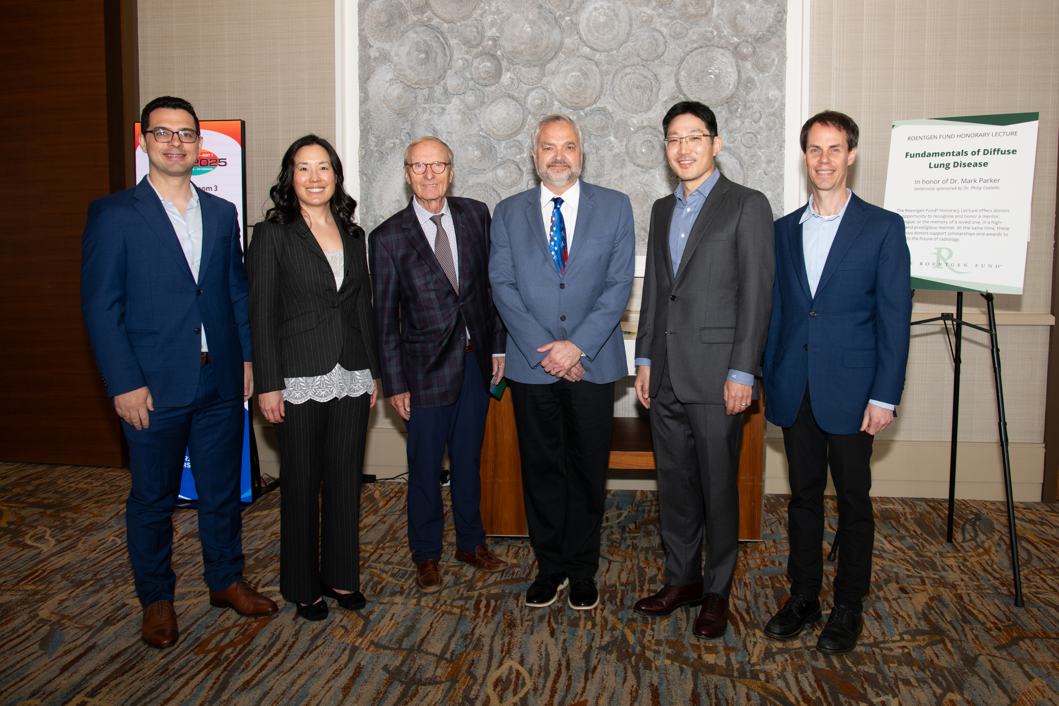
2010 Figley Fellow Dr. Jeffrey P. Kanne (far right) presented during one of the two 2025 Roentgen Fund Honorary Lectures. Sponsored by outgoing Roentgen Fund board chair Philip Costello, MD (middle left), Fundamentals of Diffuse Lung Disease honored Mark Parker, MD (middle right).
Roentgen Fund Roll Call
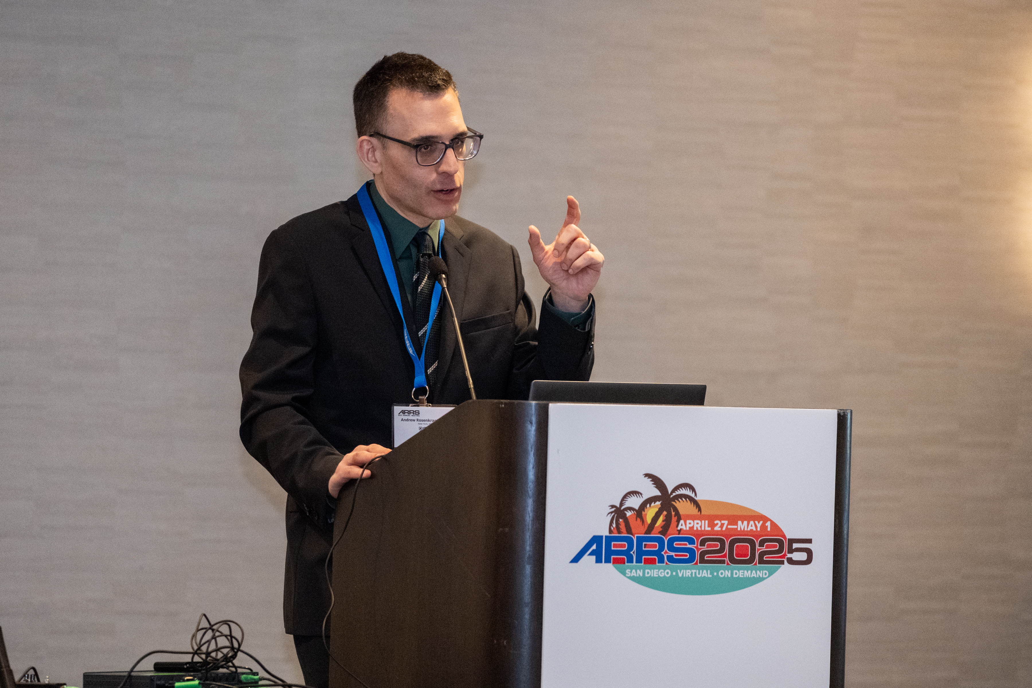
Dozens of prior awardees, like Dr. Nguyen, returned to present during all four days of the 2025 ARRS Annual Meeting in San Diego, CA. Congratulations to these trailblazing scholars and visionary educators—all of whom will continue to shape the future of our specialty:
Opportunities and Obligations in Imaging Patients with Alzheimer’s Disease with Tammie L.S. Benzinger, MD, 2008 ARRS Scholar
Critical Education for Safe, Ethical, and Successful AI in Radiology with Charles E. Kahn Jr., MD, MS, 1993 ARRS Scholar
Wellness Symposium – Creating a Positive Work Environment in Radiology with Christine B. Chung, MD, 2003 Figley Fellow
ARRS Scholar Presentation – Leveraging the “Art of Medicine” within Radiology: Investigation of Report Framing as a Digital Therapeutic To Mitigate Nocebo Effects with Steven A. Rothenberg, MD, BS, 2023 ARRS Scholar
ARRS Scholar Presentation – Deep Learning Tool to Eliminate Nephrographic Phase Images and Reduce Radiation Dose in CT Urography with Andrew L. Wentland, MD, PhD, 2023 ARRS Scholar
Quality and Safety in Radiology – The Unrelenting Pursuit of Excellence with Richard B. Gunderman, MD, PhD, 2011 Berlin Scholar
Cardiac Imaging in the Acute Setting with Jadranka Stojanovska, MD, 2015 Figley Fellow and Brett W. Carter, MD, 2016 Figley Fellow
Challenging Abdominal Cases with Perry J. Pickhardt, MD, 2002 Figley Fellow
Radiology Year in Review with Erin Alaia, MD, 2025 Figley Fellow and Domen Plut, MD, PhD, 2025 Rogers Fellow
Mastering Radiology: A Comprehensive Board Review for Residents and Refresher for Radiologists in Practice with Tanya Moseley, MD, 2025 Distinguished Educator
Categorical Course – Imaging in the ED Musculoskeletal Emergencies: Fractures and Infections with Sameer Raniga, MD, FRCR, 2018 Rogers Fellow
Gastrointestinal Imaging – Gastric Gliomas/Tumors, Bowel Ischemia and Bariatric Surgery with Benjamin Yeh, MD, 2006 ARRS Scholar
GI Post-treatment Imaging with Samuel Galgano, MD, 2022 Figley Fellow
Neuroradiology – CT Radiomics and Machine Learning with Suresh K. Mukherji, MD, 1998 Figley Fellow
Radiology Case Review – Musculoskeletal Imaging with Erin Alaia, MD, 2025 Figley Fellow
Award Winning Exhibits – Gastrointestinal Imaging with Benjamin Yeh, MD, 2006 ARRS Scholar
ABR Update: 2025 with John A. Kaufman, MD, 1996 Figley Fellow
Gastrointestinal Imaging – Hepatocellular Carcinoma with Claude B. Sirlin, MD, 2002 ARRS Scholar
Upper Extremity Challenging Cases with Christine B. Chung, MD, 2003 Figley Fellow
Categorical Course – Comprehensive Insights into Transplant Imaging: Bone Marrow Transplant with Meghan G. Lubner, MD, 2018 Figley Fellow
Neurovascular Disease with Achala S. Vagal, MD, 2012 ARRS Scholar
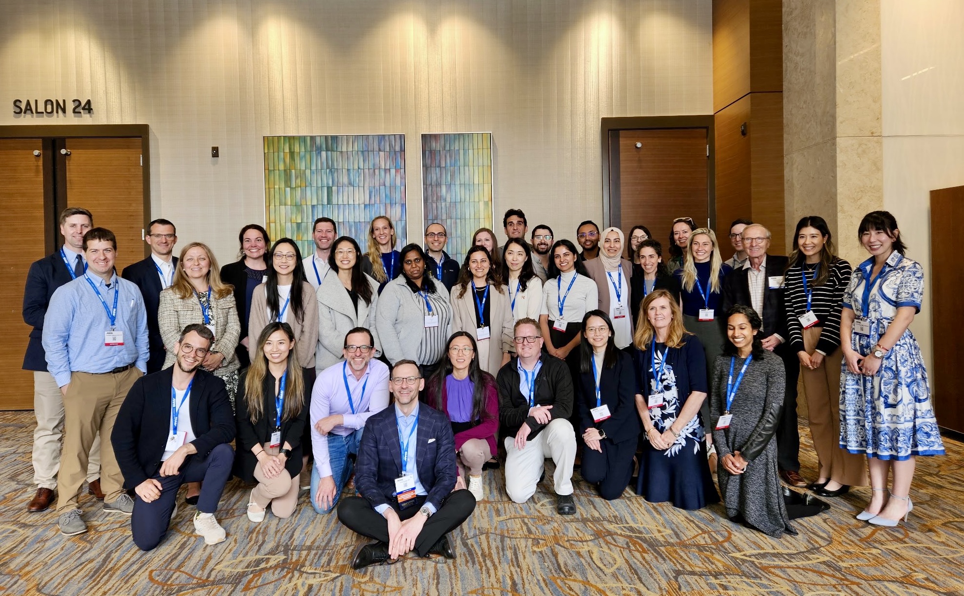
2024 Distinguished Educator David Naeger, MD (seated) presented Imaging of Lung Nodules and Lung Cancer and served as Course Director for The Roentgen Fund’s Clinician Educator Development Program on Saturday, April 26