Author: Logan Young

  • Musculoskeletal Imaging of Chest Wall Injuries in Athletes

    Musculoskeletal Imaging of Chest Wall Injuries in Athletes

    Published on May 13, 2022

    avatar

    Martin Torriani

    Department of Radiology, Division of Musculoskeletal Imaging and Intervention Massachusetts General Hospital and Harvard Medical School

    The chest wall includes a variety of osseous, cartilaginous, and musculotendinous structures that are vulnerable to athletic injuries. Lesions involving these structures usually can be divided in two main categories:

    1. lesions caused by trauma in which an external force causes direct or indirect injury to bones, cartilage, or soft tissue, leading to fractures, dislocations, and soft-tissue contusions;
    2. lesions caused by mechanical overload from powerful muscle contraction, with or without adequate balance provided by muscle antagonists, leading to myotendinous and muscular lesions and repetitive bone stress changes.

    A variety of contact and noncontact sporting activities may have mechanisms that predispose athletes to chest wall injuries. This article summarizes important aspects of athletic chest wall lesions, keeping in mind that many such lesions can occur in nonathletic endeavors, given the similarity of injury mechanisms. General technical aspects for imaging musculoskeletal athletic chest wall injuries are discussed, with most of the focus on CT and MRI, followed by discussions of injuries to the ribs, costochondral cartilage, sternoclavicular joint, and manubriosternal joint. In addition, athletic injuries to select muscles, such as the latissimus dorsi and teres major, are discussed in the context of athletic activities involving throwing.

    Technical Considerations

    Adequate imaging of chest wall injuries presents technical challenges that are specific to the scanning modality (CT vs MRI) and the targeted structure (rib or costochondral vs soft-tissue lesions). For both CT and MRI, a general recommendation is to perform at least one acquisition with bilateral FOV coverage for comparison purposes. A helpful procedure is to place MRI- or CT-visible skin markers in the area of pain or bracket a zone of discomfort. This allows the radiologist to better focus on specific structures that may be injured but are not clinically or functionally obvious at presentation. Another recommendation regarding MRI includes attempting to minimize respiratory motion by having the patient lie on the affected area (e.g., prone position, if lesion is anterior), which has the combined effect of reducing motion and keeping the structure in contact with the imaging coil. This is, of course, limited by patient comfort, so such decisions may require discretion from the technologist and attending radiologist on a case-by-case basis.

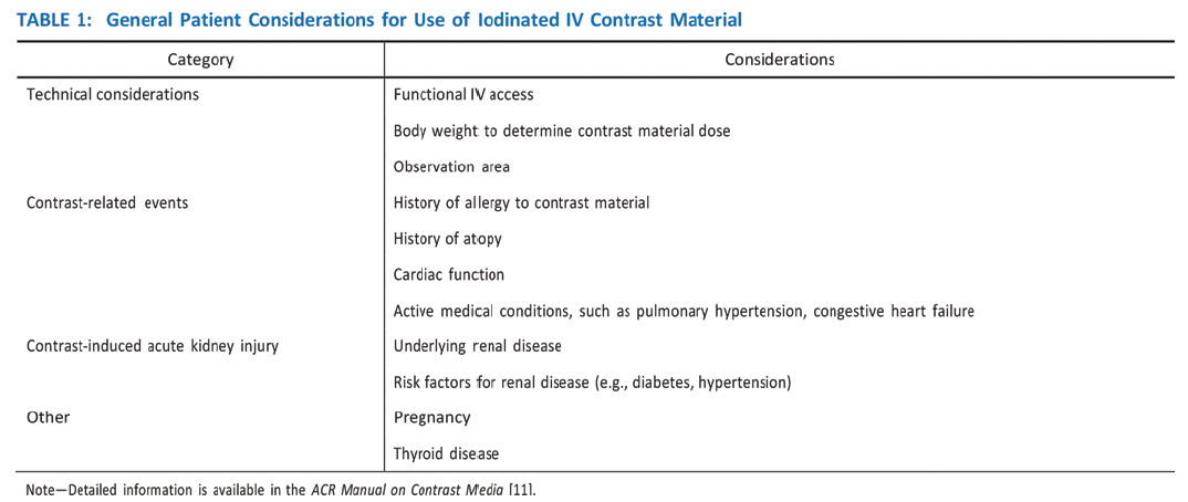
    Imaging of bone and costochondral injuries with MRI can be challenging because of the curved anatomy of the chest wall and ribs, which may limit proper visualization of small fractures if nonangled sagittal and coronal planes are used. Obtaining oblique sagittal and/or coronal MR images that are tangential to the abnormality allows identification of the lesion with adequate visualization of the adjacent rib structure and/or costochondral cartilage. Fat suppression is also difficult during chest wall MRI because achieving magnetic field homogeneity often requires the use of more robust techniques, such as STIR pulse sequences. Further, breathing motion and pulsation artifacts may cause image quality degradation, reducing imaging and diagnostic accuracy. The use of fast MRI techniques, such as T2 HASTE, PROPELLER (GE Healthcare), BLADE (Siemens Healthineers), and breath-hold pulse sequences, is desirable, as these approaches mitigate the effect of motion artifacts. On CT, bilateral FOV coverage to compare an affected area with similar contralateral anatomy and the use of thin-slice high-resolution images with bone kernel reconstructions are also recommended. CT allows rapid multiplanar and flexible reconstructions that may be useful when evaluating rib and costochondral fractures, as well as sternoclavicular and manubriosternal joint alignment. Finally, 3D reconstructions are easily obtained from CT images and may prove useful for surgical planning of sternoclavicular joint injuries.

    Chest wall muscular injuries are best imaged with MRI, including T1-weighted and T2-weighted fat-suppressed pulse sequences. In the author’s experience, the axial plane is the most important because it provides the best visualization of the pectoralis major, latissimus dorsi, and teres major tendon attachments. Oblique coronal images along the muscle planes can be obtained, though they may provide limited utility for surgical decisions and may be difficult to standardize across multiple sites in an institution. As described for osseous and cartilaginous lesions, the use of fast MRI techniques can help produce images that are less degraded by motion artifact. Bilateral FOV MRI may compromise spatial resolution and should therefore be limited to one to two acquisitions that allow comparison with the unaffected contralateral side.

    Sternoclavicular joint injuries are best imaged with CT, especially if clinical signs indicate a posterior dislocation. CT is a prompt imaging method that not only shows the degree of joint malalignment but also evaluates the integrity of upper mediastinum and adjacent great vessels. Manubriosternal joint injuries can be adequately assessed by both MRI and CT, with coronal and sagittal planes being the most adequate to examine for fractures or malalignment.

    Ultrasound may prove useful in certain situations. For example, because of the superficial nature of rib and costal cartilage, sonographic detection of fractures is possible and may represent a prompt method to evaluate focal complaints in patients [1]. In this context, the sonographer is able to place the ultrasound probe (preferably using frequencies ≥ 7 MHz) very precisely over the affected area to evaluate for focal osseous or cartilaginous discontinuities. In situations where muscle injuries are suspected, ultrasound is also a reliable imaging method [2]. However, in the author’s experience, it is common for patients with such injuries to present well-developed musculature (e.g., bodybuilders, football players), which may pose a limiting factor for adequate visualization of deep structures, such as tendon attachments. Complementary MRI is required in these cases, with ultrasound serving as a triage tool to be used at the discretion of the medical team. Finally, ultrasound is limited in its capability of assessing malalignments or dislocations of joints, such as the sternoclavicular and manubriosternal joints.

    Rib Stress Fractures

    Given that ribs are nonweightbearing bones, two main mechanisms can cause rib fractures:

    1. direct impact from an external source, commonly occurring in football, martial arts, and other contact or extreme sports;
    2. stress fractures due to muscular contraction.

    The latter mechanism most often results from strong muscular contraction that is unopposed by a counteracting fatigued muscle. In the case of rib stress fractures, the serratus anterior muscle plays a critical role [3, 4]. The serratus anterior muscle arises from the medial or ventral aspect of the scapula, with multiple slips extending anteriorly that attach to the first through tenth ribs at their middle third. The main role of the serratus anterior muscle is to protract the scapula. Importantly, this action is opposed by the scapular retractors (rhomboid muscles), which stabilize the scapula. In this situation, the serratus anterior muscle will promote an increase in the lateral diameter of the chest cavity. This mechanism can lead to abnormal stress at the middle third of the ribs in sports that involve repetitive contraction of the serratus anterior muscle, such as in elite rowing and swimming. In rowing, stress fractures are relatively common and may affect up to 12% of rowers. The peak contraction of the serratus anterior muscle occurs at the initial phase of the rowing cycle (known as “the catch”), in which the rowing oars are pulled away from the body, causing strong stabilization of the scapula by the rhomboid muscles and serratus anterior muscle. Performed repetitively, this action creates a compressive force vector directed toward the middle thirds of the ribs that can lead to stress fractures, most often affecting the fourth through eighth ribs in up to 86% of cases. If this compression is unopposed by a fatigued serratus anterior muscle, a repetitive stress injury may occur [3, 4]. Stress fractures also affect novice golfers who are still developing their technique and often strike the ground during swing strokes. Frequent ground impact force due to repetitive inaccurate strokes is thought to be transmitted to the chest wall on the leading side, causing stress fractures [5]. Additional sports in which rib stress fractures have been described include baseball, lacrosse, weightlifting, ballet, and gymnastics. MRI findings of rib stress fractures are similar to those seen in the extremities, including a well-defined linear area of low signal intensity surrounded by marrow edema, with the best contrast noted in T2- weighted fat-suppressed images (Fig. 1).

    Edema in the surrounding intercostal muscles and extrapleural space is often noted [6]. As mentioned earlier, such fractures may be better depicted on oblique sagittal or coronal images that are tangential to the fracture and provide context, better showing rib portions adjacent to the fracture itself. Healing of rib stress fractures can be monitored by MRI, showing progressive improvement of marrow edema and bony bridging; however, clinical symptoms may persist even though the fracture appears healed on imaging.

    Stress fractures affecting the first rib result from a different proposed mechanism that is a function of the rib’s anatomic features. The serratus anterior muscle attaches to the first rib’s lateral edge at its middle third, whereas the anterior and middle scalene muscles attach to the medial edge at the same level. The anterior scalene muscle attaches at a well-defined bony protuberance, behind which is a biomechanically weak zone of the first rib. This area, through which the subclavian vessels and brachial plexus travel, has been dubbed the “Achilles heel” of the first rib [7]. First rib fractures occur more commonly in throwing athletes, such as baseball players. Throughout the throwing cycle, the most prominent eccentric muscular contraction of the serratus anterior muscle occurs at the arm cocking stage, during which the scapula moves posteriorly, leading to strong opposition by the serratus anterior muscle. This motion, performed repetitively in high-performance athletes, can place undue mechanical stress on the weak zone of the first rib, leading to stress fractures. Such fractures can be treated conservatively but may take a long time to heal (6–12 months) and, if nonunion occurs with a large bone callus, may lead to symptoms of thoracic outlet syndrome. Additional activities for which first rib fractures have been described include jive dancing, basketball, tennis, and weightlifting. First rib fractures can be depicted adequately on MRI, though most studies on such fractures have used CT and occasionally nuclear medicine bone scans [7].

    Costal Cartilage Injury

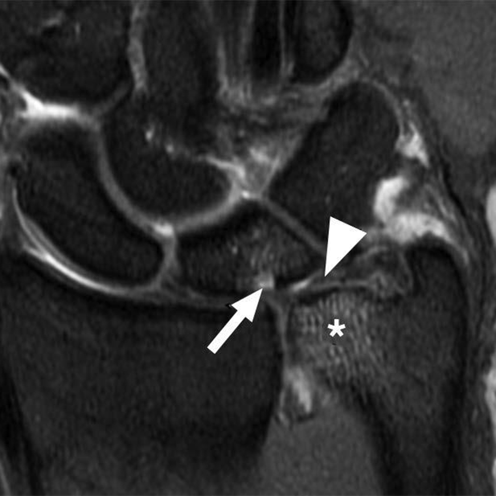
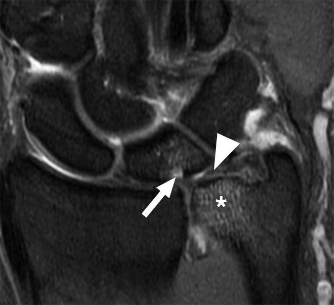
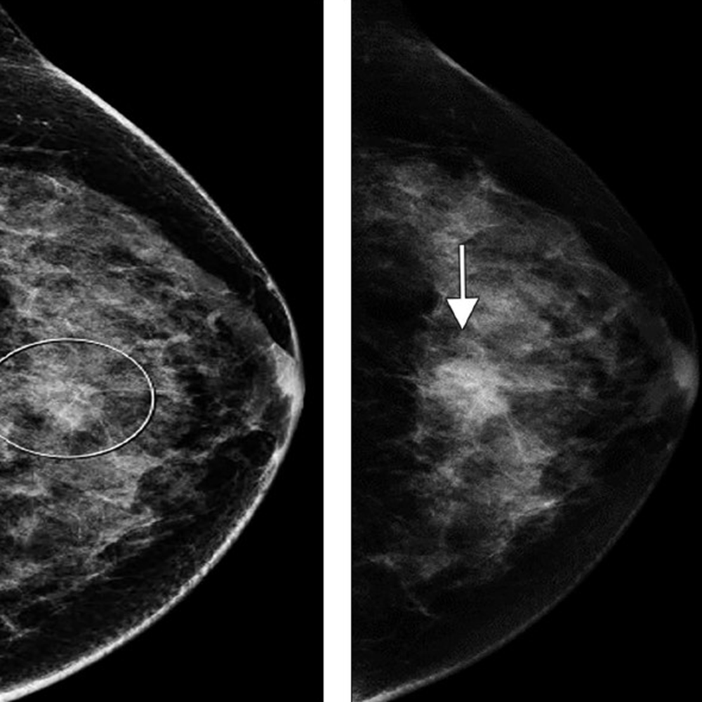
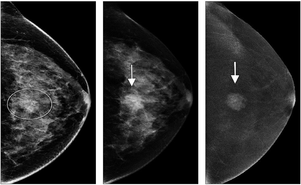
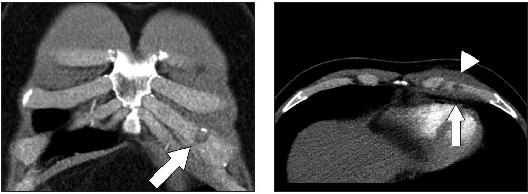
    Areas that may be affected by costal cartilage injury include the sternochondral junction, midsubstance of the costal cartilage, and costochondral junction. Most commonly, such injuries affect the relatively immobile upper rib cage (first to third costal cartilages) and are due to rotation injury and/or excessive axial loading (such as in weightlifting). Lesions affecting the lower rib cage (fourth through eighth costal cartilages) typically result from direct impacts, such as seen in contact sports (hockey, football, rugby, and martial arts) [8]. An important anatomic feature of costal cartilage is the presence of a central cavity that is best seen on CT images as a zone of lower attenuation tracking along the structure [9]. A predominance of blood vessels, loose connective tissue, macrophages, and fat exist within this area. Although this zone resembles a bone marrow cavity, no marrow cells or lineage is present. Anatomic studies suggest this central channel in costal cartilage is more akin to nutrient channels because of the dominant feature of high vascularity. The presence of a dense vascular network in this portion of costal cartilage predisposes to significant bleeding in the event of fractures. A common feature in costal cartilage fractures is the presence of an associated hematoma of the chest wall, which can lead to clinician suspicion of neoplastic masses. Scrutiny of the area using thin-slice CT may reveal a linear discontinuity of the costal cartilage adjacent to the suspected mass, increasing the likelihood of a trauma-related cause. Patient history may help direct the radiologist, as a traumatic event related to lifting or direct impact may have occurred, possibly resulting in subsequent pain and a local mass (Fig. 2).

    MRI provides excellent contrast to show costal cartilage fractures, which will appear as linear areas of high signal intensity against a background of low-signal-intensity costal cartilage [8]. Although MRI has superb spatial resolution and contrast, its relatively higher cost and susceptibility to respiratory motion artifacts decrease its effectiveness, when compared with CT. Ultrasound may also prove useful, given the superficial location of costochondral structures, and may show a focal stepoff at the fracture area [1]. As previously mentioned, costal cartilage injuries may occur at the sternochondral or costochondral junctions. At these locations, chondral and adjacent bone marrow edema may be present on MRI and represent the dominant findings related to traumatic injury. Treatment of costal cartilage injuries include NSAIDs, taping and immobilization belts, and rest for 2–3 weeks. In certain situations, such as treatment of professional athletes, pain can be mitigated by local injections of anesthetic [8]. An important differential diagnosis to consider when entertaining the possibility of a costochondral injury is Tietze syndrome. In this rare entity, inflammation of the sternochondral junction (the most common symptom) can occur spontaneously and lead to focal pain without any specific initiating event. The cause of Tietze syndrome is uncertain, and the condition usually affects younger patients (< 40 years old). On MRI, this entity may show edematous change surrounding the sternochondral junction, which is best seen on fat-suppressed T2-weighted images [10]. CT may not provide enough tissue contrast for adequate detection. Given that Tietze syndrome is usually self-limited, its treatment includes rest, NSAIDs, and local anesthetic injections in refractory cases.

    Sternoclavicular Joint

    Athletic injuries to the sternoclavicular joint may lead to anterior dislocation (more common and less concerning) or posterior dislocation [11, 12]. The latter phenomenon is less frequent, but more concerning, because the clavicular head has the potential to exert mass effect on the great vessels, trachea, esophagus, and recurrent laryngeal nerve (causing vocal cord palsy). Posterior dislocations require 50% more force to occur, compared with anterior dislocations. This is because the capsular structure of the sternoclavicular joint is more robust posteriorly [13]. Posterior sternoclavicular joint dislocations are most commonly related to direct-force injuries seen in football, rugby, martial arts, and motor vehicle accidents (typically motorcycle accidents). Sternoclavicular dislocations can be imaged by CT and/or MRI, both of which may show malalignment at the joint with associated fractures or hematomas from capsular disruption. In the case of posterior dislocations, CT provides excellent delineation of the joint alignment, and use of IV contrast material allows assessment of the integrity of adjacent vascular structures. Alignment of mediastinal structures and possible hematomas are also best seen on CT. The initial treatment of posterior sternoclavicular dislocations includes attempted closed reduction, which is usually performed under sedation as an urgent procedure. If this procedure fails to realign the joint, or if the joint remains unstable, a surgical approach involving open reduction and internal fixation may be necessary.

    Manubriosternal Joint

    The manubriosternal joint is rarely involved in athletic injuries. Usual mechanisms of injury are classified as either type 1, in which an anteroposterior force is exerted on the sternal body, which moves posteriorly relative to the manubrium, or type 2, in which the anteroposterior force is exerted against the manubrium [14]. The latter mechanism is usually related to forces being transmitted through the arms to the clavicle and sternoclavicular joints, displacing the manubrium posteriorly. In the author’s experience, such lesions are rare and show marrow edema surrounding the manubriosternal joint on MRI, suggesting local bone contusions. Both sagittal and coronal images through the sternum are optimal for this visualization.

    Latissimus Dorsi and Teres Major

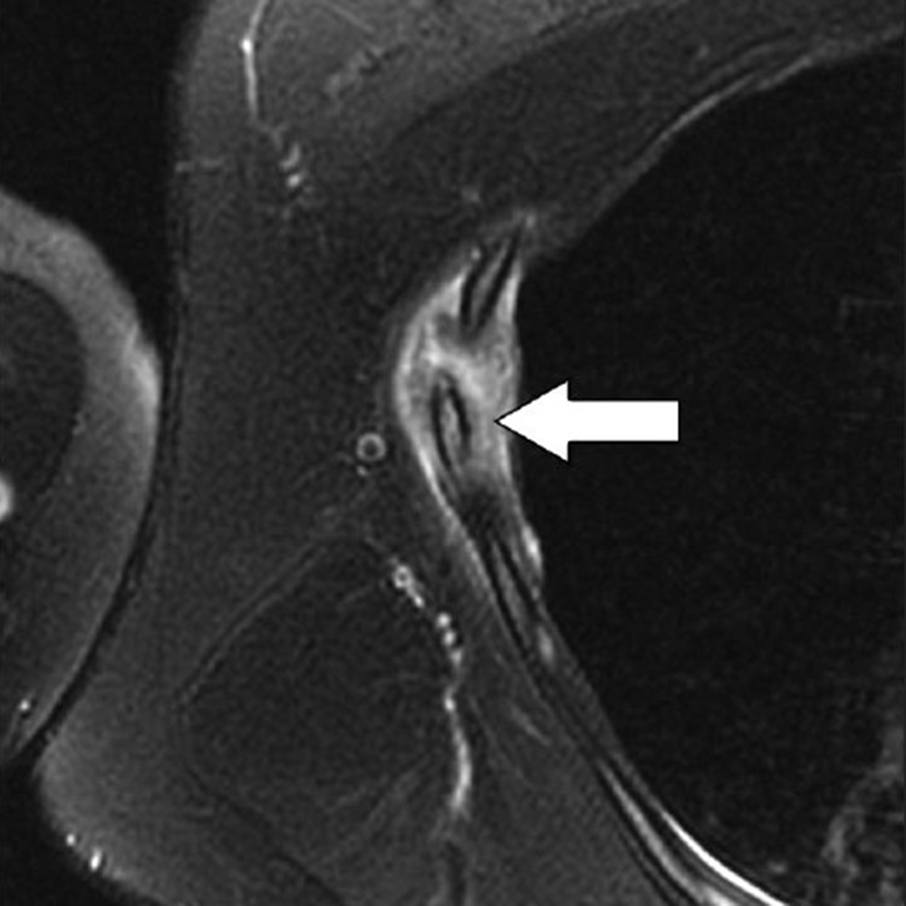
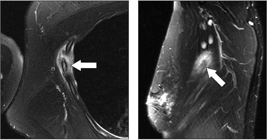
    Injuries to the latissimus dorsi and teres major muscles are most commonly seen in throwing athletes [15–18]. The latissimus dorsi muscle has a broad origin that includes, among others, multiple spinous processes of the thoracolumbar region and iliac crest; the muscle then attaches at the floor of the intertubercular groove of the humerus. The teres major muscle originates at the posterior aspect of the inferior angle of the scapula and inserts at the medial ridge of the intertubercular sulcus of the humerus. In cadaver studies, the latissimus dorsi and teres major tendon attachments were seen as a single structure in up to 83% of cases [15–18]. In the remaining cases, separate tendons attaching to the humerus were visible on axial images. One important action of these muscles is to powerfully adduct, extend, and internally rotate the humerus. During the throwing motion, the latissimus dorsi and teres major muscles are activated as a unit, with maximum levels of eccentric contraction during the arm cocking and arm deceleration stages of the throwing cycle [15–18]. Lesions of the latissimus dorsi and teres major may present along the usual spectrum of myotendinous strain injuries up to avulsions at the humeral attachments. For this reason, MRI is the preferred imaging method, as it can characterize the full gamut of possible lesions, including those affecting the more dorsal portions of the latissimus dorsi muscle. In this situation, the location of the patient’s symptoms should be considered to ensure adequate coverage on imaging. Because the latissimus dorsi covers a large area of the lateral and posterior chest wall, large FOVs may be required, especially if bilateral imaging is being performed. Another important technical point is to ensure that axial images through the humerus adequately cover the footprints of latissimus dorsi, teres major, and pectoralis major tendons, which are usually not fully viewed during standard shoulder imaging protocols. This may require obtaining axial images that extend caudally to approximately the middle third of the humerus to guarantee such coverage in all patients. Regarding treatment, avulsions at the humeral attachment, which have been described in athletes involved in waterskiing, golf, tennis, and bodybuilding, usually require surgical intervention to reattach the tendons [18]. On the other hand, most myotendinous lesions of latissimus dorsi and teres major muscles are treated conservatively.

    Imaging of athletic chest wall injuries should be performed while taking into account anatomic considerations and the advantages of specific imaging methods, including CT, MRI, and ultrasound. Because these injuries are likely to comprise a relatively smaller volume of cases at a given imaging facility, predefined workflows are important to tackle targeted anatomic locations. This may require close communication between scheduling staff, technologists, and attending radiologists, who can tailor protocols to obtain the best images of athletic chest wall injuries.

    References

    1. Malghem J, Vande Berg B, Lecouvet F, Maldague B. Costal cartilage fractures as revealed on CT and sonography. AJR 2001; 176:429–432
    2. Chiavaras MM, Jacobson JA, Smith J, Dahm DL. Pectoralis major tears: anatomy, classification, and diagnosis with ultrasound and MR imaging. Skeletal Radiol 2015; 44:157–164
    3. Warden SJ, Gutschlag FR, Wajswelner H, Crossley KM.  Aetiology of rib stress fractures in rowers. Sports Med 2002; 32:819–836
    4. McDonnell LK, Hume PA, Nolte V. Rib stress fractures among rowers. Sports Med 2011; 41:883–901
    5. Lord MJ, Ha KI, Song KS. Stress fractures of the ribs in golfers. Am J Sports Med 1994; 24:118–122
    6. Taimela S, Kujala UM, Orava S. Two consecutive rib stress fractures in a female competitive swimmer. Clin J Sport Med 1995; 5:254–256; discussion, 257
    7. Coris EE. First rib stress fractures in throwing athletes. Am J Sports Med 2005; 33:1400–1404
    8. Subhas N, Kline MJ, Moskal MJ, White LM, Recht MP. MRI evaluation of costal cartilage injuries. AJR 2008; 191:129–132
    9. Lee S, Choi YW, Jeon SC. Low attenuation areas in normal costal cartilages on CT: clinical implication and correlation with histology. Clin Anat 2012; 25:483–488
    10. Volterrani L, Mazzei MA, Giordano N, Nuti R, Ga- leazzi M, Fioravanti A. Magnetic resonance imaging in Tietze’s syndrome. Clin Exp Rheumatol 2008; 26:848–853
    11. Mirza AH, Alam K, Ali A. Posterior sternoclavicular dislocation in a rugby player as a cause of silent vascular compromise: a case report. Br J Sports Med 2005; 39:e28
    12. Galanis N, Anastasiadis P, Grigoropoulou F, Kirkos J, Kapetanos G. Judo-related traumatic posterior sternoclavicular joint dislocation in a child. Clin J Sport Med 2014; 24:271–273
    13. Spencer EE, Kuhn JE, Huston LJ, Carpenter JE, Hughes RE. Ligamentous restraints to anterior and posterior translation of the sternoclavicular joint. J Shoulder Elbow Surg 2002; 11:43–47
    14. Hayashi D, Roemer FW, Kohler R, Guermazi A, Gebers C, De Villiers R. Thoracic injuries in professional rugby players: mechanisms of injury and imaging characteristics. Br J Sports Med 2014; 48:1097–1101
    15. Schickendantz MS, Kaar SG, Meister K, Lund P, Beverley L. Latissimus dorsi and teres major tears in professional baseball pitchers: a case series. Am J Sports Med 2009; 37:2016–2020
    16. Ellman MB, Yanke A, Juhan T, et al. Open repair of an acute latissimus tendon avulsion in a Major League Baseball pitcher. J Shoulder Elbow Surg 2013; 22:e19–e23
    17. Escamilla RF, Andrews JR. Shoulder muscle recruitment patterns and related biomechanics during upper extremity sports. Sports Med 2009; 39:569–590
    18. Cox EM, McKay SD, Wolf BR. Subacute repair of latissimus dorsi tendon avulsion in the recreational athlete: two-year outcomes of 2 cases. J Shoulder Elb Surg Am 2010; 19:e16–e19
  • Resilience in Radiology

    Resilience in Radiology

    Published May 13, 2022

    avatar

    Gary J. Whitman

    2022–2023 ARRS President

    Over the last few years, we have heard a lot about resilience. What does resilience really mean?  How can we develop resilient teams and organizations? Furthermore, how can we help our patients to be more resilient? Resilience is often defined as the capacity to recover quickly from difficulties. Some equate resiliency with toughness. Another definition of resilience is elasticity, the ability to spring back into shape. Psychological resilience is characterized as the ability to cope mentally or emotionally with a crisis or to return to pre-crisis status quickly [1].

    While resilience implies bend-but-don’t-break properties, there are a couple of issues to consider:

    1. We don’t always return to our exact pre-crisis or pre-stress configuration;
    2. Resilience can allow for personal growth and help to catalyze the growth of others.

    Regarding our return to our original forms after a jolt to the system, we will never be exactly the way that we were previously. We are older, maybe heavier, maybe hungrier, and maybe more tired.  Furthermore, we probably learned something from the stress that we just endured. What we learned can lead to personal growth and the ability to share our new knowledge with others. This process of sharing and helping others has the potential to lead to more resilient teams and to help our patients become more resilient.

    Historically, we have thought of resilience in terms of personal resilience—at the individual level. There is no question that personal resilience is important, but resilience can also apply to groups and teams. It is important to consider teams, especially in radiology, as most of our activities are team-related. In fact, very few of our activities do not involve teams. Just think of the process on the front end of any imaging study, before it gets to the radiologist for interpretation: there are schedulers, front desk personnel, nurses, physicists, and technologists. How do we make our teams more resilient and more effective?

    One of the drivers for engaged, resilient teams is relational energy. Leaders with relational energy create a positive environment with higher levels of engagement, lower turnover rates, and enhanced feelings of well-being [2]. On the other hand, there are leaders who drain energy from the group, and the team members loathe working with an idea-killing, energy-sapping leader. In the book Multipliers: How the Best Leaders Make Everyone Smarter [3], Liz Wiseman discusses two types of leaders: The first group are diminishers, draining intelligence, energy, and capability from the people around them, and the second group are multipliers, leaders who employ their skills to amplify the strengths and the capabilities of those around them.

    Multipliers can have a major impact on our teams and organizations in radiology. In this ever-changing, peri-pandemic world, multipliers can make us all more resilient, by doing more with less, by attracting and developing talent, by creating a safe environment that allows for our best thinking, by challenging us to push beyond what we know, by debating decisions, and by instilling ownership and accountability. Furthermore, multipliers do not need to be great at everything. Rather, effective multipliers should have some very solid strengths and few major deficiencies. In addition, effective multipliers often choose to form teams with others who bring complementary strengths to the table [3].

    As we navigate the challenges of our topsy-turvy world with a major war in Europe, political divisiveness and the great resignation in the United States, and rising inflation, and as we try to re-equilibrate in the peri-COVID world, we need to be resilient as we continue to move our field forward and deliver top-notch care to our patients. Our patients really need us, not just to read their images and do their procedures, but to advocate for them with empathy and dignity.

    When we go to work, it may be a good day, a bad day, or a usual, non-descript day. Often, our patients are seeing us on what may be their worst day ever or what they fear will be their worst day ever. I recently spoke with a patient who was diagnosed with a cervical plasmacytoma in 1994. Soon thereafter, he was shown to have multiple myeloma involving several sites in the cervical, thoracic, and lumbar spine. Despite chemotherapy, radiation therapy, an autologous stem cell transplant, and experimental therapy, he was told that his chances for survival were less than 5%. With each imaging study came the dread that more disseminated disease would be found. Nevertheless, during those encounters, he found front desk personnel, technologists, and physicians to talk to. Even amidst a downward cycle of relapses and remissions, a radiation oncologist suggested that he consider sperm preservation (he was single at the time).

    Let’s fast forward to 2022, when the patient is a 30-year survivor of multiple myeloma, leading a foundation to help multiple myeloma patients; he is happily married, and his son is a college graduate! The patient was in a “very dark place” 25 years ago, worried that he would never see or know his son, and now, miraculously, he is a long-term myeloma survivor.

    There are countless patients who come to us every day on their journeys of resilience. We have an obligation to engage them, to treat them in a dignified professional manner, and, hopefully, what was anticipated as a very bad day may not be so bad for them.

    As we think about building resilient teams and resilient enterprises, it is important that we promote and practice empathetic, patient-centered behaviors. We need to be multipliers for our patients. 

    Recently, I performed a biopsy on a small right breast mass on a 44-year-old woman. The procedure went smoothly, and the biopsy showed evidence of a papilloma. A few days after the biopsy, the patient’s referring clinician contacted me, informing me that the patient had a 4th ventricle ependymoma resected at age 5 years, and that the patient had diminished mental capacity. Immediately after the biopsy, the patient’s mother had asked to speak to me, but she was told that I was busy and that she should check the electronic medical record. I do not know who communicated with the patient’s mother, but I certainly would have made myself available to talk to her. As we try to be multipliers for our patients, we need to do better each and every day.

    As we try to be resilient in this ever-changing world and form resilient radiology teams, we should keep in mind these words from the Cadet Prayer at West Point: “Make us choose the harder right instead of the easier wrong, and never be content with a half-truth when the whole truth can be won” [4]. We need to choose the harder right; our patients are depending on us.

    References

    1. Psychological resilience. en.wikipedia.org/wiki/Psychological_resilience. Wikipedia website. Accessed April 26, 2022
    2. Stillman J. Yale research: having this 1 trait makes leaders 4x more effective. www.inc.com/jessica-stillman/yale-research-having-this-1-trait-makes-leaders-4x-more-effective.html. Inc website. Accessed April 27, 2022
    3. Wiseman L. Multipliers: How the Best Leaders Make Everyone Smarter. Harper Business (New York); 2017
    4. Cadet prayer. www.west-point.org/academy/malo-wa/inspirations/cadetprayer.html. West Point Connection website. Accessed April 28, 2022.
  • Reimagining and Reinventing Postpandemic Radiology

    Reimagining and Reinventing Postpandemic Radiology

    Published March 31, 2022

    avatar

    Jonathan Kruskal

    2021–2022 ARRS President

    avatar

    James V. Rawson

    Senior Lecturer on Radiology
    Beth Israel Deaconess Medical Center

    Some days, it’s hard to recall what prepandemic life was like. Things have forever changed in light of this historic global event, and it’s vital to reflect and process these last three years. We’ve endured some of the most trying times of our careers, but we also have a bright future as a medical community ahead. You’re wondering what that might look like and how we can collectively “skate to where the puck is going to be, not to where it has been,” as Wayne Gretzky famously said.

    COVID-19 precipitated a fundamental change in clinical service delivery, teaching, research, staff retention, employee wellness initiatives, and communications strategies. We amended workplace safety standards and practices, stood up and resourced remote teams, recruited trainees virtually, and transformed in-person grand rounds programs into digital ones. These are just some of the many efforts that we as a specialty undertook to protect our people, uphold our missions, and keep our teams employed. And while not all changes were novel ones, the pandemic catalyzed their implementation. We now have tremendous momentum to continue innovating, especially as we begin to emerge from crisis mode together.

    Accelerating Change

    Here at Beth Israel Deaconess Medical Center, we simply couldn’t have managed this public health crisis without our highly effective, efficient, and resourceful operational surveillance systems and teams. Some of these teams were in place before the pandemic started; they rapidly responded to the initial phases of COVID-19, then swiftly transitioned into a multidisciplinary incident command structure to assess, rethink, reinvent, iterate, and communicate our health care systems and strategies on a daily, hourly, and minute-by-minute basis. This collaborative structure operated in real time and kept our trains running, on schedule and on the tracks, far more often than not. Light started to appear at the end of the tunnel, and then, the alphabet of variants arrived. It soon became clear that we would never return to prepandemic normalcy. A fundamental shift had taken place in the way we delivered our services, and some of this change represented the necessary digital transformation many had envisioned long before COVID-19 struck.

    So, what are our next steps? How can we effectively shift from a reactionary mode to one that is deliberate and purposeful? What structure will best support the necessary regrowth phase that will support our medical practices and organizations? Now is our time to be accelerators rather than incubators, to reinvent and rebrand our skills and clinical contributions, and to be thoughtful and strategic in the process. This is where the most strategic, imaginative, and operationally agile teams will lead the way and define our recovery. Those who embrace change and progress will be the best positioned to thrive. Lead the change. Be the change.

    Building COVID Recovery Hubs

    Let’s be the disruptive thinkers our field will be proud of. Define, then communicate your future radiology vision. Do this in an inclusive manner that involves all role groups. For such plans to be adopted and successful, leaders will need to continue to create forums for staff to weigh in, ask the right questions of their teams, listen to feedback, barriers, needs, and ideas, and provide other ways to share input, such as through short pulse surveys in a departmental newsletter or real-time polls during meetings. Ensure everyone’s voice is heard and incorporate major common themes into your plans.

    We suggest constructing and resourcing a formal COVID recovery hub, which can house your postpandemic mission, vision, and recovery playbook. Appoint and support a multidisciplinary team to lead and own these pioneering transformation efforts. Who have you appointed to lead your radiology recovery, reinvention, and reengineering transformation? What does your “r4” portfolio look like? By answering these questions, you will help your practice continue to keep its staff and patients safe, deliver exceptional care, manage ongoing people and supply chain shortages, support staff morale and wellness, nurture remote teams, and effectively communicate and engage with a multigenerational workforce.

    Reimagining Administrative Functions

    As part of your COVID recovery hub, consider the roles, responsibilities, and constituents of your leadership team. Are you best positioned for your recovery? Now might be a great time to reimagine these elements and challenge the traditional hierarchy, as we work to flatten authority gradients, build diverse, inclusive, and multigenerational teams, and ensure that form follows function. Additionally, these roles have likely transformed out of necessity during the pandemic and might need to evolve to meet your practice’s future needs.  

    Let’s be a little provocative: how can we identify the best person to be our “knowing exactly where the puck will be in 2030” portfolio leader? Actually, this is not a task for one person—and this is precisely why building and sustaining high-performing diverse and inclusive teams will become paramount and essential. Proudly establish your recovery hub, appoint a vice chair of recovery and reinvention to lead this effort, then establish and resource new portfolios to signal just how serious you are about recovery, regrowth, and reinvention. For example, have you considered the future of your remote teams? Based on national employee preferences, it’s clear that some level of remote work is here to stay. Perhaps, as part of a practice’s new digital innovation and transformation lab, it could create a remote workplace and team-building portfolio. Will your organization continue producing short video messages, digital newsletters, social media content, and academic webinars? What will your postpandemic communications strategy look like? Perhaps it’s a question for a newly formed digital communications and connections team. While these structures might not be novel in a large, digital-first corporate setting,they would be a progressive leap forward for many of our major academic medical practices.

    Additionally, we all know that health care delivery has progressed to incorporate population health, cost reduction improvement efforts, care coordination and integration, and customer experience, among other important factors. Simply put, our aim is to deliver the highest-quality, safest possible care and experience at the most sustainable costs. This boils down to value, and who better to drive it than an effective chief value officer?We’ve certainly been talking about this value proposition for quite some time already. On a different but equally important note, we must think about how we will continue to support the health and wellness of our staff postpandemic, especially during our nation’s mental health crisis. Is there an influential and compassionate leader on your team who could become your chief wellness leader and drive these vital efforts to aid your entire team?

    The structure of a leadership team should primarily relate to its intended function and purpose. Once you have reconfirmed your foundational core purpose, reimagined your vision and mission, and defined your annual goals, then form the team (and define their precise roles) that will help you reach your ideal future state. Energize your teams by including them in strategic brainstorming and planning sessions, imagining an exciting and successful future together.Designing a newoperational landscape is not a task for one person, which is why building and sustaining high-performing, diverse, and inclusive teams will be paramount.

    The interesting exercise that we are all engaged in now is to define that future state. Has anybody considered a leader of a recovery and reinvention portfolio? Your entire team wants to contribute! Be inclusive and build diverse teams.

    Mapping New Pathways

    This is an era of posttraumatic regrowth. Reimagining your pathway should be an inclusive, aspirational, and even inspirational process. Be thoughtful and strategic when redefining your path forward toward the new normal you and your team aspire to achieve. Reengage and revitalize your most precious resource, your workforce. Recommit to safe practices, wellness initiatives, and high-performing team building. Reconnect your teams, and work to sustain these connections. Reimagine and rethink your strategic plan and goals, and start your new journey today. Those who will flourish and thrive will do this effectively, thoughtfully, and strategically; consider the long-term goals, map out your route, and take action. As you shift from managing operations to imagining the future, try to shift your focus from keeping the trains running to considering where new rails could be built. Periodically, it’s important to pause and ponder—to consider not only how trains can be better engineered, but also to contemplate whether train travel will be a safe and efficient customer choice in the future. That’s strategic thinking.

    Let’s try to simplify. You’re done with reacting, reflecting, and responding. You’re starting to see some light at the end of this tunnel. You’re hoping that omicron is the last symbol of this pandemic alphabet. Now, more than ever before, is the time to look forward, plan your recovery strategy, and focus on building and sustaining innovation. The practices that are most likely to thrive are already thinking outside the traditional administrative oversight box. And they are moving ahead right now.

    And, finally, find ways to share your experiences of this journey. We’re all traveling new paths and learning as we go. We must learn from each other’s successes and missteps, and there will be plenty of both. As we build our departmental COVID recovery hubs, we also need to design and build collaborative teams to communicate and interact with institutional, regional, and national COVID recovery hubs, to the extent they exist. These new systems must be capable of redefining and reimagining the future, so that we can all travel along the path of progress together.

    It’s now time to be intentionally inclusive, as we commence this new journey.

  • Overuse Injuries in the Hand and Wrist: Bone and Joint Disorders

    Overuse Injuries in the Hand and Wrist: Bone and Joint Disorders

    Published March 31, 2022

    avatar

    Tetyana Gorbachova

    Department of Radiology, Sidney Kimmel Medical College
    Thomas Jefferson University Einstein Medical Center

    avatar

    Maria Grigovich

    Department of Radiology, Sidney Kimmel Medical College
    Thomas Jefferson University Einstein Medical Center

    avatar

    Micah G. Cohen

    Department of Radiology, Sidney Kimmel Medical College
    Thomas Jefferson University Einstein Medical Center

    avatar

    Yulia V. Melenevsky

    Department of Radiology University of Alabama at Birmingham Medical Center

    Overuse injuries of the hand and wrist are common in both professional and recreational athletes. These injuries, also referred to as stress injuries or repetitive strain injuries, result from cumulative microtrauma produced by a combination of abnormal force, repetitive motion, and insufficient recovery time that exceeds the tissue’s ability to repair itself. These characteristic pathologic conditions may be associated with various athletic activities and most frequently occur in racquet sports, rowing, volleyball, handball, weight lifting, and gymnastics. Certain lesions are unique to gymnastics, where, in addition to performing a wide range of movements, the upper extremity becomes a weight-bearing system. This article will review overuse injuries of the hand and wrist, focusing on pathologic conditions of the bone and joint.

    Stress Fractures

    Stress fractures in athletes typically represent fatigue fractures caused by repetitive excessive stress applied to normal bone. Overall, stress fractures are uncommon in the upper extremity. However, several sport-specific stress fractures have been described in the hand and wrist. Hook of the hamate fracture is recognized in golfers and may result from repetitive stress or a single traumatic event from the strike of a club on the ground. In sports in which a racquet is used, hamate fractures typically affect the dominant hand; in baseball, hockey, or golf, they usually occur in the nondominant hand. Metacarpal stress fractures have been described in adolescent tennis players and most commonly involve the base or shaft of the second metacarpal. Stress fractures of the scaphoid, typically occurring at the scaphoid waist, have been reported in various sports, most commonly in gymnastics, where they may be bilateral.

    Special radiographic views can be used for the diagnosis of carpal fractures. For example, to improve visualization of the hook of the hamate, the carpal tunnel view, semisupinated oblique view, lateral view with thumb abduction, and hand radial deviation view can be obtained. CT is an excellent modality for both detection of fractures and assessment of healing. MRI may depict early stress reaction manifested by bone marrow edema–like signal without a fracture line. These changes may progress to a fracture with a low-signal-intensity line or cortical break visible on MRI.

    Gymnast’s Wrist

    The wrist is the most frequently injured site in the upper extremity of female gymnasts, followed by the elbow, and is the second-most common injury location, after the shoulder, in male gymnasts. In gymnastics, the wrist is exposed to multiple types of forces, which include high-impact loading, axial compression, torsional forces, and distraction. Compressive forces in particular may amount to 16 times body weight. These stressors, combined with repetitive motion and varying degrees of ulnar and radial deviation and hyperextension, predispose the wrist to higher rates of both acute and overuse injuries.

    Stress injury of the distal radius may compromise the blood supply to the growing physis, which leads to abnormal cartilage ossification manifested with pain and, if untreated, premature closure of the physis, eventually leading to growth disturbances and secondary ulnar-sided wrist impaction syndromes; this spectrum of clinicopathologic findings is referred to as “gymnast’s wrist.” Adolescent athletes become symptomatic during peak growth velocity at 12–14 years old.

    Radiographs show widening of the distal radial growth plate, an indistinct zone of provisional calcification, and irregularity and sclerosis of the metaphysis, the latter of which sometimes give the metaphysis a beaked or a hooked appearance. These findings are frequently bilateral and involve either the entire distal radial physis or its radial and volar aspects. Concurrent radiographic abnormalities may be seen in the distal ulna in up to 20% of cases.

    MRI shows growth plate widening and periphyseal bone marrow edema–like signal. On the basis of clinical and radiographic findings, gymnast’s wrist is classified in three stages: stage I, clinical symptoms without radiographic changes; stage II, radiographic changes in the distal physis of the radius with normal radial length; and stage III, stage II with the addition of secondary positive ulnar variance. In addition to classic growth plate abnormalities, a variety of stress-related nonphyseal osseous, ligamentous, and osteochondral injuries have been described in skeletally immature gymnasts, which expand the spectrum of findings associated with the term “gymnast’s wrist”.

    Ulnar-Sided Wrist Impaction Syndromes

    Several ulnar-sided wrist impaction syndromes are recognized in athletes.

    Ulnocarpal impaction syndrome: Ulnocarpal impaction syndrome, also known as ulnar abutment, refers to the chronic impaction between the ulnar head, triangular fibrocartilage complex (TFCC), and ulnar side of the carpus [16]. This syndrome is commonly seen in gymnastics, racquet sports, and golf. Athletes are particularly susceptible to this condition when excessive ulnar loading is paired with positive ulnar variance; however, pathologic changes may occur with neutral or even negative variance.

    In gymnastics, compressive loads of the wrist are often combined with pronation, which doubles the load applied to the ulnar side of the wrist. Ulnar deviation combined with pronation, such as occurs in pommel horse or vault maneuvers, increases ulnar load from the normal 15% to approximately 40%. Positive ulnar variance in gymnasts may be congenital or may develop secondary to premature physeal closure of the distal radius.

    In comparison with acute traumatic injuries to the TFCC, which may affect various components of the complex, chronic ulnar abutment typically causes central degeneration and perforation of the triangular fibrocartilage disk proper, as outlined by the Palmer classification. The spectrum of progressive pathologic changes in ulnar abutment includes degenerative tearing of the TFCC, ulnar-sided chondromalacia, tears of the lunotriquetral ligament, and lunotriquetral instability—and, in advanced stages, osteoarthritis of the distal radioulnar joint and ulnar side of the radiocarpal compartment. The typical areas of cartilage loss and associated reactive marrow changes are localized to the ulnar head, ulnar side of the proximal aspect of the lunate, and radial side of the proximal aspect of the triquetrum (Fig. 1).

    Radiography provides the most accurate determination of the ulnar variance and cannot be substituted with other imaging modalities, particularly in the detection of subtle changes that can be determined only by standard radiographic positioning. MRI provides detailed assessment of the TFCC, bone, and articular cartilage. MRI and CT arthrography can be used to determine the integrity of the TFCC and lunotriquetral ligament.

    Ulnar styloid impaction syndrome: Ulnar styloid impaction syndrome is caused by impaction between the ulnar styloid process and the triquetral bone. It may occur as a result of congenital morphologic variations or posttraumatic and degenerative pathologic conditions of the ulnar styloid process resulting in its elongation. An ulnar styloid process is considered excessively long when the ulnar styloid process index is greater than 0.21 or when the overall length of the styloid process is greater than 6 mm. Ulnar styloid nonunion fractures may also lead to ulnar styloid impaction. This syndrome results in chronic bone contusion of the ulnar styloid and triquetrum, chondromalacia, and synovitis and can lead to lunotriquetral instability.

    Radiographs may show sclerotic or cystic changes in the triquetrum, ulnar styloid, and, in some cases, ulnar aspect of the lunate. MRI detects earlier changes of chondromalacia, reactive bone marrow abnormalities, and commonly associated degenerative tearing of the TFCC.

    Hamatolunate impaction syndrome: Hamatolunate impaction syndrome is related to a type II lunate that is defined by the presence of a separate facet along the distal surface of the lunate articulating with the proximal pole of the hamate. The repeated impingement and abrasion of these two bones in ulnar deviation of the wrist lead to chondromalacia at this accessory articulation.

    Dorsal Impingement Syndrome

    Dorsal impingement syndrome is a group of disorders encountered in sports in which repetitive dorsiflexion is accompanied by axial loading—most commonly seen in gymnasts. Impingement may result from dorsal capsulitis or synovitis with resultant capsular thickening and formation of dorsal ganglion cysts stemming from underlying ligamentous injuries and from osteophyte formation at the dorsal rim of the distal radius or dorsal aspects of the scaphoid or lunate.

    Kienböck Disease

    Kienböck disease is a condition characterized by osteonecrosis of the lunate. Although its pathophysiology is not fully understood and is likely multifactorial, the tenuous native blood supply to the bone is considered to be the leading cause of this disease. Variations in patterns of vascularity, such as a single palmar vessel, as opposed to the presence of both a dorsal and volar supply, as well as decreased intraosseous branching, may predispose to lunate ischemia. Negative ulnar variance has been traditionally implicated as a cause of lunate osteonecrosis due to increased mechanical load transmitted through the radial column of the wrist. However, studies and outcome data of newer treatments that do not alter the mechanical load on the lunate challenge the paradigm of causal relationship between negative ulnar variance and Kienböck disease. Osteonecrosis of the lunate progresses to bone collapse and mechanical failure of the proximal carpal arch, resulting in carpal instability and secondary radiocarpal and midcarpal osteoarthritis. In sports like handball, football, and gymnastics, repetitive microtrauma to the anatomically susceptible lunate may further compromise blood supply and lead to ischemia and necrosis.

    Radiographic staging of Kienböck disease is based on the presence or absence of sclerosis, lunate collapse, carpal instability, and, ultimately, osteoarthritis. MRI allows detection of early radiographically occult disease. On MRI, marrow abnormalities in Kienböck disease, in contradistinction to the ulnar-sided wrist impaction syndromes, affect the lunate more diffusely or predominantly on the radial side without involvement of the triquetrum or reciprocal findings in the ulnar head (Fig. 2).

    Pisotriquetral Joint Disorders

    Pisotriquetral joint disorders related to overuse include joint instability and osteoarthritis and conditions described as “racquet player’s pisiform”. The mechanism of injury is believed to be related to torsional stress on the pisotriquetral joint by repeated sharp pronation and supination movements when the racquet strokes originate from the wrist.

    The semisupinated oblique radiographic view may depict advanced degenerative changes in the pisotriquetral joint, manifested with joint space narrowing, osteophyte formation, erosions, and intraarticular ossified bodies, whereas MRI shows earlier cartilage abnormalities and reactive subchondral marrow changes, joint effusion, and synovial cysts. Diagnostic injection of local anesthetic into the pisotriquetral joint may be helpful in localizing the source of pain.

    Carpal Boss

    The term “carpal boss” describes an osseous protuberance at the dorsum of the wrist at the base of the second and third metacarpals adjacent to the capitate and trapezoid bones. This morphologic finding may be a result of an anatomic variant, such as an osstyloideum, or may be caused by a dorsal protuberance of the third metacarpal or capitate, osseous coalition, or acquired hypertrophy due to degenerative osteophyte formation.

    Acquired carpal bossing may be associated with posttraumatic instability at the carpometacarpal (CMC) joints, a debilitating injury prevalent among boxers. Under physiologic conditions, a lack of mobility at the second and third CMC joints stabilizes the kinetic chain when a punch is thrown. Repetitive high-energy forces transmitted from the metacarpophalangeal (MCP) joints to the CMC joints and to the wrist can result in progressive CMC instability, osseous hypertrophy, and articular degenerative changes. Chronic avulsive injury by the extensor carpi radialis brevis (ECRB) tendon on the unfused osstyloideum has been proposed as a possible mechanism of painful carpal boss in hockey players. Both congenital and acquired carpal boss may produce symptoms related to chronic mechanical irritation of the overlying structures, manifesting as ganglion cyst formation and tenosynovitis.

    Carpal boss may be depicted radiographically when implementing a modified lateral view with 30° of supination and ulnar deviation of the wrist; CT provides the most detailed evaluation of osseous anatomy. MRI is an optimal imaging modality for depicting regional osseous and soft-tissue anatomy, particularly osseous fragmentation, reactive marrow changes, and variations of the ECRB tendon insertion.

    Boxer’s Knuckle

    Boxer’s knuckle is a disruption of the sagittal band of the extensor hood with subluxation or overt dislocation of the extensor tendon. This is a closed type injury of the extensor mechanism that may occur both from acute trauma and from chronic repetitive microtrauma. The clenched-fist position, coupled with the great forces generated by punching, renders the MCP joints susceptible to injury [24, 26]. Injury can encompass an extensor hood tear, injury to the joint capsule, synovitis, and, in advanced cases, severe secondary osteoarthritis of the MCP joint [24, 26]. Sagittal band injury in boxers occurs most frequently on the radial side of the index and long fingers, resulting in ulnar subluxation of the extensor tendon; however, variability exists in the injury pattern, particularly in the index and little fingers.

    Ultrasound evaluation shows tissue swelling over the MCP joint, partial or complete discontinuity of the sagittal band, and dynamic tendon subluxation. MRI depicts morphologic changes indicative of the insufficiency of the sagittal band such as poor definition, focal discontinuity, or focal thickening, as well as arthritic changes in the MCP joint.

  • Cultural Competency in the Radiology Workplace

    Cultural Competency in the Radiology Workplace

    Published March 31, 2022

    Jennifer Hennebry, Vancouver General Hospital
    Carolynn M. DeBenedectis, UMass Chan Medical School
    Gloria J. Guzmán Pérez-Carrillo, Mallinckrodt Institute of Radiology
    Nolan Kagetsu, Icahn School of Medicine at Mount Sinai
    Daniel B. Chonde, Massachusetts General Hospital
    Juan D. Guerrero, Emory University
    Christopher P. Ho, Emory University
    Faisal Khosa, Vancouver General Hospital

    In 2000, the Association of American Medical Colleges created two standards related to cultural competence: 

    1. “The faculty and students must demonstrate an understanding of the manner in which people of diverse cultures and belief systems perceive health and illness and respond to various symptoms, diseases, and treatments.”
    2. “Medical students must learn to recognize and appropriately address gender and cultural biases in themselves and others, and in the process of health care delivery.”

    This means any patient, whether they be from Appalachia, Seattle, WA, or overseas; whether they speak English, Cantonese, a form of Sign Language, or are nonverbal; whether they identify as a man, woman, or nonbinary; whether their skin is dark, light, or in between deserve the same quality care and patient experience. For us as radiologists, many of whom spend more time interacting with other staff than patients, to provide culturally competent care to our patients, we need to be able to model those values in our own workplace and strive for a workplace culture founded on Diversity, Equity, and Inclusion (DEI). Workspaces that have embraced and showcased the principles of DEI demonstrate reduced workforce burnout and turnover, alongside improvements in employee morale, culturally competent care, and overall patient outcomes.

    With this in mind, how do we go about developing a culturally competent imaging workforce?

    Initial Steps

    First, we need to develop a culture of inclusion. This includes recognizing and minimizing the presence of microaggressions—comments or action that subtly and often unconsciously or unintentionally express a prejudiced attitude towards a marginalized group. Additionally, we must encourage team members to move from being passive bystanders to upstanders willing to name the microaggression, intervene, and act in support of the victim of the microagression. Although this may produce uncomfortable situations, there are many helpful techniques to combat microaggressions, such as “GRIT” (Gather, Restate, Inquire, Talk It Out) or the “5 Ds” (Distract, Delegate, Document, Delay, or Direct).

    Second, we need to mitigate bias in recruiting and hiring. To this end, we must minimize unconscious bias in hiring practices, increase the number of underrepresented applicants in our hiring pool, use holistic approaches to application review, and develop more objective interview metrics, like structured interviewing. Job advertisements should include terms like equal opportunity and affirmative action and, whenever possible, illustrating how the hiring institution embraces and showcases these practices. Furthermore, when inviting applicants for interviews, it is important to incorporate a clear statement about how to request accommodations, including a specific contact person. Reiterating measures that have already been taken to ensure access for interviewees would be efficacious (e.g., all interview spaces are wheelchair-accessible).

    Third, we need to encourage DEI in promotion, networking, mentorship, and sponsorship. It is imperative for progress in cultural competency. Matriculation cohorts from medical schools are increasingly diverse; however, this plurality has not translated into diversity among leadership positions or in academic ranks. Given the lack of affirmative action in faculty recruitment, promotions, and leadership, after matriculating, members of underrepresented minority groups can be discouraged from pursuing competitive academic disciplines and dissuaded from leadership roles.

    The pervasive nature and extent of gender and racial disparities have been studied in all medical disciplines—explained sometimes as a “sticky floor,” or “broken ladder,” and, at times, the “glass ceiling”. Two additional populations, which have received less attention, are gender and sexual minorities, as well as individuals with disabilities.

    Gender and Sexual Minorities

    Living in a heteronormative society, gender and sexual minorities (GSM) face multiple challenges in the workplace. While it is true that explicit hate speech and overt bias have decreased in the last 50 years since the Stonewall Movement of 1969, implicit bias and varying degrees of homophobia and transphobia are still prevalent in the medical field. In fact, according to Nama et al., 14.6% of trainees witnessed LGBTQ+ discrimination, and 31.1% witnessed heterosexism. Almost half of the trainees (41.6%) reported anti-LGBTQ+ jokes, rumors, and/or bullying by their colleagues or other members of the medical team. Addressing these adverse working conditions has multiple benefits, including greater job commitment, improved job satisfaction, and less discrimination among others.

    As a community, we need to acknowledge the impact of disparities on our LGBTQ+ members, while developing strategies and policies to address them. In a recent literature search, no demographic data could be found on GSM, neither within the imaging nor general medical literature, regarding the percentage of individuals reporting nonbinary gender identification and sexual orientation. Additionally, no peer-reviewed data is available to detail the specific challenges faced by the LGBTQ+ community within academic or private practice radiology departments. In the absence of radiology-specific literature, we can draw upon the experiences of the business world to develop a strategic plan and best practices for our institutions, including:

    1. Enact concrete protocols to protect both employment and health care rights of our LGBTQ+ cohort
      • Nondiscrimination policies for sexual orientation and gender identity
      • Identical benefits for domestic partners and same-sex spouses, including parental and FMLA leave
      • Gender-neutral restrooms
    2. Increase visibility of support for our LGBTQ+ colleagues
      • Rainbow flag lapel pins
      • Inclusivity signage in clinical spaces, like reading and conference rooms
      • Support messaging from imaging chairperson or CEO sent to all community members
      • Highlight inclusivity on organizational websites and social media
      • Cosponsor related events at home institutions or within the community
    3. Implement educational sessions for cultural competency—safe-zone training, Fenway Institute—for entire radiological department
    4. Adequately fund dedicated interest group or full-time advisor within department to support radiology trainees
    5. Become familiar with concepts of gender (pronouns, identity, expression, presentation) and sexual orientation

    People With Disabilities

    Disability is an important dimension of diversity we need to recognize as radiologists. Our practice partners and employees, just like our patients, should not face unreasonable challenges because of disability. Physicians with disabilities have a unique voice, given their dual roles as doctor and patient.

    Inclusion of radiologists with disabilities requires fostering a culture of safety, where people are free to disclose a disability, without fear of stigmatization. Use person-first language, such as “person with a disability,” and discard phrases like “handicapped,” “suffering disability,” “wheelchair-bound,” and “confined to a wheelchair.” Even better, we can ask individuals their preference: “person with a disability” (commonly used in the US) versus “disabled person” (more common across Europe and Asia).

    More importantly, practices should consider integrating someone with knowledge of disability, disability rights law, and reasonable accommodations to serve as a point person for confidential disclosure of disability and as lead for accommodations.

    There is an ancient proverb that says, “physician, heal thyself.” If we hope to understand the unique cultural challenges that our diverse patients face, we must have a workforce as diverse as the patients we hope to serve. We cannot be afraid to stumble along the way, for with each mistake, we have an opportunity to learn, do better, and best serve all our patients and colleagues alike. This conversation regarding the provision of culturally competent care must continue, so that we can protect each patient and support every provider.

  • Contrast-Enhanced Mammography: Current Applications and Future Directions

    Contrast-Enhanced Mammography: Current Applications and Future Directions

    Published March 1, 2022

    avatar

    Jasmine Gandhi

    Department of Radiology
    Beth Israel Deaconess Medical Center

    avatar

    Jordana Phillips

    Department of Radiology
    Boston Medical Center

    Contrast-enhanced mammography (CEM), also known as contrast-enhanced spectral mammography and contrast-enhanced digital mammography, is a diagnostic imaging modality approved by the US FDA in 2011. With this technique, information regarding physiologic enhancement is obtained in conjunction with density and morphologic information obtained from digital mammography.

    CEM Basics

    CEM begins with IV administration of nonionic iodinated contrast material at a dose of 1.5 mL/kg and rate of 3 mL/s. Acquisition of CEM images begins 2 minutes after the start of the injection. To perform CEM, the standard four views of a mammogram (craniocaudal and mediolateral oblique of each breast) are acquired by means of dual-energy technique, whereby low-energy images (below the K-edge of iodine) and high-energy images (above the K-edge of iodine) are performed for every imaging position. Although contrast material has already been administered, the low-energy images do not display contrast enhancement, and these images look like standard 2D mammograms. Studies have shown that low-energy images are not inferior to 2D images [1]. The high-energy images capture contrast material within the breast but are not directly interpretable. The mammography unit postprocesses the low-energy and the high-energy images to create a recombined set of images that are akin to subtraction images in breast MRI, which reveal areas of increased vascularity. The recombined images, which highlight contrast enhancement, are interpreted in conjunction with the low-energy images, which display the standard mammographic features of breast abnormalities. There is no current standard order of image acquisition, and imaging centers vary in their approaches.

    Any additional diagnostic views to be obtained with dual-energy technique, such as spot compression and lateral views, typically are acquired after the standard four projections. All images must be acquired 2–10 minutes after contrast material injection to ensure adequate opacification of any abnormality.

    A CEM report includes interpretation of both the low-energy images and the recombined images. As of this writing, there is no dedicated lexicon for CEM [2]. For this reason, low-energy images are currently described with BI-RADS mammography descriptors. Recombined images are described with BI-RADS MRI descriptors. Should either low-energy images or recombined images show suspicious features, further evaluation with diagnostic imaging or biopsy is needed. If the low-energy images reveal a concerning imaging finding, the finding should be worked up regardless of the presence or absence of enhancement, given that some cancers can, albeit infrequently, present without enhancement.

    CEM has been studied primarily in the diagnostic setting, where it has been compared with mammography, mammography combined with ultrasound, and breast MRI. Overall, the perfor­mance metrics of CEM have consistently been found better than those of standard imaging with mammography and ultrasound with improved cancer detection and a higher NPV [3–7]. CEM has been found consistently to have a cancer detection rate similar to that of breast MRI with fewer false-positive findings [8–10].

    CEM Advantages

    The main advantage of CEM is that it provides standard mammographic information, while also providing physiologic information without the need for breast MRI. However, there are multiple other advantages of CEM.

    First, there are fewer equipment, space, and personnel requirements. To perform CEM, some of the commonly used standard mammography equipment can be upgraded to allow dual-energy imaging. This includes the addition of a copper filter and software and firmware upgrades. As a result, practices across the country could begin using CEM without needing to purchase a new machine or acquiring more clinical space. In addition, mammography technologists can be trained to perform CEM, owing to its similarity to standard mammography, so no new personnel are needed.

    Second, interpretation of low-energy images is like that of standard digital 2D mammograms, and interpretation of recombined images is like that of MRI subtraction images, sequences familiar to radiologists. As a result, learning to interpret CEM images is more achievable than learning an entirely new imaging technique.

    Third, although CEM still involves radiation, the radiation does is well within the acceptable range for mammography [1, 2].

    Last, CEM can serve as an alternative modality to breast MRI at medical centers where MRI may not be available or for patients with contraindications to MRI. The advantages of CEM compared with MRI are that it is a shorter examination, is less expensive, is more accessible, and has rates of diagnostic accuracy similar to those of MRI [1]. Moreover, patients tend to prefer CEM to MRI for screening and diagnostic imaging.

    CEM Challenges

    CEM is not without its challenges. The main challenge of CEM relates to contrast administration. There is the small but real risk of a contrast material–related event, such as contrast reaction, contrast extravasation, or contrast-induced acute kidney injury (CI-AKI). Severe contrast reactions, which include both allergy-like and physiologic reactions, have been reported at a frequency of 0.04% [11]. Fatal reactions are rare; the American College of Radiology contrast material manual [11] reports a frequency of fatal reaction among 170,000 patients. Those with prior reactions to contrast material or a history of atopy in general (e.g., asthma, urticaria) are at increased risk of development of a contrast reaction. Before receiving contrast material, patients must be assessed for contrast reaction risk factors. In addition, patients need to stay in the department for 15–30 minutes after CEM is performed to ensure that a reaction does not occur.

    CI-AKI is acute renal injury caused by contrast material that develops within 48 hours of contrast administration. Recent data [11] suggest that CI-AKI is essentially nonexistent among patients with an estimated glomerular filtrate rate (eGFR) of 45 mL/min/1.73 m2 or greater and rare (0–2%) among patients with an eGFR of 30–44 mL/min/1.73 m2. As a result, some institutions have adopted a more relaxed approach to contrast administration and do not routinely measure eGFR, unless the patient has history of kidney disease or risk factors for kidney disease (such as diabetes or medically treated hypertension). Other institutions continue with a more conservative approach and measure eGFR for all patients and limit contrast use on the basis of the eGFR calculations.

    The safety assessments and care to minimize contrast-related events invariably prolong the patient’s time in the imaging department. There is the additional logistical challenge of finding time, room, and personnel for insertion of the IV line (Table 1).

    Additional challenges of CEM relate to the risk of false-positive and false-negative results. For example, fibroadenoma, pseudoangiomatous stromal hyperplasia, abscesses, and papillomas are known benign entities that may exhibit contrast enhancement. Unfortunately, it is often not possible to prospectively determine that these imaging findings are benign, and biopsy is frequently necessary. False-negative findings can be caused by limitations of the imaging modality in capturing abnormalities along the chest wall, sternum, and axilla. Moreover, the natural tendency of the breast tissue to become enhanced (background parenchymal enhancement) can limit the ability to detect abnormal enhancement related to cancer. CEM artifacts, such as scatter radiation in the breast (matrix artifact or rim artifact), can also limit the ability to detect abnormal enhancement.

    Last, CEM biopsy capability has been approved but is not universally available. As a result, when suspicious lesions are identified on recombined images only, further evaluation with standard digital mammography, ultrasound, or MRI is required for tissue sampling. This can lead to more patient imaging, which has the potential to increase patient costs and anxiety.

    Current Applications

    As of this writing, CEM has been approved by the FDA only as a diagnostic examination. For this reason, imaging practices are primarily using CEM as an alternative to MRI, when MRI cannot be performed. CEM is also used as a problem-solving tool in cases of known or suspected lesions. It is used in cases of recalls from screening; breast cancer staging (Fig. 1); evaluation of symptomatic breasts; troubleshooting complicated mammographic and ultrasound imaging, although this is rare; and treatment response to neoadjuvant chemotherapy. Some institutions are using CEM for supplemental breast cancer screening of patients who cannot undergo MRI, have dense breast tissue, or need additional screening.

    Future Directions

    Although screening mammography is associated with reduced mortality rates, it consistently underperforms in the evaluation of patients at high risk of breast cancer and those with dense breast tissue. Even with the addition of digital breast tomosynthesis, supplemental imaging with ultrasound and MRI is often recommended for these patients to improve cancer detection. However, ultrasound and MRI have their own sets of challenges. Ultrasound is operator dependent, time-consuming, and has a high false-positive rate. Similarly, MRI is time-consuming, has a high false-positive rate, is expensive, and is not readily available worldwide.

    CEM has the unique advantage of functioning at the level of MRI without the associated limitations. For this reason, there is interest in using CEM for breast cancer screening, particularly in the subset of women at intermediate and increased risk of breast cancer. A few studies of CEM for screening have been conducted. One [12] showed improved performance of CEM compared with mammography; CEM showed an additional 13.1 breast cancers per 1,000 women screened. Pilot studies comparing CEM with breast MRI [9, 10] also had promising results. Moving forward, the multisite prospective Contrast-Enhanced Mammography Imaging Screening Trial will compare CEM with tomosynthesis for breast cancer screening. Additional areas of interest include improved understanding of the value of CEM for diagnosis. A study comparing the accuracy, feasibility, and cost of CEM compared with standard diagnostic imaging workup of patients recalled from breast cancer screening is currently underway. [5]. Other research is being conducted in areas of contrast-enhanced tomosynthesis, radiomics and artificial intelligence, and whether the CEM enhancement pattern can be predictive of cancer subtypes and treatment success.

    A multimodality review—everything from routine ultrasound and mammography to the latest DBT and AI applications—ARRS’ Breast Tumor Imaging Online Course delivers the interpretive, technical, and systems knowledge that practicing radiologists need to provide quality breast cancer screening. Additional lectures address pathology, the BI-RADS lexicon, and even the history and economics of breast cancer, all critical for improving overall care disparities and patient outcomes.

    Read faculty excerpts from the abbreviated breast MRI sessions on InPractice.

  • A Recipe for Resilience: 10 Key Ingredients to Add to Your Mix

    A Recipe for Resilience: 10 Key Ingredients to Add to Your Mix

    Published March 1, 2022

    avatar

    Jonathan Kruskal

    2021–2022 ARRS President

    “This pandemic is really getting me down… I’m not sleeping well… Small things worry me constantly… My concentration drifts while interpreting studies… Antacids are taking care of my epigastric symptoms… Alcohol has become a necessary crutch to help me sleep… Everybody seems so needy around me… The media is driving me insane… The sense of loss overwhelms me at times… I cannot bear the thought of more Zoom meetings…”

    Resilience. It’s a concept that predates the pandemic and one that we’ve heard about in personal development books, TED Talks, and leadership courses many times before. The word conjures a sense of unshakeable inner strength that’s impermeable to outside forces, like a giant African baobab tree—also known as the continent’s “tree of life”—during a torrential storm. You might define resilience as the capacity to recover and bounce back from adverse circumstances, such as those many of us are currently experiencing, as illustrated by the sampling of comments above.

    It often feels like the pandemic swiftly derailed the pre-2020 tools and strategies we had introduced to our organizations to identify and combat employee burnout and support the collective health and wellness of our teams. While stressors have expanded and amplified, the concepts that were leading us on a path to healthier workplaces are still valid and valuable, particularly when it comes to resilience. With intention, practice, patience, and persistence, resilience can be learned, sustained, and strengthened; with resilience, we can emerge from our proverbial emotional basements, even during the most turbulent of weather.

    Opening the Cookbook

    While it’s not quite as simple as following a step-by-step recipe for your favorite meal, several key ingredients can help you develop resilience. Let’s explore 10 of them here.

    1. Take care of yourself, first and foremost: If you’re a leader, remind yourself of the airline analogy to put on your own oxygen mask first. Learn to practice mindfulness to slow down and reduce anxiety. Learn to focus on being intensely aware of your senses and feelings in the moment, without interpretation or judgment. Be mindful, too, that you may be using unhelpful coping solutions. Try to eat healthily, sleep to rejuvenate, and exercise as best as you can, wisely. Doing so should boost your capacity for physical resilience. Consider strategies to boost your mental resilience, as well. How do you reignite your energy and creativity after challenging situations? Are you able to effectively disconnect? Build time into your schedule to recharge. Develop coping skills to help you manage stress, so that it doesn’t compound. One example of a valuable coping mechanism is laughter, which can reduce anxiety and increase our intake of fresh oxygen. Try to find ways to laugh each day, as part of your self-care practice. You can even find laughter yoga exercises on YouTube.
    2. When something is not quite right, recognize, acknowledge, and call it what it is: Stress. Anxiety. Overwhelm. Depression. PTSD. Whether it is a formal diagnosis from a care provider or a gut instinct that you have, it’s OK not to be OK. The pandemic is amplifying our national mental health crisis. Recognize and mourn your losses, no matter how big or small you think they are. Communicate openly and honestly about your current state of mind; don’t minimize or ignore your symptoms until they become intolerable. Share your concerns with your primary care provider, a licensed therapist, a trusted family member or friend, or a 24/7 hotline. If you are in a potentially life-threatening situation, call 911, or go to your nearest emergency room. Opening up and asking for help can be terrifying, but you are worth it. No one is alone here. Seek the support and care that you deserve and need.
    3. Find your sense of purpose: Develop your personal W-H-Y? Find intentional ways to connect to your larger life purpose and learn to savor them. What are your volunteer efforts? What does your charitable giving list look like? Altruism drives a sense of purpose and is a recognized trait of resilient individuals. Try to integrate your work and life effectively for you. Strive to be a realistic optimist and, rather than focusing on the negative, hone in on what you can contribute to your community, region, state, or country.
    4. Get connected: Establish and nurture a supportive social network. Who comprises your safety net? Whose safety net are you in? Help others to support and nourish you by building a social resilience community. Never be afraid to lean on your support systems, even if virtually. How did you build your support group? Do you have an online community? Develop positive and trusting relationships in which you can work together to endure and recover from stressors. By listening and hearing, we can be kind and compassionate to others when they need it most. Do a proverbial mitzvah!
    5. Find your resilience role models: On a personal level, I derive such joy and inspiration experiencing the resilience of my immediate family members. As a South African, it will also never cease to amaze me when I consider the remarkable resilience shown by Nelson Mandela. His endurance and persistence in the face of severe adversity were coupled with his ability to show emotional regulation, empathize, build connections, demonstrate self-efficacy, and stick to his guiding moral compass through authenticity. His favorite poem was “Invictus,” written by William Henley, which ends with the powerful line, “I am the master of my fate / I am the captain of my soul.”  
    6. Seek to constantly learn and improve: Be coachable and seek feedback that you learn from and act upon. Seek this feedback from those sources most likely to be helpful to you. Recognize that change can be good, however inconvenient or uncomfortable. View so-called “failures” as learning and improvement opportunities and embrace them; activate your action plan, rather than dwelling on what might have been.
    7. Know what emotional intelligence looks like: Practice self-awareness by knowing your stress levels and noticing your emotions. Train your brain—build emotional intelligence, moral integrity, and physical endurance. To boost your emotional resilience, work on understanding, appreciating, and regulating your emotions, while consciously choosing your feelings and responses to avoid being reactive. Learn to become self-aware. This includes recognizing what drives your stressors. What pushes your buttons? Finding and sticking to your moral center may aid this journey.   
    8. Find ways to relax and decompress that work for you: Some examples include spending time with friends, pursuing hobbies, cooking, meditating, and listening to music. Each of these can be enjoyed in groups or individually, depending on what you prefer. As one example, photography is an art that can be practiced in mindful ways, shared with colleagues, and even used as a communication and connection tool. It might even influence your choice of travel locations and online connections. Surround yourself with positive energy. Misery doesn’t love company—find new ways to manage or even avoid adversities and adversaries. Have an executable plan to eliminate your blockages.
    9. Practice gratitude and self-compassion: Hardwire this into your daily activities list; it will help you to feel content. This might simply include journaling things that you are grateful for. You already possess a series of resiliency tools and have likely overcome adverse situations that you learned from. Your journey has already begun, and you have endured 100% of your worst days. Congratulate yourself for this.
    10. Reflect: This can go hand-in-hand with journaling. Simply put, sit quietly with the events and feelings of the day and see what comes up. Committing to creating the time for reflection allows one to build and increase self-awareness (an important component of emotional intelligence), encourages learning, and opens doors to being more adaptable. For events that occur, consider what happened, how it made you feel, and what lessons or new approaches you learned from the experience.

    Sharing the Recipe

    As a leader, your resilience impacts your performance, as well as the performance and engagement of your teams. Stressed leaders engage in fewer positive leadership behaviors, such as enunciating optimistic visions, setting and overseeing goals, communicating confidence, clarifying roles, showing genuine appreciation, and recognizing performance. Stressed leaders can become passive—they step in only when needed, tend to avoid decision-making, and can be emotionally absent. These attributes get noticed and impact teams. Resilient leaders can keep calm under pressure and develop additional skills (a component of posttraumatic growth) in the face of adversity. Through self-reflection and feedback, resilient leaders have a keen sense of the main components of emotional intelligence.

    Resilient leaders can also regularly assess their leadership effectiveness and styles, more readily responding to change and unexpected situations. Striving to learn and grow continuously, resilient leaders are often purpose-driven individuals—they can visualize their work effort as being meaningful. Resilient leaders cultivate relevant and helpful relationships in their internal and external work environments that support them through tough times.

    Why Is Resilience at Work Important?

    Resilience shapes the way employees respond to the stress of change. It also relates to work engagement, job satisfaction, and organizational commitment. Resilience is inversely related to the frequency and manifestations of burnout and can improve organizational and employee performance.

    How Do We Recognize Resilient Behaviors in Others?

    A spectrum of characteristic behaviors and skills is recognized under the resilience rubric. Many of these are also included under a larger umbrella of effective leadership behaviors. A person who manifests resilient behavior communicates clearly, thoughtfully, and consistently. Moreover, effective leaders may design a strategy for communicating and managing change that accounts for different stakeholders and their communication preferences. Resilient individuals are coachable, regardless of their position in a hierarchy, and many seek opportunities for learning and improvement. They are willing to embrace change, and, ideally, they’re skilled at managing it. Resilient individuals are comfortable saying, “I don’t know” (and “I would like to learn”). They know how and when to take bold risks or when to initiate new ideas. Similar to effective leaders, resilient individuals are willing to and do invest in the development and advancement of others.

    Those with high levels of resilience are better equipped to cope with stressful situations. They tend to see change as an opportunity, are optimistic, adaptable, and realistic about realities, and engage colleagues for support. Resilient individuals possess emotional regulation skills and don’t allow stress to impede their functioning. They practice self-compassion to reduce harsh self-criticism, soothe difficult emotions, and find sources of motivation. Resilient individuals show cognitive agility, a difficult skill to develop, which entails shifting how one thinks about negative situations.

    Let’s face it: It’s really difficult learning to become resilient. It takes time, persistence, effort, commitment, energy, and a drive to succeed. We do know that resilient teams are best served by resilient leaders. Now more than ever before, we need our imaging teams to function effectively. Our teams should be equipped with resilience to face ever-changing challenges and unanticipated adversities, and whether they are or not begins with us as leaders.

    What do you think? Tell us about your favorite resilience strategies and other interests. How do you nourish your mind and combat fatigue? How do you create mental breaks during the day? I’d love to hear from you: jkruskal@bidmc.harvard.edu.

  • Least to Last—Imaging the Cranial Nerves

    Least to Last—Imaging the Cranial Nerves

    Published March 1, 2022

    avatar

    Ashok Srinivasan

    Clinical Professor of Radiology, Division of Neuroradiology
    Associate Chair of Operations, Quality, and Safety
    University of Michigan

    Presented as a featured Sunday Session at the 2022 ARRS Annual Meeting, “Cranial Nerve Imaging: From the Least to the Last” is specifically designed to explore the approaches to evaluating different symptomatology arising from cranial nerve pathologies.

    Our sense of olfaction is a vital contribution to how we experience the world, both as the sense of smell and as a strong provider to the experience of “taste.” This session will investigate olfaction from sensory bodies in the olfactory mucosa to the olfactory cortex, along with an evaluation of the myriad causes of anosmia and dysosmia.  

    Hearing loss can be caused by diseases of the external, middle, or inner ears leading to either a conductive, sensorineural, or mixed dysfunction. Imaging plays a central role in the management of these conditions by providing important diagnostic clues and detailed information to help medical and/or surgical treatment. This session offers an overview of these disease conditions and discusses important points that assist diagnosis. 

    The facial nerve can be affected by a number of central and peripheral pathologies that result in weakness or paralysis of the facial musculature. Detailed knowledge of the normal course of the facial nerve is essential to recognize various pathologies. During “Cranial Nerve Imaging: From the Least to the Last,” we will review the complex imaging anatomy of the facial nerve in the brainstem, temporal bone, and extracranial soft tissues and review the characteristic imaging findings of common pathologies affecting the facial nerve. 

    Hoarseness is a common clinical problem with a lifetime prevalence of 30%. Initial evaluation requires clinical assessment, with endoscopy reserved for hoarseness persisting more than 2 weeks in duration without benign etiology. Imaging is important to characterize and stage laryngeal masses and to investigate other structural lesions causing hoarseness, usually from vocal cord paralysis. Reviewing anatomy relevant to the workup of hoarseness, this session will also present classic imaging examples of pathology.

    After attending this Sunday Session—presented in partnership with the American Society of Head and Neck Radiology—radiologists will have a more thorough understanding of both common and uncommon conditions affecting the cranial nerves, as well as updated insights regarding their imaging findings and treatment options.

  • Your Dream Imaging Team—Fairy Tale or Reality?

    Your Dream Imaging Team—Fairy Tale or Reality?

    Published March 1, 2022

    avatar

    Victoria Chernyak

    Director of Oncological Imaging; Abdominal Radiology
    Beth Israel Deaconess Medical Center

    avatar

    Kathryn J. Fowler

    Associate Professor of Radiology
    University of California San Diego Health

    Most of us spend more than half of our waking hours at work, where we interact with peers, report to supervisors, and, potentially, manage teams. While work is gratifying to many of us in this specialty, only a minority of radiologists report being happy. In a Medscape survey conducted in 2017, only 25% of radiologists claimed to be happy at work, 47% reported that they experienced burnout and/or depression, and 50% of respondents attributed long work hours as the cause for their burnout. Unhappiness and burnout can lead to downstream impacts. Those experiencing burnout and/or depression report higher levels of exasperation with their team members, as well as decreased levels of engagement. This can lead to a dysfunctional team environment, which, in turn, can further negatively impact professional satisfaction in the workplace. 

    In contradistinction, being a respected and productive member of a high-functioning team contributes substantially to one’s happiness at work. However, few of us are lucky enough to join a team that naturally functions with reliable precision and success. Creating and maintaining a highly functional team may even seem as elusive as magic.

    Magic may not be needed, but skills and effort are required to create a cohesive team. By working to improve social and emotional intelligence, and maintaining a culture of clear and open communication, one can cultivate a hopeful, supportive environment. This, in turn, fosters a growth mindset that allows us to learn from, and not fear or create conflict with, opinions that diverge from our own. Ultimately, with appropriate skills and insights, we can manage adversity and succeed.

    One of our favorite examples of managing adversity can be seen in the popular show, Ted Lasso. Ted, an American football coach, is recruited to England to coach a professional soccer team, AFC Richmond. In addition to his ignorance of the game of “proper football,” he faces severe adversity from the team and its inherent dysfunction, as well as deliberate sabotage, in some instances, from superiors.  Yet, in the end, Ted triumphs against all odds. Over the course of two seasons, Ted builds a team we all dream of having: driven, united, inclusive, diverse, cohesive, and effective. Ted’s team is one where each member’s unique perspective and talents are valued, where each member is given an opportunity to become the best version of themselves, where each member is supported and lifted up. While such a team sounds too good to exist outside of a TV show, Ted’s off-the-chart emotional intelligence skills, his kindness, his mindset of hope and optimism—if implemented in real life—can bring the environment of our own teams closer to that of AFC Richmond.

    The concept of emotional intelligence (i.e., emotional quotient or EQ) is a relatively recent one, yet it is crucial to personal and professional success. Emotional intelligence constitutes several key soft skills (i.e., skills related to one’s function in a team) for understanding and managing emotions of self and others. By recognizing emotions, both positive and negative, and understanding their meaning, you can interpret them as data to help inform actions and ensure your intentions translate appropriately to others. In addition to emotional awareness, empathy and social skills contribute to high EQ.

    You may be thinking, “I’m just not good at this stuff!” In response, we will quote Ted Lasso: “Well, when I was a baby, I wasn’t good at walking and talking, but I stuck with it, and look at me now.” Soft skills are crucial to professional success, yet are rarely formally taught. Our 2022 ARRS Annual Meeting Sunday Session, “Sharpening Teamwork and Communication Skills,” will provide a framework to start honing the many soft skills that are important to the success of both individuals and teams, including:

    • High-yield understanding of the intricacies of team dynamics
    • Leveraging the psychology of interpersonal communication
    • Pro tips for sending and receiving effective emails
    • Acknowledging the interdependence of communication styles and leadership abilities
    • Expert strategies for combating imposter syndrome
    • Practical advantages of inclusivity

    Following this featured Sunday Session, participants will have a solid understanding of the broad range of soft skills needed to facilitate effective leadership and membership in a team environment. To quote our favorite coach, “Success is not about the wins and losses; it is about [players] becoming the best versions of themselves, on and off the field.”

  • Musculoskeletal Imaging, Meet Computational Science

    Musculoskeletal Imaging, Meet Computational Science

    Published March 1, 2022

    avatar

    Oganes Ashikyan

    Associate Professor
    Department of Radiology, Musculoskeletal Imaging Section
    UT Southwestern Medical Center

    avatar

    Hillary W. Garner

    Assistant Professor of Radiology
    Mayo Clinic Florida

    Some of you may remember the time when your voice recognition stubbornly transcribed “pulmonary edema,” even though you clearly said “bone marrow edema.” One of the co-moderators of our 2022 ARRS Annual Meeting Sunday Session, “Practical Applications of Computational Science in Musculoskeletal Imaging,” was a second-year resident when voice recognition was first implemented in his department. His faculty and co-residents were divided on whether to welcome or resist this new technology. The issue was not about young versus old, or tech savvy versus not, but about whether or not the technology was ready for prime time. Initially, voice recognition was not as accurate as advertised. It took time for the software to mature to its current, more robust form.

    Years later, the moderator was taking an online introductory artificial intelligence (AI) course, during which the technology behind voice recognition was explained. Contrary to his early assumption, the software was not trying to transcribe phonemes into the exact words. Instead, it was listening to chunks of speech within sentences and assigning probabilities to what was being said, ultimately displaying the highest probability word(s) related to the overall context. Large amounts of voice and context data were required for the software to be able to achieve high accuracy and allow for appropriate probability models for various users in different subspecialty settings.

    Today, we hear about AI and other computational technologies achieving unbelievable feats. We are approached by salespeople who already have FDA-approved software that can perform tasks that were only achievable by the human mind just a few years ago. Some of these computer and data science solutions will be able to stand the test of time. Other solutions will seem incredibly promising but inevitably fail. Regardless of which solutions persevere, very few people in health care have a firm understanding of how these technologies work and what limitations they may have. As physician leaders, we need to narrow this knowledge gap to help contain costs and to better serve and protect our patients. To achieve these goals, we need to improve our ability to judge available solutions, including their potential benefits and drawbacks, as well as their probability of wide adoption and success. Having a basic understanding of how things work in computational science is therefore becoming as necessary as being able to read, write, and do basic arithmetic in today’s rapidly advancing high-tech world.   

    https://arrs.org/ARRSLIVE/Education/OnlineCourses/OC_CSMK22.aspx

    During “Practical Applications of Computational Science in Musculoskeletal Imaging,” you will hear from fellow physicians on various topics that integrate computer science and musculoskeletal imaging, including automated evaluation of arthritis, use of AI in speeding up MRI acquisitions, automating measurements of bone loss, and performing fat versus muscle mass measurements. Furthermore, we hope to answer some popular questions in the realm of musculoskeletal AI, such as where we are today regarding a completely automated solution in fracture detection on radiographs and what it means for a software solution to be FDA approved, as opposed to FDA cleared. You will also hear about potential pitfalls and biases that AI may introduce. We hope that you will join us in learning more about this exciting and continually evolving aspect of musculoskeletal radiology.

  • Everything You’ve Wanted to Know About Temporal Bone Imaging (But Were Afraid to Ask!)

    Everything You’ve Wanted to Know About Temporal Bone Imaging (But Were Afraid to Ask!)

    Published February 16, 2022

    avatar

    Richard Wiggins, MD

    Associate Dean of CME 
    Professor of Radiology and Imaging Sciences
    University of Utah Health Science Center

    The temporal bone (TB) is a complex structure within the lateral skull base, often with difficult pathologies, including a wide range of congenital, infectious, inflammatory, vascular, and neoplastic processes. To correctly evaluate TB imaging, it is critical to understand TB anatomy—and then, how to use logic and rationality to objectively evaluate cross-sectional imaging findings of the TB. The correlation between clinical presentation and physical examination findings significantly alters TB imaging interpretation. Also, CT and MRI are complementary imaging modalities for evaluating TB abnormalities.

    On Friday, March 4, 2022, “Temporal Bone Imaging Made Easy: Basic to Advanced (Everything You Ever Wanted to Know, But Were Afraid to Ask)” will offer imaging professionals a unique opportunity to interact with a diversity of expert head and neck radiologists from across North America. Relevant for a broad audience—practicing head and neck radiologists, neuroradiologists, otolaryngologists, neurologists, as well as allied residents and fellows—this ARRS Virtual Symposium offers up to 4 CME and 4 SA-CME credits during and after the live event, through March 3, 2023.

    The symposium will begin with a complete review of TB anatomy (Fig. 1), care of Dr. Laura Eisenmenger from the University of Wisconsin School of Medicine, followed by an examination of the facial nerve by Dr. Luke Ledbetter from UCLA’s David Geffen School of Medicine.

    Next, we will address the critical anatomy and pathology leading to both conductive hearing loss (CHL) and sensorineural hearing loss (SNHL) from Dr. Blair Winegar at the University of Utah Health Science Center and Dr. Kalen Riley from Indiana University Health, respectively. When imaging a patient with hearing loss, the clinical examination does not always include an accurate history of the hearing loss itself (unilateral, bilateral, slow or fast onset, conductive, sensorineural, or mixed) or the patient’s medical history, which may be significant. In general, we want to begin a CHL case using CT with focused imaging of the inner ear, middle ear, and external auditory canal, whereas an SNHL case may begin with MRI focusing on the inner ear, internal auditory canal, and cerebellopontine angle.

    Following the break, Dr. Remy Lobo from University of Michigan Medicine will review imaging findings of pulsatile tinnitus pathologies. Infectious and Inflammatory processes of the TB will then be discussed by Dr. Nick Koontz from Indiana University School of Medicine. Thin-section CT and MRI provide complementary information in evaluating these TB pathologies (Fig. 2).

    I will then analyze complex postoperative imaging findings of TB cases, pointing out key details for consideration. And for the final lecture, Dr. Kelly Dahlstrom from University of Kansas Health Systems will present interactive cases reviewing the top points from all our earlier presentations. As we will do before the break, we’ll end the symposium with another Q&A session, allowing faculty to address individual questions, comments, and concerns.

    A thorough understanding of the TB’s anatomical complexities and cross-sectional imaging of pathologies can facilitate expert interpretation of these difficult cases. Because clinical presentation and physical examination findings can significantly change our interpretation of TB imaging studies, communication with referring health care providers is crucial to providing the best patient care.

  • Practical Implementation of a CT Colonography Service

    Practical Implementation of a CT Colonography Service

    Published on December 21, 2021

    avatar

    Cecelia Brewington

    Professor of Radiology
    Vice Chair, Clinical Operations
    Department of Radiology
    University of Texas Southwestern Medical Center

    avatar

    Bradley Strout

    Assistant Professor of Radiology
    Community Radiology Division
    University of Texas Southwestern Medical Center

    avatar

    Vasantha Vasan

    Associate Professor of Radiology
    Abdominal Imaging Division
    University of Texas Southwestern Medical Center

    CT colonography (CTC) is an abdominal/pelvic CT exam with the colon insufflated with room air or CO2, following a bowel preparation to optimize visualization of the mucosa. Postprocessed images of the colon include both 2D and 3D images for interpretation with or without a three-dimensional flythrough of a “virtual colonoscopic” view similar to optical colonoscopy. The American College of Radiology (ACR) appropriateness criteria list indications for diagnostic CTC exams in symptomatic patients. Importantly, CTC is also listed by the ACR appropriateness criteria and the United States Preventive Services Task Force (USPSTF) recommendations as an indicated exam for colorectal cancer (CRC) screening in asymptomatic people of an appropriate age. The sensitivity for detection of polyps 6 mm or greater in size and for advanced neoplasia equates to that of the optical colonoscopy (OC), which is the only other available direct visualization test of the entire colon that can lead to prevention of CRC, not just detection. This is an important distinction, as prevention of CRC can be achieved by finding precancerous polyps with the use of direct-visualization tests, enabling subsequent surgical removal. Considering CRC is the second-leading cause of cancer death in the United States, and it is mostly preventable through screening, the importance of prevention with CTC is amplified. Additional CRC screening options include stool-based tests, which have high cancer detection rates, but less sensitivity for detecting precancerous lesions.

    Because CTC provides high sensitivity for polyp detection and doesn’t require anesthesia, it is an ideal service for radiology practices. Establishing a CTC service requires a simple, but organized plan that can be easily followed by those involved in the project, as well as understood by those who have financial responsibility. A practical starting point is finding a program champion who can drive three simple phases: visioning, analysis, and implementation.

    Visioning Phase

    During this aspirational phase, the radiologist champion collaborates with other interested parties to establish a vision, mission, and set of objectives for the endeavor. A clearly stated vision keeps everyone aligned to the goals in establishing a CTC service. For example, “By 2025, we will establish a CT colonography program to screen patients for colorectal cancer, inclusive of follow- up evaluations per recommendations by the USPSTF.” The champion is also responsible for identifying stakeholders, such as hospital administrators, ACO administrators, colleagues from within the practice and from external referring services, and, importantly, patients who value such an initiative.

    Analytical Phase

    During the analytical phase, the radiologist or project champion decides whether the project can or should be proceed. The project champion might consider conducting a “SWOT” analysis to determine current strengths and weaknesses, as well as potential opportunities and threats:

    Strengths

    • New multislice CT scanner
    • CT scanner underutilized
    • OC appointments backlogged
    • Gastrointestinal (GI) service supports CTC initiative

    Weaknesses

    • One radiologist trained in CTC
    • Insufflation device needed
    • CT technologists untrained

    Opportunities

    • CTC technologists’ career advancement
    • Federal incentive programs for hospitals include increased colon cancer screening
    • Understaffed GI service
    • Low CRC screening compliance

    Threats

    • Hospital aggressively recruiting new GI physicians
    • Family practice MD training to perform OC
    • Stool-based tests to resolve backlog, though less accurate for detecting precursor polyps

    Another consideration is an assessment of the market using “Porter’s Five Forces”—a method for analysis taking into consideration the competition (other radiology practices, or medical specialties); suppliers of colon evaluation (gastroenterologists and colorectal surgeons); service purchasers (patients and insurance companies); and threats of substitutes (stool-based tests, fecal immunochemical tests, and blood tests). For example, “Are screening and diagnostic CTC already offered by gastroenterology or colorectal surgeons in the health system or nearby?” Asking such questions will drive an analysis of whether a successful CTC service can be achieved in the face of competition or whether it cannot be achieved due to market saturation or other forces. Moreover, can the CTC service synergize with current programs, resulting in an ideal collegial opportunity, with more specific direction of high-yield colonoscopies and improved resource allocation?

    An added benefit to performing such an evaluation is the production of a financial analysis, which may guide stakeholders, such as hospital administrators or practice partners, in decision making. Such an analysis can be a simple income statement weighing the projected revenues, compared to the expenses. A financial analysis also shows downstream revenues to the affiliated hospital (e.g., increased early-stage as opposed to late-stage CRC surgeries, which may be a benefit to academic and private health systems and, ultimately, the patients).

    “Heavy Lifting” Phase

    Once the project has been approved by the various stakeholders, it is time to act. The first step is to identify gaps between the current and future states. Common tools include developing a timeline for implementation, creating a checklist, and determining milestones that need to be achieved to stay on target:

    • 8 Months Out—Establish training plan for radiologists and CT technologists
    • 7 Months Out—Purchase all equipment (insufflator device, rectal catheter, oral tagging solution)
    • 6 Months Out—Create digital and paper patient instructions
    • 5 Months Out—Create order sets in hospital EMR and version to send providers outside system
    • 4 Months Out—Set scheduling rules
    • 3 Months Out—Draft memo on “Go Live” date for marketing
    • 2 Months Out—Complete training for radiologists and technologists
    • 1 Month Out—Test “Go Live” with three pilot patients
    • “Go Live”—First day of program launch

    For many practices, defining the current state will be straightforward (i.e., where no CTC program exists, begin with whatever CT services are offered). It is also possible that the practice has no other competing or complimentary services that have surfaced during the analysis phase. The next step is to determine how a future state will look based on that vision. Finally, set up benchmarks that must be met within the desired time frame. This can be done using a Gantt chart or a simple timeline. Start by creating a task list:

    • Inventory equipment
      • Identify location(s) and CT machine(s) that will be used for performance of procedure
      • Purchase CO2 insufflator and CO2 tank supply
    • Establish imaging protocol
    • Create bowel preparation protocol with patient instructions
    • Obtain needed supplies, such as tagging solution and balloon-tip rectal catheter
    • Perform information technology- and billing-related tasks, such as creating order form or electronic orderable, setting fees, and building dictation template
    • Hire and train patient navigator
    • Draft standard patient letters (normal, follow-up required, referral to specialist)
    • Create scheduling template
    • Train radiologists

    Once these tasks and milestones are complete, a CTC service can begin, thus providing an excellent opportunity for patients to avoid a potentially preventable cancer through successful screening. The system will also be effective for necessary added value diagnostic CTC exams, benefiting patients who are unable to undergo OC for assessment.

    The Next-Level Goal

    Once a successful CTC service has been established, a next-level goal is to launch a multidisciplinary program in collaboration with gastroenterologists, surgeons, and oncologists to provide options for screening candidates and to provide swift follow-up action for abnormal findings. Prior to 2021, the age of recommended initial screening for those at average risk was 50. Approximately one-third of eligible screening candidates remained unscreened, and yet, there were access resource constraints. Now that the age for initial screening has been lowered to 45, access challenges to screening tests have continued to increase. The use of CTC as part of a multidisciplinary program, with options made readily available to screening candidates at first contact, may be more successful in reaching those who otherwise remain continually unscreened. As experts commonly say, “the best test is the one the candidate will do.”

    Establishing a CTC service is not an insurmountable task, but one that can be more easily implemented with detailed organization, as is common to most initiatives in radiology involving cross-sectional imaging. This article is meant to provide one guiding template to establishing a CTC service, with room for variation as needed by the project champion. Establishing a CTC service is an excellent opportunity for radiologists to provide and demonstrate added value to a partner hospital or health system in today’s health care market, which is changing into a “fee for performance” model focused on quality outcomes. Metrics, such as the percentage of a population managed within a given health system that has undergone CRC screening, are valuable to hospitals competing for federal incentives (e.g., Health Care Effectiveness Data and Information Set metrics for CRC screening). Therefore, CTC programs are valuable to radiology practices in demonstrating added value to such institutions. The ability to provide a CTC service can serve as a unique bargaining chip for private radiology practices vying for new contracts, or for academic radiology practices in need of quality contributions meaningful to population health. Moreover, establishing a CTC service can provide a more convenient option for patients in need of CRC screening, which can result in fewer deaths from CRC.

    CTC and COVID-19

    A discussion of establishing a CTC service would be incomplete without discussing that service in the setting of the coronavirus disease (COVID-19) pandemic. The benefits of a CTC service in that context are multifold. CTC can be performed with less use of PPE, which may be in sparse supply; the risk of exposure for the radiologist and technologist are lower, compared to an anesthesiologist and colonoscopist in a positive pressure room; fewer health care workers are exposed per screening candidate, and those health care workers can remain socially distanced from respiratory exposure throughout the procedure. CRC screening should be considered a necessary ongoing service during the pandemic, given the high likelihood of increased cancer mortalities, if screening is not ongoing.