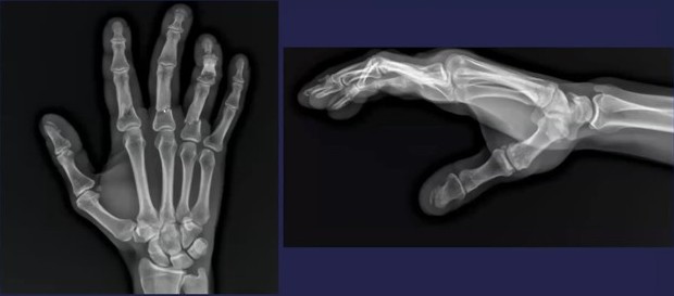
During March’s AJR Live webinar on the increasing (albeit varying) attrition of our nation’s rad workforce, AJR Section Editor for Policy, Quality, and Practice Management Jonathan L. Mezrich, MD, relayed the following point of order from the audience to presenter Eric W. Christensen, PhD:
“Increased corporatization of academic practices more focused on clinical productivity may also be contributing to academic attrition. The practices are no longer the place some may have signed up for.”
Crowded House: Many rads choose academic paths specifically for the opportunity to teach and conduct research. That’s baked into the gig. And as Dr. Christensen acknowledged, when clinical demands start supplanting such pursuits, the job can become significantly less appealing.
The Distinction: Dr. Christensen drew a clear line between the two distinct types of movement he’s seeing in the specialty:
Attrition—completely leaving the rad workforce
Turnover—switching from one practice to another
The Numbers:Additional AJR research from Christensen et al. would indicate that the pain threshold for academic rads might be lower than for those in non-academic practices.
At lower clinical workload levels, academic rads are more likely to turnover than their non-academic peers.
Higher work volumes often interfere with the primary reasons they entered academia in the first place: instruction and investigation.
Bottom Line: While the nature of the work is different, academic rads aren’t necessarily doing less work; rather, increasing clinical burdens are undermining their raison d’être and driving practice instability.
Lymphoma is the most common malignant tumor of the mesentery, occurring in 30% to 50% of patients with non-Hodgkin’s lymphoma. Because it often mimics sclerosing mesenteritis, identifying specific imaging clues is critical for an accurate diagnosis.
Bread & Butter Tip: The “sandwich sign“occurs when mesenteric masses involve both leaves of the mesentery while relatively preserving the central fat and vasculature. This creates a layered appearance, kinda resembling a sammy.
Key Differentiators for Lymphoma:
Lack of Calcification: Untreated lymphoma typically doesn’t calcify. If calcification is present within mesenteric masses, lymphoma can generally be eliminated from the differential.
Multifocal Disease: Lymphoma often presents with involvement beyond the mesentery, frequently extending into the retroperitoneum.
Node Size: A short-axis dimension of 10 mm or more is a major red flag that requires further workup.
💯 Percent:Recent research indicates that the presence of just one of these worrisome features, significant node size or extra-mesenteric lymphadenopathy, yields 100% sensitivity and 100% NPV for malignancy.
Bottom Line: Whereas lymphoma can share features with mesenteric panniculitis (e.g., relative mass effect, tumoral pseudocapsule delineation, the fat ring sign), the latter is dominated by fat necrosis and inflammation—rather than the bulky, “sandwiching” masses seen in malignant disease.
Primary sclerosing cholangitis (PSC) is a rare, chronic disease that causes inflammation and scarring of the bile ducts. When left untreated, PSC can lead to severe complications like cholangitis and hyperbilirubinemia, as seen in this 25-year-old male—lost to follow-up for a whopping 13 years!
Background: With hyperbilirubinemia way up in the 30s, the patient underwent endoscopic retrograde cholangiopancreatography (ERCP) with plastic stent placement. But as Christopher R. Bailey, MD, of Johns Hopkins Hospital presented during the ARRS Annual Meeting’s Vascular/Interventional Radiology Review, ERCP didn’t really intervene too terribly much.
A stark example of what PSC can look like for a patient out in the world untreated for more than a decade, take note of key cholangiographic features, including:
Multifocal strictures—Numerous short, irregular narrowings along intrahepatic and extrahepatic bile ducts.
Beaded appearance—Alternating pattern of strictures and dilations creates a “string of beads” look.
Pruning—Loss of smaller peripheral ducts; distal branches become fewer and less visible.
Irregular walls—Bile ducts often have rough, irregular contours (not smooth like normal ducts).
Dominant strictures (sometimes)—A single, especially tight narrowing in a major bile duct, often the common bile duct or hepatic ducts.
Bottom Line: Whereas ERCP with plastic stent remains common, it may not always be an effective intercession, especially in advanced cases. In this instance, percutaneous biliary tube placement proved to be a successful alternative to bridge the patient to liver transplant.
Left to right: ARRS Executive Director Dale West, CAE; American College of Radiology CEO Dr. Dana Smetherman; U.S. House Representative Suhas Subramanyam (VA-10th); Society of Breast ImagingSenior Director, Membership & MarCommKesha Willis, CNP, CAE; Dr. Vivek Yedavalli
The Academy for Radiology & Biomedical Imaging Research (the Academy) and your American Roentgen Ray Society (ARRS) joined forces with a dozen other leading radiological societies on Tuesday, March 24 to radvocate for the future of the specialty during this year’s Medical Imaging Research Hill Dayin Washington, D.C.
As ARRS Executive Council member and Membership Committee chair Darcy J. Wolfman, MD, noted, imaging is the “crux of medicine,” yet unpredictable federal funding makes long-term research planning nearly impossible. Dr. Wolfman and ARRS Executive Director Dale West, CAE, both contended that a “unified voice” remains essential to ensuring lawmakers understand that breakthroughs in imaging research improve patient lives and our health care ecosystem writ large.
The Big Picture: Rads of all practice types and at every level of training united with allied scientists for a multi-day radvocacy blitz designed to secure robust federal investment for fiscal year (FY) 2027.
Debate Prep: Medical Imaging Research Hill Day kicked off the day before with intensive radvocacy best practices at Le Méridien Hotel to prepare participants for high-stakes meetings.
Bicameral FaceTime: Radvocates then held face-to-face meetings with members from both chambers of the 119th Congress and their staff on Capitol Hill, sharing personal stories of research impact.
#MedTech26: Later, in the Rayburn Foyer, the Academy and partner societies hosted a hands-on technology showcase demonstrating cutting-edge advances like custom 3D-printed solutions, opportunistic cardiac screening, AI-driven stroke recovery, and even wearables for neuroradiology.
Dale West and ARRS Executive Council member Dr. Darcy Wolfman at the Sam Rayburn House Office Building
By The Numbers: The day’s radvocates presented three critical asks to lawmakers for FY 2027:
$51.3 billion for National Institutes of Health (NIH)
$1.7 billion for Advanced Research Projects Agency for Health
$500 million for National Institute of Biomedical Imaging and Bioengineering
The Academy’s radvocacy extended into Wednesday with a Research Roundtable, focused on two existential challenges for the field:
AI Integration: Preparing infrastructure and governance alike for AI-forward imaging.
Talent Pipeline: Developing strategies to support early-career investigators, who are all too often the most vulnerable to funding shifts.
For emerging researchers, especially, Medical Imaging Research Hill Day is more than mere lobbying; it is vital background work that safeguards the funding upon which they rely. Participation promises invaluable liaising with the Department of Health and Human Services, as well as agency introductions to NIH, Food and Drug Administration, and Centers for Disease Control and Prevention, while learning the political process that powers scientific discovery.
Left to right: Dr. Wolfman; GE Healthcare Senior Director, Political Advocacy & Government Affairs Betsy Tower; Dr. Clifford Weiss; Dr. Yedavalli
Presented during the ARRS Annual Meeting’s Musculoskeletal Radiology Review, this case utilizes CT (and an arboreal aid) to highlight a benign lipoma with cortical attachment, emphasizing key differentiators from aggressive lesions.
Imaging: Two views of an unusual surface lesion coming from the femur. Bone exostosis is evident. On the right, it doesn’t appear there’s any communication with the marrow space. Looking at soft tissues, you can make out a large, low-density mass—sort of draped below that little piece of bone coming off of the cortex.
CT shows the bone exostosis corresponds with this lesion coming from the surface of the cortex. It does *not* involve the medullary space; it doesn’t extend through the cortex. That low-density mass corresponds with this very fatty mass atop the bone.
Sure, there are a few septations, but no other suspicions.
DDX? This combination of a benign fatty mass attached to the underlying bone surface is a parosteal lipoma.
“Trunk”—The bony outgrowth emerging from the femoral cortex.
“Branches”—That large, low-density soft-tissue mass draped over the bone.
Bottom Line: Differentiating benign surface lesions from aggressive bone tumors is critical for avoiding unnecessary biopsies and patient anxiety. And if the “trunk” (i.e., bone) and “branches” (i.e., fat) are present sans marrow involvement, it’s likely just a lipoma.
This 32-year-old female patient was beaten by her boyfriend, using his fists, knees, and feet. Imaging showed a liver laceration, facial fractures and contusions—which are target injuries—as well as left AC joint separation and left elbow dislocation, which may have been sustained in defense.
For radiologists to flag intimate partner violence (IPV) in the clinical setting, the key lies in identifying specific patterns of physical injuries that increase the likelihood of abuse. By observing combinations of target and defensive injuries—especially when they lack a consistent trauma history—rads can play a frontline role in early detection.
As Ellen X. Sun, MD, from the Trauma Imaging Research Innovation Center at Brigham and Women’s Hospital/Harvard Medical School outlined during the ARRS Web Lecture Emergency Radiology III, synchronous injuries in multiple locations without a history of major trauma are giant red flags. Common combinations include:
Dr. Sun presented a case study of a 24-year-old female patient who sustained both blunt and penetrating trauma from a partner she met on a dating app.
Craniofacial: CT revealed multiple lacerations and facial fractures, including fracturing of the right orbital floor.
Defensive: Radiography revealed a right-sided fifth middle phalangeal fracture, characteristic of defensive positioning.
Chronic Injuries: Injuries of different ages—such as the acute periorbital hematoma alongside a chronic nasal bone fracture below—are highly suspicious for repeated cycles of abuse.
Recurrence: Chronic fractures of the extremities and nasal bones are significantly more frequent among victims of IPV, compared to matched controls.
Consistency: Suspicion increases when old and new injuries involve the same target or defensive side, such as the midface, hands, feet, or ankles.
Reporting Dilemma: Rads face the challenge of documenting suspicion without compromising patient safety, especially since aggressors may access federally mandated online portals. When IPV is suspected, the recommended workflow is to inform the referring physician to facilitate a private, caring conversation with the patient. If she or he confirms abuse, clinicians then need to follow institutional protocols; if patients deny it, they should still be pointed toward actionable resources.
Bottom Line: Rads can, indeed, make a difference by recognizing the high imaging use of IPV victims, injury location and imaging patterns specific to IPV, injuries inconsistent with the provided history, as well as old injuries that may be caught on physical exam.
Faced with a difficult-to-access lesion, Eric Aaltonen, MD, shows how IR ingenuity offers a safe, effective alternative to surgery for liver tumor ablation.
The Patient: During his Vascular/Interventional Case Review from the ARRS Annual Meeting, Dr. Aaltonen introduced us to a 68-year-old male with HCC, post-TACE x2, who presented with a new segment 7 lesion at the hepatic dome.
The Question: Given the obstructions, which approach would work best for microwave ablation (MWA)?
A. Bone
B. Bone
C. Lung
D. Heart (and cartilage)
…E. Call surgery?
Honestly, none of the above are particularly great. Bone is going to be hard, of course. No one wants to drill through the rib. You probably don’t want to go through the heart with an ablation probe either. You’ve got cartilage there, too.
The Solution: Instead of going through the lung—or, worse yet, calling surgery!—Aaltonen et al. opted to drop it. And by intentionally inducing a pneumothorax, they created a safe path to the lesion.
Even Better? After positioning the probe, performing MWA, reinflating the lung (and making sure it stayed up), you can see that they got their lesion…
12-year, 6-month–old boy with second episode of acute right scrotal pain within 4 days. Sagittal color Doppler image shows spermatic cord twisting (thin arrow) into whirlpool sign. Pseudomass (thick arrow) is present just below twisted cord.
For a patient in acute scrotal pain, the clock really is ticking. Differentiating between a surgical emergency, à la testicular torsion, and a medically managed condition is critical, as salvage rates for the testis drop from nearly 100% within 6 hours to a mere 20% just 12 hours on.
Due to so much discomfort, alas, physical exams provide precious few details. Thus, as Shweta Bhatt, MD, of Mayo Clinic Jacksonville illustrated during the most recent session of ARRS’ GU Longitudinal Course, color Doppler US becomes your gold standard.
Pool Side: One of the most reliable indicators of torsion is the whirlpool sign, which represents the spiral twisting of the spermatic cord. Finding this sign above the testis (indicating at least a 360-degree twist) has been reported in the literature to have as much as 100% sensitivity and specificity for torsion.
Complete—Typically presents with absent or significantly reduced blood flow in the affected testicle, compared to the contralateral side.
Intermittent—Defined as sudden unilateral pain that resolves spontaneously, this one is notoriously difficult to diagnose because blood flow may appear normal, or even increased, during examination.
Beware the Mimics: A common pitfall in scrotal imaging is misdiagnosing intermittent torsion as epididymitis. Following spontaneous detorsion, the ischemic tissue may respond with vasodilation, leading to hyperemia that mimics the appearance of infection. Additionally, twisting of the cord can create a heterogeneous “pseudomass” below the twist, composed of the congested epididymis and vascular bundle. Without careful evaluation of the cord’s proximal spiral twist, this can easily be mistaken for an inflamed epididymis.
Check the Cord: Always perform longitudinal and transverse imaging of the spermatic cord vasculature—not just the testis, itself.
“Boggy” Cord? In cases of intermittent torsion where a whirlpool isn’t obvious, an abnormal or thickened cord is a significant finding that supports the diagnosis.
Differentiate by Resistance: Whereas epididymitis shows high flow and a low resistive index, torsion often presents with high resistive flow or absent diastolic flow.
Another 12-year-old boy with partial torsion of left testis. Sonograms with color Doppler flow of left spermatic cord reveal “torsion knot” and whirled pattern of its accompanying vessels (arrows) in transverse (left) and sagittal (right) planes.
Bottom Line: Even if flow seems normal today, repeat episodes of scrotal pain can’t be dismissed. A careful search for the whirlpool sign or an extratesticular pseudomass can be the key to saving a testis from future infarction.
The math for pancreatic cancer screening has always been a tough pill to swallow. With an annual incidence of just 13 per 100,000 in the general population, traditional screening tests simply can’t function effectively at such a low prevalence. Fortunately, a new strategy is emerging: risk enrichment.
During the AJR Forum on Opportunistic Screening, Michael Rosenthal, MD, PhD, of Dana-Farber Cancer Institute outlined how we can utilize AI to flip the script on these stats. Instead of searching for a needle in a haystack, first, let AI shrink the haystack.
High-Risk Filter: The goal? Move the screening population from a risk of 13 per 100,000 more toward a risk profile of 0.5 to 1 per 100—a hundred-fold intensification. This mirrors the risk levels seen in known familial and genetic cohorts, where screening is already proven to save lives. Dr. Rosenthal describes a multi-layered “filter” approach:
Top Layer (low cost/low risk)—using both EMR and opportunistic imaging analysis to filter out low-risk individuals at the average-risk pool level
Deepening the Filter—as the pool narrows, clinicians can move toward more intensive and specific tools
Targeted Surveillance—final groups identified then receive direct surveillance: blood testing, stool testing, active imaging
AI Advantage: AI thrives in this “top-of-the-funnel” environment. And by analyzing vast amounts of data from existing records and imaging, it can provide the insight needed to identify high-risk subgroups…minus the cost and invasiveness of primary screening.
Bottom Line: In harnessing AI to identify these high-intensity risk groups within gen pop, finally, we’ll make active pancreatic cancer screening a clinical reality.
It’s the age-old CXR dilemma: “I don’t want to go to CT unless I have to!” But when a potential finding appears, how do chest rads decide if it’s really real or simply a summation of shadows?
Here, we do see a nodule in the right lung. And it certainly looks discreet. Could it be an abnormality within the overlapping rib? Perhaps. Could it be something on the patient’s skin? Maybe, but probably not.
According to Travis S. Henry, MD, professor of radiology and chief of cardiothoracic imaging at Duke, his approach to focal lung disease is decidedly inquisitive.
Is there volume loss?
Is there lymphadenopathy?
Is the finding chronic/recurrent?
The first line of questioning here, though? “Is there anything else?”
Finding associated abnormalities can turn a “maybe” into a “must-scan,” and in this case from Dr. Henry’s ARRS Web Lecture, a closer look reveals a huge subcarinal lymph node.
Clinical Context: The diagnostic weight of these findings increases significantly when combined with the patient’s history. This individual had a remote history of epithelioid angiosarcoma. While there was no known metastatic disease at the time, the combination of a new nodule and significant lymphadenopathy is highly concerning.
CT Correlation: Indeed, follow-up CT confirmed what the radiograph found:
The presence of that right lung nodule.
The massive subcarinal lymph node is clearly visible, showing exactly why the azygoesophageal interface was obscured.
Bottom Line: Rads mustn’t look at any nodule in isolation. Always search for secondary signs or associated findings to guide your next step.
When evaluating an intramedullary spinal cord lesion in a pediatric patient, your first instinct may be to consider ependymoma. In adults, that reflex is often correct; in children, the picture is more nuanced.
As Neil Lall, MD, noted during the ARRS Annual Meeting Radiology Case Review, ependymomas are the most common intramedullary spinal tumors overall, but they rank third in frequency in peds patients, making them less common in this age group than many trainees expect.
MRI Clues: In Dr. Lall’s case, the lesion demonstrates several classic characteristics:
Expansile intramedullary mass
Heterogeneously T2 hyperintense signal
Enhancing solid components
Well-demarcated margins
Short-segment spinal cord involvement
These features support the diagnosis of spinal ependymoma. The well-circumscribed margins are particularly helpful, reflecting the tumor’s relatively defined growth pattern compared with more infiltrative pediatric spinal tumors.
Peds Context: While ependymomas do occur in children, their presence should prompt consideration of an underlying genetic syndrome. One of the strongest associations is neurofibromatosis type 2 (NF2). Need a mnemonic? MISME syndrome captures the hallmark tumors associated with the disorder…
M—Multiple
I—Inherited
S—Schwannomas
M—Meningiomas
E—Ependymomas
Diagnostic Focus: In this case, attention should remain on the cord lesion, rather than incidental degenerative changes. Distraction via unrelated findings (e.g., disc degeneration in an adolescent) can lead to misses, if the primary abnormality is not carefully evaluated.
Clinical Takeaway: Spinal ependymomas in children are uncommon but important to recognize. When an intramedullary mass appears expansile, well-defined, and enhancing, ependymoma should remain in the differential—and the possibility of NF2 should be considered.
Bottom Line: In pediatric spinal imaging, recognizing the pattern is only the first step. Understanding the syndromic context can be just as important as identifying the tumor, itself.
Well over a decade since ACR’s last lexical update, BI-RADS® v2025 Manual—née Atlas—truly reflects the tech, integrating the nomenclature of digital breast tomosynthesis (DBT), automated breast ultrasound (US), and contrast-enhanced mammography (CEM). Sure, adopting this latest manual can feel like “learning a new language,” but one of v2025’s standout features is modality-neutral terminology.
Informal, food-based descriptors for mammographic calcifications have retired, too. Out: “popcorn-like” and “milk of calcium.” In: precise descriptors, such as coarse and layering. Across all modalities, mass shapes are standardized: oval, lobulated (which has been reintroduced!), round, irregular. The mammographic descriptor “microlobulated” has been removed to prevent confusion with the shape descriptor “lobulated.” It’s replaced with indistinct.
Presentation—A screening mammogram revealed subtle calcifications in the lateral aspect of the breast (yellow box). After magnification and stereotactic needle core biopsy, the pathology returned as atypical ductal hyperplasia (ADH), suspicious for ductal carcinoma in situ (DCIS).
Management Dilemma—Typically, surgical consultation and excision are recommended for this pathology. However, in this case, patient and surgeon alike opted for surveillance, instead of excision.
Reporting the Follow-Up—When a patient with an unresected high-risk lesion returns for surveillance (e.g., 6 months to 1 year later), your goal is active monitoring. Step 1: identify the titanium biopsy marker (yellow box) . . .
Step 2? Check for changes. Specifically, you’re looking for residual calcifications or any new areas of calcification.
II vs. IV—If the biopsy site and any residual calcifications remain stable over a designated period (as defined by your practice), you can safely assign a BI-RADS 2. Post-biopsy changes from a vacuum-assisted device are acceptable here, as long as you are comfortable with their stability.
If you see more calcifications on follow-up, you must assign a BI-RADS 4. This requires either repeating the needle biopsy or sending the patient back for a surgical excision consultation.
Cool To Be Rind: It’s worth noting that BI-RADS v2025 terminology now aligns more closely with contrast-based modalities via descriptors for nonmass lesions. Additionally, “echogenic rind”—a feature long known to be associated with malignancy—is officially included, with specific guidance for inclusion in your lesion measurements.
MRI & CEM Tweaks:
No “focus”for MRI; these findings should now be categorized as either small masses or background parenchymal enhancement (BPE).
“Multiple regions” is similarly removed for nonmass lesions, also better described as BPE.
Give the rise of ultrafast MRI, “early phase” replaces “initial phase.”
A new descriptor for T2-hyperintensity has been added as well.
Staging & Reporting:
BI-RADS 6 Extent-of-Disease: This Cat 6 definition allows for additional “close findings” (within 2 cm of a known malignancy) to be included if they would not change patient management.
Method of Detection: You are highly encouraged to document MOD to help track outcomes, demonstrate the benefits of early detection, and improve communication.
Temporal Shift: Finally, in a move toward purely morphologic assessment, descriptors that explicitly incorporate temporal change (i.e., “developing asymmetry”) have been removed. In AJR, Drs. Seely and Bissell warnthat this risks underemphasizing the diagnostic importance of temporal changes, so be sure to acknowledge such in your reports for appropriate management.