Author: Logan Young

  • Contrast-Enhanced Mammography–Guided Intervention—What Next?

    Contrast-Enhanced Mammography–Guided Intervention—What Next?

    83-year-old woman with invasive lobular carcinoma in left breast and enhancing right breast mass. No mammographic or ultrasonic correlate to right breast. Contrast-enhanced mammography–guided intervention localization per surgeon’s request.

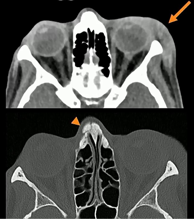
    At some point of inflection, every new imaging modality will require its own image-guided biopsy capabilities. Why? Because as Haydee Ojeda-Fournier, MD, of UC San Diego Health reminded us in her ARRS Quick Byte video, suspicious findings may be occult on all other systems.

    Yes, and … Contrast-enhanced mammography (CEM) is transitioning from diagnostic instrument to a functional intervention platform—addressing Dr. Ojeda-Fournier’s “what next?” query for findings seen only on recombined images.

    Percentage Points:

    • 62% of CEM-detected lesions can be identified during a second look at digital breast tomosynthesis (DBT).
    • 76% of CEM findings have an ultrasound correlate; these correlates are more likely to be malignant.
    • ~60% of MRI-guided biopsies could potentially be performed via CEM, providing a faster, lower-cost alternative for impacted MR units.

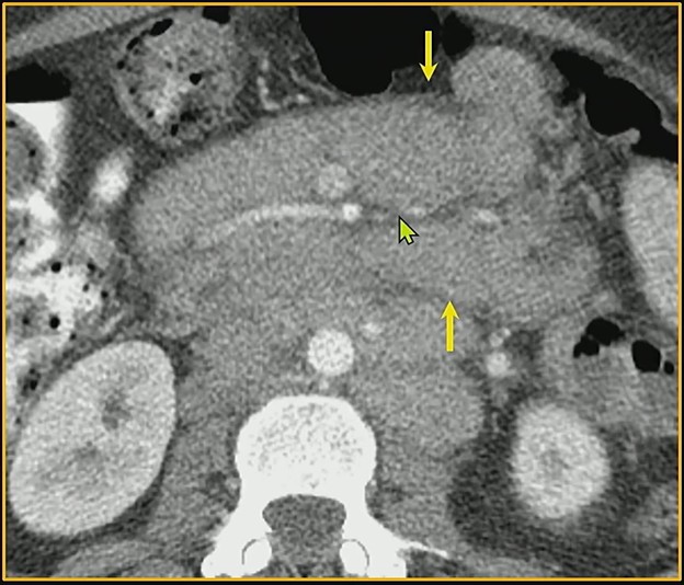
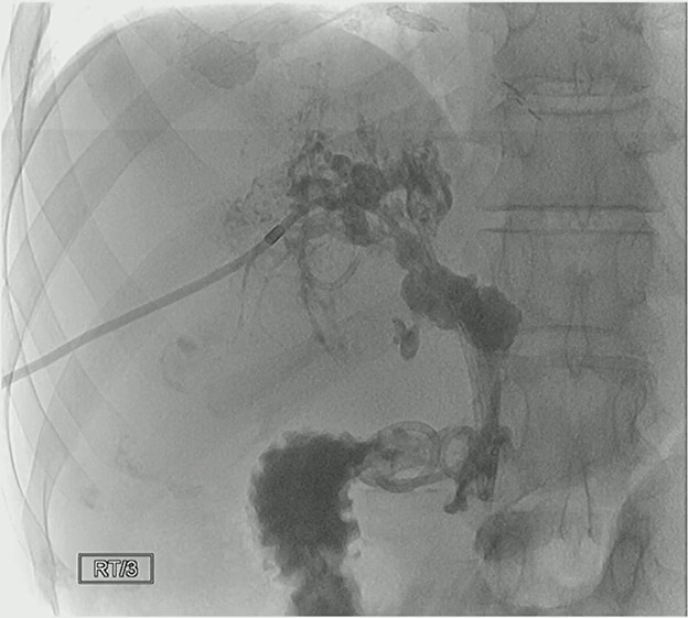
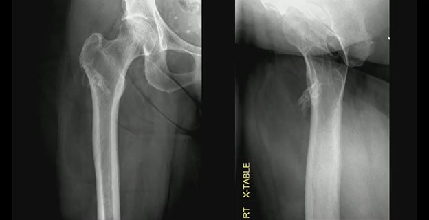
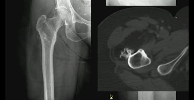
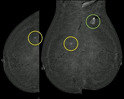
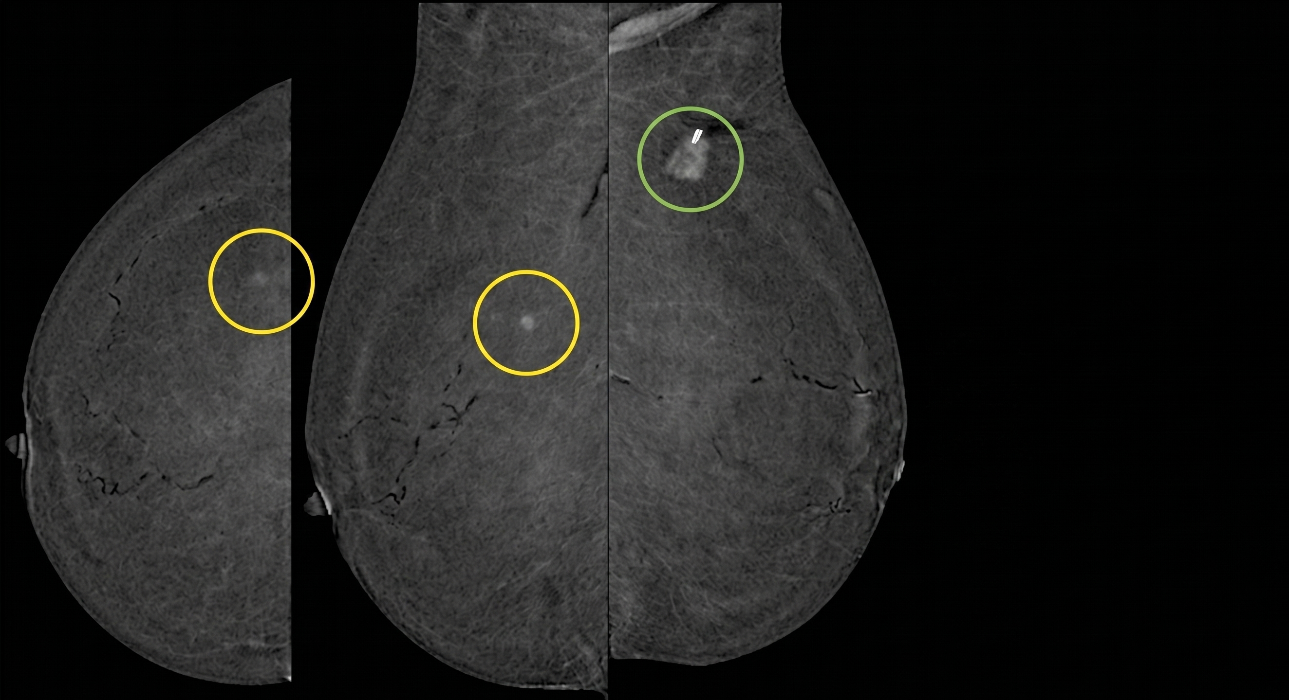
    Case in Point: CEM-guided intervention remains a game-changer for patients who cannot tolerate MRI. For example, in the 83YO patient above with invasive lobular carcinoma who was not an MRI candidate, CEM-guided localization successfully targeted a contralateral mass that was mammographically and sonographically occult.

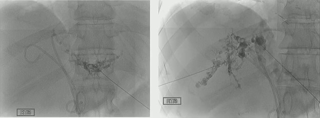
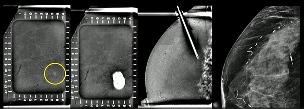
    In the projection below, you’ll notice some artifact from the needle. And after Dr. Ojeda-Fournier’s team deployed their wireless localizing device (based on geography), pathology came back benign. She’ll need a follow up, regardless.

    Final Points of Order:

    • Search in Teams: Using a combined scout (CEM+DBT) provides the most flexibility for choosing the optimal guidance modality.
    • Needle Up! While both horizontal and vertical approaches are possible, typically, the vertical needle approach is preferred because it samples a larger tissue volume in the z-axis and is less reliant on precise depth estimation.
    • Singular Sensation: A successful biopsy is defined as obtaining tissue within one procedure under a single compression, whether guided by recombined CEM, DBT, or stereotactic techniques.
  • Tears of the Pancreas: Why Cyst Stability Isn’t Enough

    Tears of the Pancreas: Why Cyst Stability Isn’t Enough

    More than isolated findings, pancreatic cystic lesions can be a cry for help from the entire organ. In fact, the field defect suggests that the presence of a cyst indicates an increased risk of developing pancreatic ductal adenocarcinoma elsewhere in the gland.

    Look Around You! Rads frequently focus on the stability of a cystic lesion, but malignancy can arise in the surrounding pancreatic parenchyma—even when the cyst, itself, remains unchanged for months, even years.

    • Crying Metaphor: In his ARRS Quick Byte video, Atif Zaheer, MD, of Johns Hopkins dubs these lesions “tears of the pancreas,” warning of synchronous or metachronous cancer development.
    • Surveillance Trap: Whereas some guidelines suggest stopping surveillance for stable cysts after 5 years, Dr. Zaheer’s research indicates a continued risk of malignancy beyond the 5–10 year mark.
    • What’s the Protocol Here? Abbreviated MRI protocols designed for cyst follow-up must include sequences that specifically evaluate the parenchyma to avoid missing cancers arising in the uncinate process of the pancreatic head.

    Under New Management: Managing pancreatic cysts is shifting from a disease of technology toward a more personalized, multi-faceted approach, including:

    • Beyond Imaging: Effective risk stratification now involves clinical data, biochemical markers, and cyst fluid DNA sequencing for mutations in genes like PIK3CA and TP53.
    • AI Edge: Emerging deep-learning algorithms and radiomics are helping to create automated risk-prediction models to differentiate high-risk lesions from benign ones.
    • Patient Empowerment: With the 2020 Cures Act, patients have immediate access to their reports, increasing the need for structured, recommendation-based reporting that tracks imaging features over time.

    Bottom Line: Don’t just look at the cyst. Evaluating the health of the entire pancreatic gland is critical to detecting early invasive cancer that the field defect may be denying.

  • True vs. False Lumen in Acute Aortic Dissection

    True vs. False Lumen in Acute Aortic Dissection

    In matters of the heart, truth matters all the more. And identifying the real lumen remains critical for planning endovascular repairs, ensuring branch vessels are correctly supplied to prevent end-organ ischemia. Sure, continuity with the normal aorta remains the gold standard for sussing things out. But as Ferco Berger, MD, pinpointed during the ARRS Online Course “Imaging in the Emergency Department,” tracing it can be difficult in cases involving the aortic root or when only limited abdominal scans are available.

    False Witnesses:

    • Beak Sign: Acute angle formed where dissection flap meets outer wall, it’s the most useful and reliable indicator (present in nearly 100% of acute and chronic cases).
    • Larger Size: A false lumen typically distends and has a larger cross-sectional area than the real deal.
    • Cobweb Sign: Thin, linear defects representing remnants of media layer. Albeit rare, they are highly specific to false lumen.

    Truth Tellers:

    • Calcification: Generally, hardening along outer wall or eccentric flap is indicative of true lumen. [N.B. In chronic cases, false lumen can occasionally calcify.]
    • Wraparound: In transverse aortic arch, if one lumen appears to wrap around another, that inner lumen is invariably the truer one.
    • Density: In arterial phase, true lumen frequently show higher contrast density.

    Supply Chain: Differentiating the supply side here helps predict which organs are at risk:

    • True Lumen: Usually supplies celiac trunk, superior mesenteric artery, right renal artery.
    • False Lumen: Often supplies left renal artery and more prone to thrombus formation.

    Bottom Line: When continuity is unclear, look for the beak sign and larger caliber to identify the false lumen; double-check the arch wraparound and calcification patterns to confirm the true lumen.

  • The Buford Complex—Anatomy or Abnormality?

    The Buford Complex—Anatomy or Abnormality?

    Occurring in less than 2% of your shoulder patients, apropos, there are just two things you need to ID the Buford complex. As Robert J. French, Jr., MD, enumerated for the ARRS Online Course “Expanding the Field: Imaging of Sports-Related Injuries Beyond the Knee,” this normal anatomic variation is defined by an absent anterior superior labrum and a thickened middle glenohumeral ligament (MGHL).

    The Details:

    • Location: Labrum is missing in the 1- to 3-o’clock position (right shoulder) or 9- to 11-o’clock (left shoulder).
    • Reconstitution: Typically, labrum reappears at or below glenoid midpoint.
    • MGHL: Hypertrophied, it often appears as a discrete structure—similar in size to long head of biceps tendon.
    • Stability: Unlike pathologic lesions, biceps anchor and contiguous superior and anterior inferior labrum remain normal.

    Closed for Repairs: Mistaking this variation for a labral avulsion is a common pitfall. In fact, in one surgical error documented in AJR, a cordlike MGHL was unfortunately attached to the glenoid as a “fix,” resulting in severely restricted shoulder motion for the patient.

    The Planes:

    • Axial: Look for absence of low-signal-intensity labral tissue along anterior superior glenoid rim.
    • Sagittal: MGHL can be seen as a linear structure separate from the subscapularis tendon, inserting into biceps anchor.

    Bottom Line: If the superior labrum and biceps anchor are intact, yet the anterior superior labrum is missing alongside a thickened MGHL, think Buford complex—not a labral tear.

  • Decoding Dementia: The Lewy Body Problem

    Decoding Dementia: The Lewy Body Problem

    Dementia with Lewy bodies (DLB) mimics Alzheimer’s disease, indeed. And as RadNet neuroradiologist Suzie Bash, MD, discussed during the ARRS Online Course “Opportunities and Obligations in Imaging Patients with Alzheimer’s Disease,” specific multimodal patterns—occipital lobe involvement, particularly—can help hone your DDX.

    Big Pics: 73 y/o male presenting with visual hallucinations, resting tremors, and frequent falls (but only minimal memory loss) illustrates the clinical profile of classic DLB. Those bodies are defined by alpha-synuclein, sure, but imaging often reveals a much more mixed bag:

    • MRI: Showed atrophy in the hippocampi, as well as occipital and parietal lobes.
    • Amyloid PET: Positive, confirming beta-amyloid plaques can be present in DLB cases.
    • Tau PET: Revealed deposition in posterior cingulate gyrus and occipital/parietal lobes.
    • FDG PET: Confirmed hypometabolism in same posterior regions, a hallmark of the disease.

    Bigger Numbers:

    • 1 million: Americans living with DLB, compared to 6.7 million with Alzheimer’s.
    • 80%: Frequency of visual hallucinations as a presenting symptom in DLB.
    • 5–8 years: Typical survival rate following diagnosis.

    Bottom Line: Rads should pay closer attention to the occipital lobe. Whereas Alzheimer’s and DLB share temporal and parietal overlaps, the addition of occipital atrophy and hypometabolism is a strong indicator of Lewy body pathology.

  • What Lies Between—Navigating the Osseous and Membranous Labyrinth

    What Lies Between—Navigating the Osseous and Membranous Labyrinth

    No matter what happens here, suffice it to say that it almost always begins in the scala chambers. Whether identifying pathology or planning for surgery, as Amy Juliano, MD, of Massachusetts Eye and Ear Infirmary and Harvard Medical School delineated in her ARRS Quick Byte, rads need to lend their own ear toward the scala tympani.

    Three Examples: Understanding the compartmental anatomy of the osseous and membranous labyrinth allows rads to differentiate between tumors, inflammatory changes, and surgical targets.

    • Cochlear schwannoma: More often than not, these space-occupying, enhancing masses originate within the scala tympani. Contralateral comparison is key for detection.
    • Labyrinthitis ossificans: This pathological calcification of the lumen often follows meningitis (frequently bilateral), trauma/fracture, or prior surgery. It predominantly affects the scala tympani. Working theory? White blood cell recruitment from specific vasculature predisposes this chamber to ossification.
    • Cochlear implant: The surgical goal is to thread the electrode array into the scala tympani. Placing the implant in the scala vestibuli is a no-no.

    Bottom Line: Look first and frequently at the scala tympani, as that’s where clinical and pathological significance alike most likely lies.

  • What Is Academic Radiology’s Pain Threshold?

    What Is Academic Radiology’s Pain Threshold?

    During March’s AJR Live webinar on the increasing (albeit varying) attrition of our nation’s rad workforce, AJR Section Editor for Policy, Quality, and Practice Management Jonathan L. Mezrich, MD, relayed the following point of order from the audience to presenter Eric W. Christensen, PhD:

    “Increased corporatization of academic practices more focused on clinical productivity may also be contributing to academic attrition. The practices are no longer the place some may have signed up for.”

    Crowded House: Many rads choose academic paths specifically for the opportunity to teach and conduct research. That’s baked into the gig. And as Dr. Christensen acknowledged, when clinical demands start supplanting such pursuits, the job can become significantly less appealing.

    The Distinction: Dr. Christensen drew a clear line between the two distinct types of movement he’s seeing in the specialty:

    • Attrition—completely leaving the rad workforce
    • Turnover—switching from one practice to another

    The Numbers: Additional AJR research from Christensen et al. would indicate that the pain threshold for academic rads might be lower than for those in non-academic practices.

    • At lower clinical workload levels, academic rads are more likely to turnover than their non-academic peers.
    • Higher work volumes often interfere with the primary reasons they entered academia in the first place: instruction and investigation.

    Bottom Line: While the nature of the work is different, academic rads aren’t necessarily doing less work; rather, increasing clinical burdens are undermining their raison d’être and driving practice instability.

  • Spot the Sandwich: Refining Your Mesenteric Differential

    Spot the Sandwich: Refining Your Mesenteric Differential

    Lymphoma is the most common malignant tumor of the mesentery, occurring in 30% to 50% of patients with non-Hodgkin’s lymphoma. Because it often mimics sclerosing mesenteritis, identifying specific imaging clues is critical for an accurate diagnosis.

    Bread & Butter Tip: The “sandwich sign“occurs when mesenteric masses involve both leaves of the mesentery while relatively preserving the central fat and vasculature. This creates a layered appearance, kinda resembling a sammy.

    Key Differentiators for Lymphoma:

    • Lack of Calcification: Untreated lymphoma typically doesn’t calcify. If calcification is present within mesenteric masses, lymphoma can generally be eliminated from the differential.
    • Multifocal Disease: Lymphoma often presents with involvement beyond the mesentery, frequently extending into the retroperitoneum.
    • Node Size: A short-axis dimension of 10 mm or more is a major red flag that requires further workup.

    💯 Percent: Recent research indicates that the presence of just one of these worrisome features, significant node size or extra-mesenteric lymphadenopathy, yields 100% sensitivity and 100% NPV for malignancy.

    Bottom Line: Whereas lymphoma can share features with mesenteric panniculitis (e.g., relative mass effect, tumoral pseudocapsule delineation, the fat ring sign), the latter is dominated by fat necrosis and inflammation—rather than the bulky, “sandwiching” masses seen in malignant disease.

  • The Glaring Reality of Untreated Primary Sclerosing Cholangitis

    The Glaring Reality of Untreated Primary Sclerosing Cholangitis

    Primary sclerosing cholangitis (PSC) is a rare, chronic disease that causes inflammation and scarring of the bile ducts. When left untreated, PSC can lead to severe complications like cholangitis and hyperbilirubinemia, as seen in this 25-year-old male—lost to follow-up for a whopping 13 years!

    Background: With hyperbilirubinemia way up in the 30s, the patient underwent endoscopic retrograde cholangiopancreatography (ERCP) with plastic stent placement. But as Christopher R. Bailey, MD, of Johns Hopkins Hospital presented during the ARRS Annual Meeting’s Vascular/Interventional Radiology Review, ERCP didn’t really intervene too terribly much.

    A stark example of what PSC can look like for a patient out in the world untreated for more than a decade, take note of key cholangiographic features, including:

    • Multifocal strictures—Numerous short, irregular narrowings along intrahepatic and extrahepatic bile ducts.
    • Beaded appearance—Alternating pattern of strictures and dilations creates a “string of beads” look.
    • Pruning—Loss of smaller peripheral ducts; distal branches become fewer and less visible.
    • Irregular walls—Bile ducts often have rough, irregular contours (not smooth like normal ducts).
    • Dominant strictures (sometimes)—A single, especially tight narrowing in a major bile duct, often the common bile duct or hepatic ducts.

    Bottom Line: Whereas ERCP with plastic stent remains common, it may not always be an effective intercession, especially in advanced cases. In this instance, percutaneous biliary tube placement proved to be a successful alternative to bridge the patient to liver transplant.

  • Safeguarding the Imaging Research Pipeline

    Safeguarding the Imaging Research Pipeline

    Left to right: ARRS Executive Director Dale West, CAE; American College of Radiology CEO Dr. Dana Smetherman; U.S. House Representative Suhas Subramanyam (VA-10th); Society of Breast Imaging Senior Director, Membership & MarComm Kesha Willis, CNP, CAE; Dr. Vivek Yedavalli

    The Academy for Radiology & Biomedical Imaging Research (the Academy) and your American Roentgen Ray Society (ARRS) joined forces with a dozen other leading radiological societies on Tuesday, March 24 to radvocate for the future of the specialty during this year’s Medical Imaging Research Hill Day in Washington, D.C.

    As ARRS Executive Council member and Membership Committee chair Darcy J. Wolfman, MD, noted, imaging is the “crux of medicine,” yet unpredictable federal funding makes long-term research planning nearly impossible. Dr. Wolfman and ARRS Executive Director Dale West, CAE, both contended that a “unified voice” remains essential to ensuring lawmakers understand that breakthroughs in imaging research improve patient lives and our health care ecosystem writ large.

    The Big Picture: Rads of all practice types and at every level of training united with allied scientists for a multi-day radvocacy blitz designed to secure robust federal investment for fiscal year (FY) 2027.

    • Debate Prep: Medical Imaging Research Hill Day kicked off the day before with intensive radvocacy best practices at Le Méridien Hotel to prepare participants for high-stakes meetings.
    • Bicameral FaceTime: Radvocates then held face-to-face meetings with members from both chambers of the 119th Congress and their staff on Capitol Hill, sharing personal stories of research impact.
    • #MedTech26: Later, in the Rayburn Foyer, the Academy and partner societies hosted a hands-on technology showcase demonstrating cutting-edge advances like custom 3D-printed solutions, opportunistic cardiac screening, AI-driven stroke recovery, and even wearables for neuroradiology.
    Dale West and ARRS Executive Council member Dr. Darcy Wolfman at the Sam Rayburn House Office Building

    By The Numbers: The day’s radvocates presented three critical asks to lawmakers for FY 2027:

    • $51.3 billion for National Institutes of Health (NIH)
    • $1.7 billion for Advanced Research Projects Agency for Health
    • $500 million for National Institute of Biomedical Imaging and Bioengineering

    The Academy’s radvocacy extended into Wednesday with a Research Roundtable, focused on two existential challenges for the field:

    • AI Integration: Preparing infrastructure and governance alike for AI-forward imaging.
    • Talent Pipeline: Developing strategies to support early-career investigators, who are all too often the most vulnerable to funding shifts.

    For emerging researchers, especially, Medical Imaging Research Hill Day is more than mere lobbying; it is vital background work that safeguards the funding upon which they rely. Participation promises invaluable liaising with the Department of Health and Human Services, as well as agency introductions to NIH, Food and Drug Administration, and Centers for Disease Control and Prevention, while learning the political process that powers scientific discovery.

    Left to right: Dr. Wolfman; GE Healthcare Senior Director, Political Advocacy & Government Affairs Betsy Tower; Dr. Clifford Weiss; Dr. Yedavalli
  • When Bone Grows Branches—Interpreting the Tree Sign

    When Bone Grows Branches—Interpreting the Tree Sign

    Presented during the ARRS Annual Meeting’s Musculoskeletal Radiology Review, this case utilizes CT (and an arboreal aid) to highlight a benign lipoma with cortical attachment, emphasizing key differentiators from aggressive lesions.

    Imaging: Two views of an unusual surface lesion coming from the femur. Bone exostosis is evident. On the right, it doesn’t appear there’s any communication with the marrow space. Looking at soft tissues, you can make out a large, low-density mass—sort of draped below that little piece of bone coming off of the cortex.

    CT shows the bone exostosis corresponds with this lesion coming from the surface of the cortex. It does *not* involve the medullary space; it doesn’t extend through the cortex. That low-density mass corresponds with this very fatty mass atop the bone.

    Sure, there are a few septations, but no other suspicions.

    DDX? This combination of a benign fatty mass attached to the underlying bone surface is a parosteal lipoma.

    • “Trunk”—The bony outgrowth emerging from the femoral cortex.
    • “Branches”—That large, low-density soft-tissue mass draped over the bone.

    Bottom Line: Differentiating benign surface lesions from aggressive bone tumors is critical for avoiding unnecessary biopsies and patient anxiety. And if the “trunk” (i.e., bone) and “branches” (i.e., fat) are present sans marrow involvement, it’s likely just a lipoma.

  • Radiologic Red Flags: Combinations of Intimate Partner Violence

    Radiologic Red Flags: Combinations of Intimate Partner Violence

    This 32-year-old female patient was beaten by her boyfriend, using his fists, knees, and feet. Imaging showed a liver laceration, facial fractures and contusions—which are target injuries—as well as left AC joint separation and left elbow dislocation, which may have been sustained in defense.

    For radiologists to flag intimate partner violence (IPV) in the clinical setting, the key lies in identifying specific patterns of physical injuries that increase the likelihood of abuse. By observing combinations of target and defensive injuries—especially when they lack a consistent trauma history—rads can play a frontline role in early detection.

    As Ellen X. Sun, MD, from the Trauma Imaging Research Innovation Center at Brigham and Women’s Hospital/Harvard Medical School outlined during the ARRS Web Lecture Emergency Radiology III, synchronous injuries in multiple locations without a history of major trauma are giant red flags. Common combinations include:

    • Defensive Upper Extremity (UE) + Target Craniofacial Injuries
    • Defensive UE + Target Abdominal Injuries

    Dr. Sun presented a case study of a 24-year-old female patient who sustained both blunt and penetrating trauma from a partner she met on a dating app.

    • Craniofacial: CT revealed multiple lacerations and facial fractures, including fracturing of the right orbital floor.
    • Defensive: Radiography revealed a right-sided fifth middle phalangeal fracture, characteristic of defensive positioning.

    Chronic Injuries: Injuries of different ages—such as the acute periorbital hematoma alongside a chronic nasal bone fracture below—are highly suspicious for repeated cycles of abuse.

    • Recurrence: Chronic fractures of the extremities and nasal bones are significantly more frequent among victims of IPV, compared to matched controls.
    • Consistency: Suspicion increases when old and new injuries involve the same target or defensive side, such as the midface, hands, feet, or ankles.

    Reporting Dilemma: Rads face the challenge of documenting suspicion without compromising patient safety, especially since aggressors may access federally mandated online portals. When IPV is suspected, the recommended workflow is to inform the referring physician to facilitate a private, caring conversation with the patient. If she or he confirms abuse, clinicians then need to follow institutional protocols; if patients deny it, they should still be pointed toward actionable resources.

    Bottom Line: Rads can, indeed, make a difference by recognizing the high imaging use of IPV victims, injury location and imaging patterns specific to IPV, injuries inconsistent with the provided history, as well as old injuries that may be caught on physical exam.