Category: Latest Posts

  • Midgut Malrotation and Volvulus in Children: US or Upper GI?

    Midgut Malrotation and Volvulus in Children: US or Upper GI?

    In smaller hospitals, choosing between ultrasound or upper GI for suspected malrotation can feel high-stakes. During the latest AJR Live Webinar, Jonathan Dillman, MD, and HayThuy Nguyen, MD, tackled a key question:

    Should community hospitals perform both ultrasound and upper GI—or is one enough before transferring a child?

    The Big Picture

    Even though ultrasound for midgut volvulus performs incredibly well in published studies, your institutional comfort and consistency matter. Smaller centers may see fewer neonates, and that affects how confident technologists and radiologists feel with real-time sonographic anatomy.

    Dr. Nguyen’s advice: Start with both. Build confidence. Then taper.

    Key Takeaways

    Ultrasound + Upper GI Can Be Complementary, At First

    • Even at large centers, both exams are often paired early on.
    • Not because ultrasound underperforms, but because every institution needs their own “local data.”
    • Once your team demonstrates consistently accurate ultrasound performance, you can safely drop routine upper GI.

    See a Whirlpool Sign? Call Surgery!

    A positive whirlpool sign on ultrasound is highly specific. No need to wait for additional imaging; direct referral to pediatric surgery is appropriate.

    No Whirlpool, but Symptoms Persist → Consider Upper GI or Transfer

    Ultrasound may still be equivocal in some infants. Upper GI remains a helpful confirmatory test when the diagnosis is uncertain but suspicion stays high.

    Don’t Forget: CT or MRI Can Make the Diagnosis for Older Kids

    Dr. Dillman emphasized that adolescents or older children being scanned for unrelated reasons may still show:

    • SMV/SMA reversal
    • Swirling mesentery
    • Engorged mesenteric vessels
    • Collaterals

    Even a noncontrast CT for renal stone can incidentally reveal the vascular swirl. MRI—especially rapid MR protocols—can also depict abnormal vascular orientation.


    Bottom Line

    Start with both ultrasound and upper GI, if your institution needs to build confidence. Once your team demonstrates reliable ultrasound performance, ultrasound alone is often sufficient…and a positive whirlpool sign should trigger immediate surgical evaluation.

  • Scimitar Syndrome with Horseshoe Lung: Key Imaging Clues

    Scimitar Syndrome with Horseshoe Lung: Key Imaging Clues

    Scimitar syndrome represents a distinctive form of right lung partial anomalous pulmonary venous return. It is classically associated with right lung hypoplasia, abnormalities of the right pulmonary artery, and a characteristic anomalous vein draining into the systemic venous system. As Abbey J. Winant, MD, MFA, illustrates in “Pediatric Thoracic Vascular Disorders: Congenital to Acquired Pathology,” CTA plays a central role in defining venous anatomy and identifying associated anomalies.

    What Defines Scimitar Syndrome?

    Scimitar syndrome (RLL PAPVR) with R Lung hypoplasia

    The hallmark is a right lower lobe pulmonary vein draining anomalously—most often into the inferior vena cava, but occasionally into the inferior right atrium. This vein produces the classic “scimitar” appearance on chest radiography and cross-sectional imaging. Children often have concurrent right lung hypoplasia, which alters airway and vascular proportions.

    Scimitar Syndrome (“Scimitar Vein”)

    Recognizing Associated Findings

    In addition to partial anomalous pulmonary venous return, scimitar syndrome often presents with:

    • Hypoplastic right lung
    • Hypoplastic right pulmonary artery
    • Systemic arterial supply to portions of the right lung
    • Bronchial abnormalities, including bronchiectasis

    Scimitar Syndrome with Horseshoe Lung: Hypoplastic right lung, PAPVR to RA, horseshoe lung, R lung bronchiectasis

    One of the most notable associations is horseshoe lung, seen in approximately 80% of cases. Horseshoe lung consists of a parenchymal isthmus connecting both lungs across the midline, usually posterior to the heart. When present, it further reinforces the diagnosis and alerts the radiologist to search for additional congenital anomalies.

    Additional Congenital Abnormalities to Consider

    Although not seen in every case, associated developmental abnormalities may include:

    • Extralobar sequestration
    • Vertebral anomalies
    • Diaphragmatic defects
    • Cardiac malformations

    Their presence can significantly influence management, operative planning, and follow-up.

    Why Does CTA Matter?

    CTA provides the most comprehensive view of the venous drainage pattern, systemic arterial contributions, and bronchial architecture. It allows precise localization of anomalous veins and helps differentiate scimitar syndrome from other types of partial anomalous pulmonary venous return.

    Bottom Line

    Scimitar syndrome is more than an anomalous pulmonary vein. Its constellation of findings—right lung hypoplasia, anomalous venous return, and frequent association with horseshoe lung—requires careful, structured evaluation. CTA remains the best tool to clarify anatomy and guide clinical management.

  • Open-Source AI for Radiology Reporting: Barriers and Practical Workarounds

    Open-Source AI for Radiology Reporting: Barriers and Practical Workarounds

    Large language models (LLMs) are reshaping radiology, but their integration into the reading room is far from straightforward. 2026 ARRS Annual Meeting Categorical Course Director Yee Seng Ng, MD, outlines the most significant barriers to adoption and why the solutions are more complicated than they appear.

    Security Slows Adoption

    Most widely available LLMs live on proprietary cloud platforms. Sending protected health information (PHI) outside a hospital network creates immediate compliance issues, and current guidelines from major organizations explicitly prohibit using public LLMs for protected patient information. Even if models claim not to store or reuse data, rads cannot verify how patient information is handled, refined, or monetized.

    Private Solutions, But Not for All

    Institutions can build private LLM instances behind their firewall, but this requires substantial infrastructure, IT support, and vendor partnerships—resources generally limited to large academic centers. Local installations using open-source models (via interfaces such as webUI) avoid cloud exposure but introduce new challenges: maintenance, computing requirements, and accessibility across the department.

    Where Is NLP Already Helping?

    Even though LLMs aren’t widely used at the point of care, rads are already benefitting from improved natural language processing (NLP) embedded in commercial reporting tools, including

    • Automated impression generation from narrative text
    • Converting freeform dictation into structured reports
    • Organizing sentences under correct headings

    These features accelerate reporting and reduce cognitive load without exposing PHI externally.

    Error Prevention Still Matters

    Simple NLP tools remain some of the most valuable. PowerScribe’s laterality and gender checks prevent avoidable mistakes that can undermine confidence in a report. Tools that flag mismatched anatomy—such as referencing a prostate in a female patient—provide immediate, low-friction safety nets that rads consistently appreciate.


    Bottom Line

    Security and workflow realities remain the biggest obstacles to adopting LLMs for radiology reporting. Until private, institution-controlled LLMs become practical and widely available, rads will continue to rely on integrated NLP tools that improve.

  • Orbital Trauma Reporting: Why Markowitz and Manson Matter

    Orbital Trauma Reporting: Why Markowitz and Manson Matter

    When evaluating orbital trauma, one detail rads should address is the Markowitz and Manson (M&M) classification. As Blair A. Winegar, MD, explains, this system focuses on the degree of comminution in the region of the lacrimal fossa and helps predict whether the medial canthal tendon is likely to be injured.

    Left: Type I, Intact medial canthal tendon connected to single large fracture fragment; Center: Type II, Intact medial canthal tendon connected to single comminuted fracture fragment; Right: Type III, Disrupted medial canthal tendon with severe comminution about the lacrimal fossa

    What M&M Describes

    The key question is whether the lacrimal fossa remains intact or is significantly fragmented.

    • Intact lacrimal fossa: Low likelihood of medial canthal tendon injury.
    • Heavy comminution in the lacrimal fossa: Higher suspicion for medial canthal tendon disruption, which may require surgical repair.

    Why Does It Matter?

    The medial canthal tendon anchors at the anterior lacrimal crest. When that region is fractured extensively, the surgical team needs to prepare for possible tendon repair. Including this observation in your rad report sets appropriate expectations and guides planning for reconstruction.

    Left: Type I; Right: Type II

    Practical Approach

    On CT, evaluate the anterior lacrimal crest and adjacent lacrimal sac fossa.

    Describe whether this region is intact, minimally displaced, or extensively comminuted. Explicitly link substantial comminution to the potential for medial canthal tendon involvement.


    Bottom Line

    In orbital trauma, reporting the Markowitz and Manson classification provides actionable information. Identifying comminution in the lacrimal fossa helps surgeons anticipate medial canthal tendon repair and improves communication between rads and the operative team.

  • Keeping Remote Radiologists Connected

    Keeping Remote Radiologists Connected

    The promise of remote radiology is reading from anywhere. The reality? It can feel like you’re reading from an island. In a talk at the ARRS 2025 Wellness Symposium, William Moore, MD detailed NYU’s seven-year mission to end isolation for its 60-person remote radiology team.

    Why it matters: Disconnected radiologists are far more likely to leave their jobs, which can lead to staffing issues and a loss of institutional knowledge.

    Cracking the Engagement Code

    Every journey has moments of discovery, NYU’s came when they figured out what truly connected people.

    • They found the magic formula for meetings. Useful, can’t miss education sessions, like interesting case conferences or non-punitive peer learning sessions provided opportunities for meaningful connection.
    • Go big or stay home. Remote staff would skip out on virtual mixers, but made time to attend department parties and CME conferences that built camaraderie.
    • Fast, flawless technology. To make remote work successful, you must invest in hardware and software that makes the remote experience as seamless as being on-site.

    Battles Still Being Waged

    Dr. Moore highlighted several ongoing issues:

    • Solutions can create new problems. A move to split teams into remote and in-person divisions solved one conflict but created organizational silos.
    • Old tensions linger. Conflicts over goals and roles are still a challenge, showing that policies alone don’t solve turf wars.

    The takeaway: The answer to remote isolation isn’t one static fix; it’s a dynamic work culture. Teams thrive by making sure no one is left behind.

  • The Koala in the Reading Room

    The Koala in the Reading Room

    Why do we need a complex algorithm to tell us an image’s texture? In a recent R3 Author Interview, Hyun Ko, MD, lead author of an R3 article on radiomics, explained that our eyes can be misleading.

    Her perfect example: a koala. Most people imagine a koala as a cute, fluffy animal. But its actual texture is rugged like a doormat.

    Why it matters: This is precisely what radiomics is intended to address. It’s designed to identify when the look of a lesion doesn’t match its underlying texture.

    • We see the forest, but radiomics can analyze the individual trees.
    • Human eyes miss the detailed interpixel relationships that are beyond the limits of perception.
    • This hidden texture could be the key to “characterizing lesions, predicting behavior, and detecting mutations” in ways simple size or attenuation metrics can’t.

    The bottom line: The promise of radiomics is seeing what the human eye can’t. To make this promise a reality, strategic shifts are needed to produce meaningful evidence to show it’s ready for clinical adoption.

  • Riding the Wave: How Data-Driven Scheduling Can Transform Your Radiology Practice

    Riding the Wave: How Data-Driven Scheduling Can Transform Your Radiology Practice

    At the ARRS 2025 Wellness Symposium, William Moore, MD, delivered a compelling presentation on a strategy that is reshaping radiology workflows for the better: wave scheduling. While the traditional definition involves scheduling multiple patients at the top of the hour, Dr. Moore explained how he has adapted this concept to create a more balanced, efficient, and predictable environment for radiologists. The core idea? To move away from a reactive “feast or famine” workday and towards a steady, manageable flow of cases.

    What is Wave Scheduling in Radiology?

    In the context of a radiology department, wave scheduling isn’t about grouping patients, but about building systems that create a “wave” of studies ready for interpretation. This ensures that when radiologists begin their day, a backlog of cases is already waiting for them.

    One of the most significant pain points this system addresses is the morning lull. “When [physicians] show up in the morning and there’s nothing on the list, you want to hear some angry people,” Dr. Moore noted. To combat this, his outpatient centers continue scanning late into the evening, well after the radiologists have finished reading for the day. This ensures a robust worklist is available first thing in the morning, allowing physicians to be productive from the moment they arrive.

    A similar logic is applied to the inpatient and Emergency Department (ED) settings. Overnight readers are instructed to leave non-critical ICU cases, such as follow-ups for line placement, for the morning team. This provides an immediate queue of work, while urgent reads are, of course, handled immediately overnight.

    Letting Data Drive the Schedule

    The successful implementation of wave scheduling is impossible without one critical component: data. Dr. Moore stressed the need for analytics to understand workflow patterns and make informed decisions. By analyzing historical data, his department uncovered predictable peaks and troughs in case volume, both throughout the week and across the year.

    Weekly Trends: The data revealed a clear pattern: Mondays are consistently the busiest day of the week, with case volume tapering off towards Friday.

    • The Problem: An evenly staffed week would leave radiologists overwhelmed on Monday and underutilized on Friday.
    • The Solution: “We have anywhere between one to two extra people on a Monday,” Dr. Moore explained. As the week progresses, more radiologists are allocated academic or administrative time. This matches staffing levels to the actual workload, balancing the week for the entire team.

    Seasonal Trends: Analysis of yearly data showed predictable lulls and surges. For instance, August is consistently a slow month, while volume explodes in September and October before dipping again in December.

    • The Problem: Strict vacation policies can lead to burnout and retention issues, especially when staff want time off during busy periods.
    • The Solution: By identifying August as a reliably slow period, the department can confidently approve vacation requests during this time. “We have the opportunity using our data to schedule our physicians in a meaningful way so that they can get time off with their family and try to keep them,” said Dr. Moore. This data-backed flexibility is a powerful tool for improving physician wellness.

    The Four-Part Plan for Success

    Dr. Moore concluded by boiling the process down to a clear, iterative cycle:

    1. Make a Plan: Use data to design a workflow and staffing model that anticipates patient volume.
    2. Execute the Plan: Implement the schedule and the technology to support it.
    3. Tweak It (Endlessly): This is not a “set it and forget it” solution. Continuously adjust distribution rules, staffing levels, and schedules based on performance and new data.
    4. Get Feedback: The most crucial step. “You must get feedback from your radiologists,” Dr. Moore urged. “If you don’t, you can’t possibly understand what you’re going to do.”

    By embracing wave scheduling, departments can create a system that smooths out the chaotic peaks and valleys of the workday. It’s a data-driven, flexible approach that not only enhances efficiency but also directly contributes to radiologist wellness by creating a more predictable and manageable work environment.

  • Strategies To Retain Your Workforce

    Strategies To Retain Your Workforce

    With more than 1,700 radiology job openings nationwide and many practices actively hiring, retaining rads has become a growing concern. During the ARRS Wellness Summit, Dr. Jay Parikh of University of Texas MD Anderson Cancer Center emphasized that this challenge predates the COVID-19 pandemic. Turnover rates were already rising due to increasing imaging volumes, workflow changes, and mounting operational pressures.

    Burnout Drives Turnover: Burnout, recognized by the World Health Organization as a consequence of chronic, poorly managed workplace stress, remains a central factor. Studies estimate burnout prevalence in radiology between 37% and 80%, depending on subspecialty. Dr. Parikh highlighted research showing that rads experiencing burnout are twice as likely to consider leaving their jobs compared with those who are not. This direct link makes burnout a retention issue, not just a wellness concern.

    Fulfillment as Counterbalance: Dr. Parikh dubbed professional fulfillment as the most effective antidote to burnout. Fulfillment is shaped by cultural wellness, efficient practice environments, and personal resilience. He challenged the notion that burnout stems from insufficient physician resilience, noting evidence that physicians often demonstrate higher resilience than the general population. Instead, system-level issues—workload intensity, organizational culture, and operational inefficiencies—play a larger role.

    Operation and Leadership Roles: Operational decisions strongly influence rad wellbeing. While managing workload and improving efficiency are essential, Dr. Parikh cautioned against “over-efficiency,” which removes the slack time necessary for reflection, creativity, and thoughtful interpretation. Flexible scheduling, adequate staffing, and financial stability are critical, but so is recognizing radiologists as human capital rather than interchangeable labor.

    Leadership quality also has a measurable impact. Leaders who model self-care, promote psychological safety, and support professional growth can reduce burnout and improve job satisfaction across teams.

    Training Works, Really: Dr. Parikh cited data showing that structured leadership training—focused on emotional intelligence, resilience, and burnout awareness—improves teamwork and reduces work–life conflict. These programs benefit not only individual leaders but also the departments and cultures they shape.

    Bottom Line: Rad retention depends less on individual toughness and more on culture, leadership, and system design. Addressing burnout requires intentional investment in professional fulfillment, operational balance, and leadership development. Practices that prioritize these areas are more likely to build stable, engaged radiology teams. And keep them.

  • Fall: A Time for Renewal

    Fall: A Time for Renewal

    Fall is my favorite season, a time of change that invites us to slow down, reconnect, and nurture our wellbeing. As the air turns crisp and the leaves shift to rich hues, the season offers a unique opportunity to embrace balance and self-care.

    The cooler temperatures make outdoor activities more inviting. Whether it’s a brisk morning walk or a weekend hike through the changing foliage, spending time outdoors in fall can improve mood and reduce stress. Nature’s beauty in this season also inspires mindfulness—being present in the moment, whether it’s during a walk or while enjoying a hot cup of tea.

    Fall is also a season of nourishment. With harvests of pumpkins, apples, and squash, it’s a perfect time to incorporate warm, hearty meals that fuel both body and soul. Seasonal produce supports immunity and helps prepare us for the cooler months ahead. (See here for my favorite butternut squash soup recipe!)

    As the days grow shorter, it’s natural to embrace rest. Fall is ideal for creating or refining evening routines that promote relaxation, such as reading, meditation, or enjoying a calming tea before bed. Prioritizing sleep and rest during this season help to restore energy and prepares us for winter.

    Finally, fall encourages us to let go, just as the trees shed their leaves. It’s a time for reflection, to release stress or habits that no longer serve us, and to set new intentions as we approach the year’s end.

    By aligning with the rhythm of the season, we can nurture our wellbeing and find peace in the transition that fall brings.

    Lily M. Belfi, MD, FACR

    Professor of Clinical Radiology

    Director of Medical Student Education

    Division of Emergency/ Musculoskeletal Radiology

    Weill Cornell Medicine

    In “Words of Wellness” on www.radfyi.org/, members of the ARRS Wellness Subcommittee share what “wellness” and “wellbeing” mean in their own clinical practices, research focuses, and everyday lives.

    Dr. Belfi’s ARRS “Sound of Wellness” Playlist Selection:

    Carolina In My Mind

    You may also be interested in
    https://www.radfyi.org/2023/09/20/words-sounds-of-wellness-dr-sherry-wang/
  • Burnout, Wellness, and More in Residency Training

    Burnout, Wellness, and More in Residency Training

    The term “burnout” dates as far back as 1974. Coined by psychologist Herbert J. Freudenberger in a Journal of Social Issues article entitled “Staff Burnout,” he discussed job dissatisfaction precipitated by work-related stress.

    Presently, burnout is included in the World Health Organization’s (WHO) 11th Revision of the International Classification of Diseases (ICD-11)—as an occupational phenomenon, however.

    Burnout is not classified as a medical condition.

    In the WHO’s chapter on factors influencing health status or contact with health services, the agency includes reasons for which people contact health services that are not classed as illnesses or health conditions.

    And in its definition of burnout as a syndrome, the WHO identifies three key components that contribute to chronic stress associated with work:

    1. Feelings of energy depletion or exhaustion;
    2. Depersonalization, feelings of cynicism, negativity;
    3. Reduced professional efficacy.

    Burnout During Residency Training: A Literature Review

    Distress during medical school and residency can lead to burnout—which, in turn, can result in negative consequences as a working physician. Prevalent in medical students (28%–45%), residents (27%–75%, though specialty dependent), and in practicing physicians (63%), burnout’s psychological distress and physical symptoms impact both work performance and patient safety. Specific contributors of said burnout include the following: time demands, lack of control, work planning and organization, as well as inherently difficult job situations and interpersonal relationships.

    Fortunately, there are several workplace interventions for mentors to mitigate burnout with in-training physicians, such as wellness workshops, workload modifications (e.g., increased diversity of work duties), and better stress management education or appropriate emotional intelligence training.

    As individuals, we have our own behavioral interventions to make: meditation, counseling, etc. Social interventions matter, too, especially when promoting our professional relationships. We can’t forget the importance of exercise and other physical activity either.

    If not addressed, the risks of burnout are myriad. In addition to increased cardiovascular disease and inflammatory biomarkers, burnout elevates rates of depression and suicidal ideation. Thankfully, plans and attempts in burnout states do tend to decline with recovery.

    Importantly, clinician depersonalization is associated with lower patient satisfaction and longer post discharge patient recovery time. So, we need to be able to identify elements of burnout—in ourselves and in others.

    Physical symptoms:

    • Insomnia
    • Change in appetite
    • Fatigue
    • Colds or flu
    • Headaches
    • Gastrointestinal distress

    Psychological symptoms:

    • Low or irritable mood
    • Cynicism
    • Decreased concentration
    • Can negatively affect productivity and rapport

    Additional elements:

    • Daydreaming
    • Procrastination
    • Increased alcohol or drug use

    Recommended Reading:

    The Moral Crisis of America’s Doctors | New York Times

    Back from Burnout: Confronting the Post-Pandemic Physician Turnover Crisis (mgma.com)

    Addressing Health Worker Burnout: U.S. Surgeon General’s Advisory on Building a Thriving Workforce (nih.gov)

    A Blueprint for Organizational Strategies To Promote the Well-being of Health Care Professionals | NEJM Catalyst

    Estimating the Attributable Cost of Physician Burnout in the United States – PubMed (nih.gov)

    Preventing a Parallel Pandemic — A National Strategy to Protect Clinicians’ Well-Being | New England Journal of Medicine (nejm.org)

    Physician Well-being 2.0: Where Are We and Where Are We Going? – Mayo Clinic Proceedings

    Ralph Drosten, MD

    Professor, Department of Medical Imaging, University of Arizona
    Tenured Professor, Creighton University Medical School

    Dr. Drosten’s ARRS “Sound of Wellness” Playlist Selection:

    Delibes

    Tchaikovsky

    Mendelssohn

    You may also be interested in
    https://www.radfyi.org/2023/09/20/words-sounds-of-wellness-dr-sherry-wang/
  • Why We Miss Things: The Science of Perception

    Why We Miss Things: The Science of Perception

    Medical errors are common and can affect overall patient care. Radiology is integral in many aspects of overall patient care, and radiologists play a critical role. As such, radiologists can affect patient morbidity and mortality as a consequence of diagnostic error. Radiologists must recognize common forms of bias and become familiar with methods (both internal and external) to minimize them. 

    Diagnostic errors account for a significant cause of patient morbidity and mortality and are an understandable source of anxiety for patients, clinicians, and radiologists alike. The contribution of cognitive bias to diagnostic errors within radiology is well de- scribed, with Garland [1] first discussing differences in interpretations of chest radiographs. Since then, research has delved into the potential causes of diagnostic error and provided insight and a framework for understanding the basis of these errors and potential avenues for mitigation [2].

    Cognitive Processes

    Kanehman’s [3] Nobel prize-winning work first described critical concepts to understand cognition. In this framework, decision making can be divided into type 1 thinking (heuristics) and type 2 thinking (logic). Type 1 thinking is quick and involves mental shortcuts [4]; it is the muscle memory or gut reaction thinking necessary to accommodate the flood of millions of bits of sensory information processed by the brain at any given moment. Type 1 thinking allows one to make split-second decisions using limited available information, often based on experience, but it is also highly susceptible to cognitive bias. Type 2 thinking is slower and more deliberate. It is often used in completely novel situations. In radiology, an analogy would be the amount of time spent re- viewing a head CT study for the first time by a 1st-year radiology resident. The student would spend a significantly longer time reviewing the study, looking slowly and intentionally for each structure (type 2 thinking), potentially with an inefficient search pattern. Compare this to the amount of type spent by an experienced attending radiologist reviewing the same head CT study. Search patterns in this practitioner have become automatic (type 1 thinking) with attention to high-yield areas for pathologic entities and common blind spots that is based on experience. This muscle memory interpretation is what allows speed and efficiency, but it may also open the door to cognitive errors in diagnosis. A further challenge is that type 1 thinking becomes more common as an individual gets older, as more and more processes become compartmentalized [4]. Although this shift allows greater efficiency, it also creates greater opportunity for cognitive error.

    Errors can occur at any time in the process, from initial perception to final image interpretation. In addition to internal fac- tors, systemic sources can also contribute to diagnostic errors in medicine [4, 5]. In this post, common errors along the path from initial perception to final interpretation will be reviewed and potential means for mitigating diagnostic errors will be discussed.

    Perceptual Error

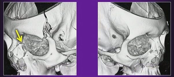
    Errors in perception account for a large majority of interpretive errors in radiology. A number of factors contribute to errors in perception such as overall lesion conspicuity, including degree of contrast and border demarcation from adjacent soft tissue [5, 6].

    Interpretive Error

    More than 30 types of cognitive bias have been described [7]. The most commonly encountered forms of bias in diagnostic im- aging include anchoring bias, confirmation bias, framing bias, availability bias, premature closure, inattentional blindness, and hindsight bias. 

    Anchoring Bias

    Also known as focalism, anchoring bias refers to the common human tendency to place undue influence or anchor on an initial diagnostic impression, despite later information to the contrary [5, 8, 9]. A radiologist’s initial gut reaction to a case, possibly made with limited initial information, can be difficult to deviate from and can potentially lead to useful information being disregarded. 

    Confirmation Bias

    Conceptually related to anchoring bias is confirmation bias. In this case, data supporting an initially suspected diagnosis are sought, and contrary information is given less significance [8, 9]. As a result, diagnoses can be delayed, and potentially unnecessary procedures can be performed [10]. Further, this type of bias may also be encountered in the academic setting with attending radiologist review of preliminary reports by radiology trainees [11].  

    Framing Bias

    In framing bias, different final diagnostic impressions can be made with the same information depending on the presentation of initial clinical information. In clinical context, different conclusions can be drawn from the same imaging study depending on the provided clinical history [10, 12]. Preliminary clinical history can be limited and potentially misleading [13, 14]. Further, the specialty of the referring physician may also be an influencing fac- tor [10].  

    Availability Bias

    In cases of availability bias, recent in- formation is given undue influence in di- agnostic decision making [15]. Recently missed diagnoses may linger in the mind of a radiologist and allow him or her to attribute a rare diagnosis in a case that they may otherwise have not. For example, a radiologist labels a case as “septic arthritis with osteomyelitis” on elbow MRI, only later to find that the case was acute lymphoblastic leukemia. This error might lead the radiologist to diagnose leukemia on more routine cases of osteomyelitis, even with confirmatory laboratory and clinical findings sup- porting that diagnosis [9]. On the opposite end of the spectrum is the concept of non- availability bias; that is, diagnoses that are rarely encountered are rarely considered [9]. A variation of this bias is alliterative error, or satisfaction of report, commonly encountered in radiology as a repeat of a prior report’s impression, even if this might not have been interpreted in the same way de novo. This error has been reported as the fifth most common cause of diagnostic errors by Kim and Mansfield [16].  

    Premature Closure

    Premature closure, the interpretation of initial conclusions as being final, is the overall most common type of error within clinical medicine [12, 17]. This er- ror includes the concept of satisfaction of search, in which an interpretive process is considered finished once an initial abnormality or finding is identified.

    Inattentional Blindness

    In the case of inattentional blindness, findings may be missed owing to their un- expected nature or their location at the periphery of the image. Corner shot findings on a radiograph or findings on the final im- ages of a cine clip of an ultrasound are examples of potential causes of inattentional blindness [16, 18–20].  

    Hindsight Bias

    Hindsight bias is described as the tendency to de-emphasize the difficulty in making an initial diagnosis after the fact. This bias can occur in group settings including tumor boards, clinical conferences, and medicolegal settings and can prevent realistic assessment of challenges faced with complex initial diagnoses [9, 21].

    External Factors

    Interruptions are a common occurrence in a busy practice with visiting clinicians, telephone interruptions, and technologist requests. In the face of these interruptions it is easy for radiologists to lose their trains of thought and potentially deviate unknowingly from their typical search patterns. These interruptions have been shown to lengthen interpretation times and reduce accuracy in abnormal cases [22, 23].

    Methods of Mitigation

    Metacognition

    A potential means of partially addressing cognitive bias is the concept of meta- cognition; that is, an individual can evaluate one’s own thought processes [22]. Metacognition involves introspection of one’s thought processes and seeking out- side perspectives.  

    Minimizing Interruptions

    Although radiologists must balance pro- viding high-level service to referring clinicians with efficient use of their time, methods for minimizing interruptions are critical [5, 16]. Employing reading room assistants to field and triage calls can provide a first line of screening for telephone calls to aid in reducing interruptions [24]. Further use of text messaging services can also allow radiologists to communicate findings efficiently and document exact conversations [25].

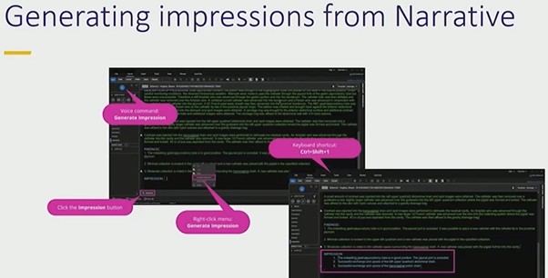
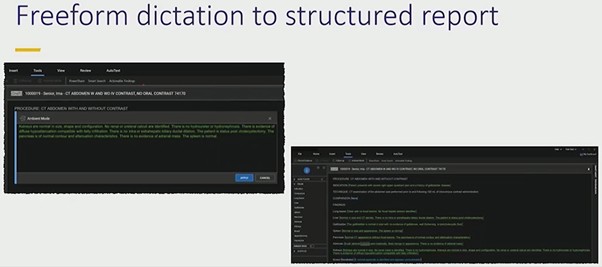
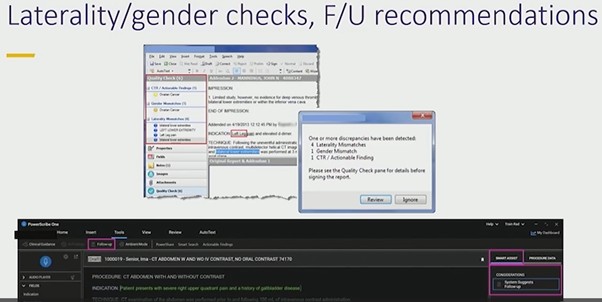
    Structured Reporting

    Structured reporting provides a check- list-style framework for reporting that al- lows reminders for interpreting radiologists to review all relevant anatomy. For trainees, this process also allows the development of desired interpretive search patterns [26].  

    Radiologic-Pathologic Review

    Follow-up on challenging cases either through quality-control conferences, tumor boards, or personal review of cases is critical for improving and expanding radiologists’ interpretive skills. Supportive and educationally oriented environments can allow meaningful discussion and review of diagnostically challenging cases.  

    Computer-Aided Diagnostics

    Use of increasingly powerful means of computer-aided image interpretation pro- vides another potential tool for radiologists to improve diagnostic accuracy and increase confidence. The current effective- ness of computer-aided detection within areas such as mammography has not been shown to be improved over interpretation without computer-aided detection [27]. However, there is growing potential for ap- plications in multiple other areas with use of neural network–based approaches [28].

    The impact of bias in radiologic interpretation can be substantial, with potential implications in patient outcomes. Better understanding the forms of bias, related to both internal and external pressures, can allow radiologists to implement methods for mitigating these biases.

    REFERENCES

    1. Garland LH. Studies on the accuracy of diagnostic procedures. Am J Roentgenol Radium Ther Nucl Med 1959; 82:25–38
    2. Degnan AJ, Ghobadi EH, Hardy P, et al. Percep- tual and interpretive error in diagnostic radiology: causes and potential solutions. Acad Radiol 2019; 26:833–845
    3. Tversky A, Kahneman D. Judgment under un- certainty: heuristics and biases. Science 1974; 185:1124–1131
    4. Durr T. Thinking, fast and slow by Daniel Kahneman. (book review) Am J Educ 2014; 120:287–291
    5. Waite S, Scott J, Gale B, Fuchs T, Kolla S, Reede D. Interpretive error in radiology. AJR 2017; 208:739–749
    6. Patel SH, Stanton CL, Miller SG, Patrie JT, Itri JN, Shepherd TM. Risk factors for perceptual-versus- interpretative errors in diagnostic neuroradiology. AJNR 2019; 40:1252–1256
    7. Rosenkrantz AB, Bansal NK. Diagnostic errors in abdominopelvic CT interpretation: characteriza- tion based on report addenda. Abdom Radiol (NY) 2016; 41:1793–1799
    8. Croskerry P. The importance of cognitive errors in diagnosis and strategies to minimize them. Acad Med 2003; 78:775–780
    9. Croskerry P. Achieving quality in clinical decision making: cognitive strategies and detection of bias. Acad Emerg Med 2002; 9:1184–1204
    10. Busby LP, Courtier JL, Glastonbury CM. Bias in radi- ology: the how and why of misses and misinterpre- tations. RadioGraphics 2018; 38:236–247
    11. Nanapragasam A, Bhatnagar P, Birchall D. Trainee radiologist reports as a source of confirmation bias in radiology. Clin Radiol 2018; 73:1052–1055
    12. Lee CS, Nagy PG, Weaver SJ, Newman-Toker DE. Cognitive and system factors contributing to diag- nostic errors in radiology. AJR 2013; 201:611–617
    13. Gunderman RB, Phillips MD, Cohen MD. Improving clinical histories on radiology requisitions. Acad Radiol 2001; 8:299–303
    14. Loy CT, Irwig L. Accuracy of diagnostic tests read with and without clinical information. JAMA 2004; 292:1602–1609
    15. Dumitrescu A, Ryan CA. Addressing the taboo of medical error through IGBOs: I got burnt once! Eur J Pediatr 2014; 173:503–508
    16. Kim YW, Mansfield LT. Fool me twice: delayed diag- noses in radiology with emphasis on perpetuated errors. AJR 2014; 202:465–470
    17. Graber ML, Franklin N, Gordon R. Diagnostic er- ror in internal medicine. Arch Intern Med 2005; 65:1493–1499
    18. Drew TH, Võ ML, Wolfe JM. The invisible gorilla strikes again. Psychol Sci 2013; 24:1848–1853
    19. Beanland V, Pammer K. Gorilla watching: effects of exposure and expectations on inattentional blind- ness. In: 9th Conference of the Australasian Society for Cognitive Science. Sydney, Australia: Macquarie Centre for Cognitive Science, 2010:12–20
    20. Drew TH, Vo ML, Olwal A, Jacobson F, Seltzer SE, Wolfe JM. Scanners and drillers: characterizing ex- pert visual search through volumetric images. J Vis 2013; 13:3
    21. Gunderman RB. Biases in radiologic reasoning. AJR 2009; 192:561–564
    22. Flavell JH. Metacognition and cognitive monitoring: a new area of cognitive-developmental inqui- ry. Am Psychol 1979; 34:906–911
    23. Wynn RM, Howe JL, Kelahan LC, Fong A, Filice RW, Ratwani RM. The impact of interruptions on chest radiograph interpretation: effects on reading time and accuracy. Acad Radiol 2018; 25:1515–1520
    24. Ngo  JS,  Maxfield  CM,  Schooler  GR.  The  current state of radiology call assistant triage programs among US radiology residency programs. Acad Radiol 2018; 25:250–254
    25. Torres A, Milov DE, Melendez D, Negron J, Zhao JJ, Lawless ST. A new approach to alarm manage- ment: mitigating failure-prone systems. J Hosp Adm 2014; 3:79–83
    26. Marcovici PA, Taylor GA. Structured radiology re- ports are more complete and more effective than unstructured reports. AJR 2014; 203:1265–1271
    27. Lehman CD, Wellman RD, Buist DSM, Kerlikowske K, Tosteson ANA, Miglioretti DL. Diagnostic accuracy of digital screening mammography with and with- out computer-aided detection. JAMA Intern Med 2015; 175:1828
    28. Taylor AG, Mielke C, Mongan J. Automated detection of moderate and large pneumothorax on frontal chest X-rays using deep convolutional neural networks: a retrospective study. PLoS Med 2018; 15:e1002697

    Jesse Courtier, MD

    Department of Radiology

    UCSF Benioff Children’s Hospital

  • Auto Sapiens: My New Assistant

    Auto Sapiens: My New Assistant

    Artificial intelligence (AI) has been likened to a new species, “Auto Sapiens.” I know this is wild, but bear with me—it may actually help us “get along,” “collaborate,” and “lead” AI to improve radiology practice.

    A recent Harvard Business Review article by Jeremy Heimans and Henry Timms explained Auto Sapiens: AI is able to act autonomously, make decisions, learn from experience, and operate without continuous human supervision, hence “Auto.” Also, AI possesses knowledge and the ability to make judgments in context, hence “Sapiens.” It is hard to think of Auto Sapiens when it’s software running on your data, but it is valid considering this terminology when thinking of AI used in humanoid robots.

    As a thought exercise, let’s think of radiology AI as an Auto Sapiens, and a coworker in the role of an “assistant.” Is this assistant going to take our jobs? Will it (they?) help us in our jobs, and could it make radiology practice more profitable? I say, no, to taking our jobs. And, yes, to helping us change our practice for the better.

    https://www.radfyi.org/2023/09/08/the-workplace-revolution

    Here is how: I believe that AI will not displace radiologists. Somebody needs to be liable for mistakes made by AI. Medical malpractice can be established when physicians deviate from the profession’s standard of patient care. If a radiologist uses an AI-enabled medical device for diagnosis or treatment of a patient, and their use deviates from an established standard of care, the physician could be liable for improper use of that AI medical device. As of now, the radiologist must independently review the AI’s recommendations, applying the standard of care in treating the patient regardless of the AI’s output. After all, AI is an assistant needing supervision, right?

    Holding AI developers liable is quite difficult. One would have to prove that the AI was defective at the time of product purchase by the user and did not become corrupt as it continued to train itself on user data. There are currently no sufficient industry standards to address this.

    I doubt that insurers would take liability; they lack the expertise to minimize liability should an AI application go awry. Radiologists are the ones assuring AI performs consistently in accordance with their intended purpose and scope, as well as at the desired level of precision. Insurers do not have the expertise to check on radiology AI applications’ correctness, relevance, robustness, or interpretability. Radiologists will be the stewards of quality assurance for AI.

    I do, however, wonder about a threat to reimbursements. It is conceivable that AI can evolve to perform better than radiologists—faster and with fewer errors. In that event, insurers could cut physician fees. We need to think about reimbursements in the AI era. Will there be a new component to the fee schedule, such as “AI supervision,” which entails auditing and supervising AI? We will need to continuously “invest” in our AI assistants to make sure they are trained up to the latest technological standard.

    OK, now that I’ve argued how AI will not replace radiologists, let’s see how our new assistants will help us. First, Auto Sapiens, like a good assistant, will happily do all the stuff many of us like less about our jobs, such as reading endless chest radiographs, scrutinizing CT images for lung nodules, measuring lesions and transcribing measurements into reports, and so many other things. Yes, I want this assistant, like, now!

    Additionally, Auto Sapiens will also help us decrease errors of perception and interpretation and delays for reporting incidental critical results, such unexpected intracranial hemorrhage on a nonemergent head CT. What is not to like about this type of assistant? Maybe liability insurance payments will even come down?

    And all of this can result in a more profitable business? Sure, as soon as AI enables radiologists, technologists, and imaging equipment to handle larger volumes, there could be a massive increase of imaging orders. Dream on, though, if you think that decision support will help us control imaging utilization. Imaging is already being used in lieu of a thorough clinical exam. In fact, Dr. Joseph Alpert called the physical exam “an ancient ritual” in 2019.

    So, the AI assistant can help us grow our business and focus on work we enjoy, like making a diagnosis and providing excellent services to physicians and patients. However, this model relies on us being proper supervisors to our AI assistants. As Curtis Langlotz, MD, PhD, once put it: “AI won’t replace radiologists, but radiologists who use AI will replace those who don’t.”

    The time to learn about AI is now, and I am excited about it!

    https://www.radfyi.org/2023/01/06/bye-bye-work-life-balance-welcome-work-life-integration

    Nadja Kadom, MD

    Director for Quality, Department of Radiology, Children’s Healthcare of Atlanta
    Interim Director for Quality, Department of Radiology and Imaging Sciences, Emory Healthcare
    Professor, Emory University School of Medicine