Category: Latest Posts

  • Little Leaguer’s Elbow: Clinical Catch-All

    Little Leaguer’s Elbow: Clinical Catch-All

    Participation in youth sports is surging, and elbow injuries have nearly doubled since the 1970s, with 31% of youth baseball players now experiencing pain annually. Because pediatric growth plates are weaker and less elastic than tendons or ligaments, they are the primary site of failure when subjected to the repetitive torque of overhead motions.

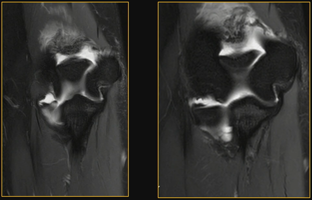
    Full Count:Little League elbow” is a clinical catch-all for a spectrum of overuse injuries. The most common manifestation is medial epicondyle apophysitis, an injury occurring during the acceleration phase of pitching when valgus stress creates significant medial traction.

    During his own ARRS Annual Meeting presentation on upper extremity trauma in peds populations, Vivek Kalia, MD, chair and medical director of radiology at Scottish Rite Children Hospital in Dallas, TX, noted the following key clinical presentations:

    • Gradual Onset: Typically, athletes report a steady decrease in pitch velocity and localized pain over the medial epicondyle that worsens with activity.
    • Overuse Mechanism: It’s heavily aggravated by inadequate rest (i.e., high pitch counts).
    • Acute or Chronic? Whereas apophysitis is a chronic stress injury, a single high-force event can lead to an acute medial epicondyle avulsion fracture.

    Additionally, Dr. Kalia’s play calling here prioritized comparing the symptomatic elbow to the contralateral side to identify subtle abnormalities.

    • X-ray: Look for a widened physis, sclerosis, and subcortical lucency of the medial epicondyle.
    • MRI: This modality is more sensitive, showing diffuse marrow edema within the apophysis and potential fluid within the growth plate.
    • UCL: In peds patients with apophysitis, usually, the ulnar collateral ligament remains intact, as the bone fails before the ligament.

    RadFYI: Early recognition is critical to prevent long-term sequelae, like osteoarthritis. While rest and physical therapy are the mainstays of treatment, prevention through strict activity limitations, as well as pitch count guidelines, remains the most effective strategy.
  • Expert Witness: The Rad’s Side Hustle

    Expert Witness: The Rad’s Side Hustle

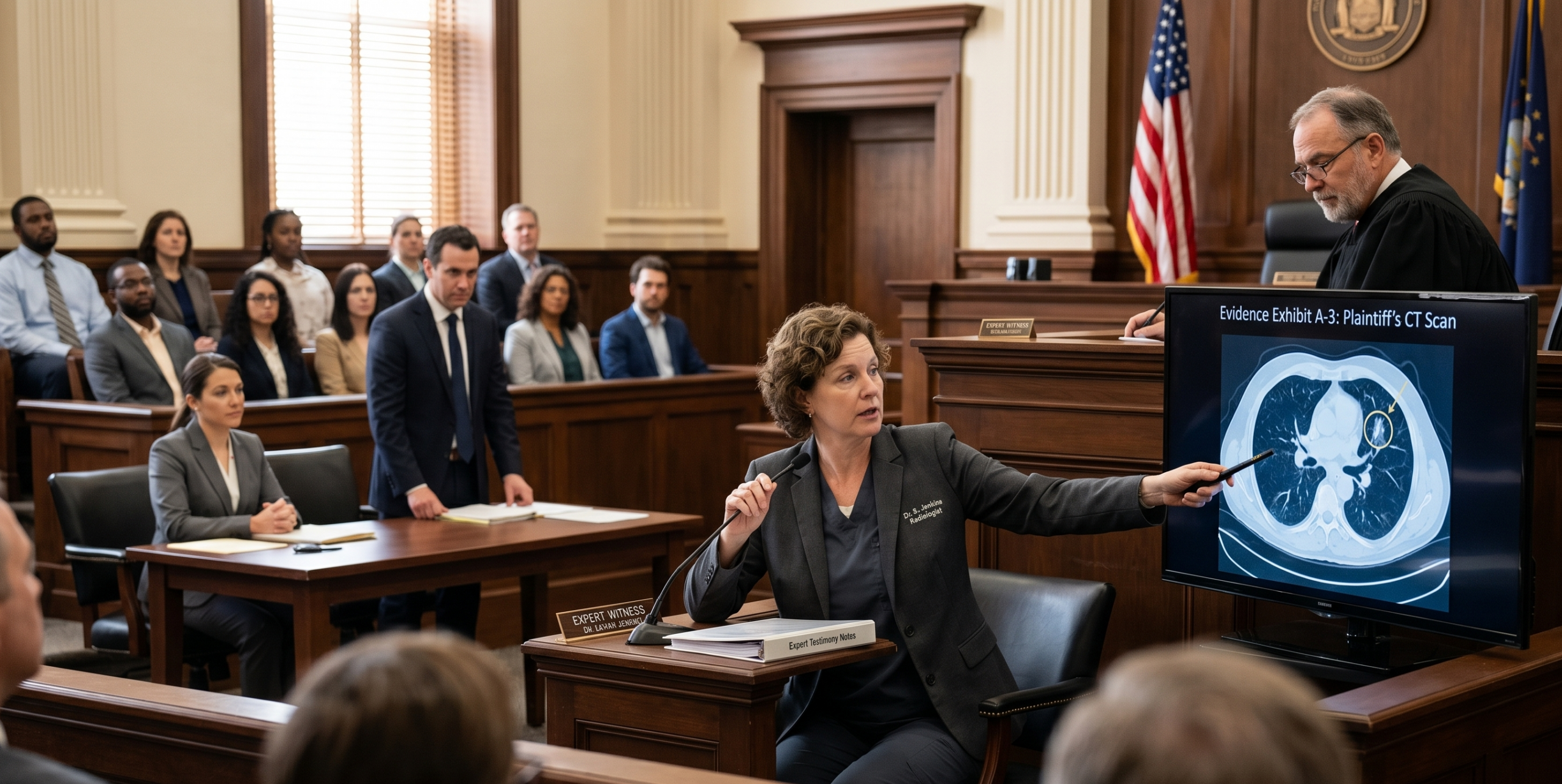
    When done correctly, legal consulting offers rads a significant path to income expansion, all the while providing a frontline look at how to protect their practices from, well, malpractice. For starters, as Seth Kligerman, MD, said during Financial Wellness for Practicing Radiologists and Beyond at ARRS 2026, the gig pays “very well.”

    Case Review: Assessing litigation not only helps avoid both the everyday pitfalls and those liabilities you didn’t even see coming, there are moral obligations here, too. Expert witnesses often address egregious medical mistakes and can defend colleagues when care more than meets the standard. Dr. Kligerman’s playbook for expansion also includes:

    • Rep > Ads: While many rads do use third-party companies, relying on word-of-mouth builds a more sustainable, higher-quality offering.
    • Both Sides: Indeed, expert witnesses must be willing to review both plaintiff and defense cases.
    • Peer Learning: Success requires the ability to explain the complexities of modern imaging, medical physics, advanced anatomy, etc. not for peer review, but instead to a jury of peers.
    • Claptrap: Alas, there are “morally ambiguous” witnesses who’ll say whatever they’re paid to.

    Fact vs. Expert: As the late Leonard Berlin noted, legally, rads remain required to testify as fact witnesses for their own cases (frequently for a nominal fee), but the expert witness role is voluntary and involves negotiated rates. Most courts won’t compel rads to provide opinions against their will, unless they possess “unique” knowledge that no other witness can provide.

    RadFYI: Expert witnessing is a lucrative way to leverage specialized knowledge, but it requires a high standard for what constitutes malpractice, as well as one’s own willingness to navigate ethical and legal entanglements alike.

  • The Thin Black Line—Split Scar Sign on Rectal MRI

    The Thin Black Line—Split Scar Sign on Rectal MRI

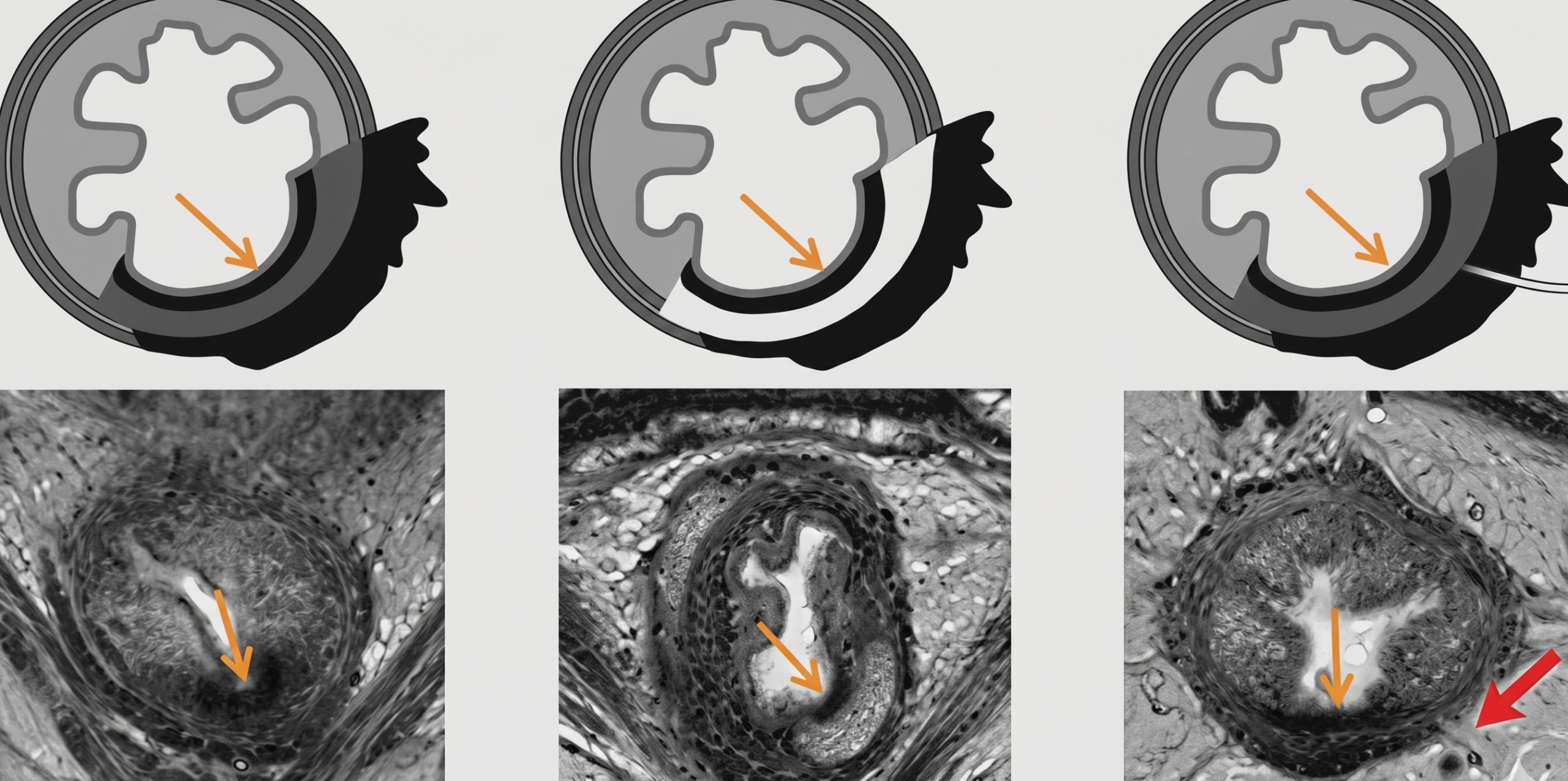
    The split scar sign, first described in 2020, is a powerful predictor for sustained complete clinical response and pathological complete response in rectal cancer patients following treatment.

    Bio Marker: The split scar sign helps rads identify patients who have responded exceptionally well to therapy—often preventing the overcalling of residual tumor. And as Professor Kirsten Gormly of the Royal Australian and New Zealand College of Radiologists (RANZCR) illustrated during the ARRS 2026 Global Partner Society Course, to determine if a split scar sign is positive, simply use the continuous line rule:

    • Positive—If you can draw a continuous thin inner black line over the scar with a marker (or “texter” as it’s called down under) without lifting it off the image, the sign is present.
    • Negative—If you have to lift that marker texter, or if the scar is so thick it requires a bigger texter, the sign is in absentia.

    Additional features from ARRS and RANZCR’s Rectal MRI: Best Practices from Ocean to Ocean to further set straight the split scar sign:

    • Inner Winner: The most critical component is the continuous thin inner scar.
    • Optional Outer: Whereas this sign is so named for its split look, you don’t always need an outer scar, especially if the original tumor was not T3.
    • Virtue Signals: Look for intermediate signal in the central muscle and the potential for mucinous degeneration (i.e., higher signal) or perforating vessels to be present within the layers.
    • Black Mass: The black layers on MRI represent submucosal collagen and fibrosis merging with muscle cells.

    RadFYI: Misinterpreting these thin scars as residual tumor is a common pitfall. Mastering this T2 assessment allows for more accurate posttreatment restaging and better informed clinical management.

  • What Radiation Oncologists Want from Diagnostic Radiologists

    What Radiation Oncologists Want from Diagnostic Radiologists

    Here in our personalized cancer care era, accurate staging and imaging are foundational for optimal treatment recommendations. Radiation oncologists are increasingly dependent upon diagnostic rads for precise identification and enumeration of metastatic disease to guide that therapy.

    The Magic Word: As Christopher Straka, MD, noted in the ARRS Online Course Radiation Oncology for the Diagnostic Radiologist, many patients are managed primarily by medical oncologists—who may only trigger a referral to a rad onc if they see the specific term: oligometastatic.

    So, to open up the black box of rad onc and improve outcomes, Dr. Straka shared some suggestions:

    • Enumerate: Provide a clear count of metastatic sites in your impression.
    • Prostate-Specific: For PSMA PET scans, specifically enumerate the number of extra-axial metastases.
    • Resolve Equivocals: If a lesion is uncertain, don’t just call it equivocal; please suggest the next modality that would provide more certainty.
    • Breadcrumbs: Use arrows, annotations, and slice numbers to help rad oncs quickly locate targets.

    RadFYI: Effective communication between rads and treating oncs has never been more important for metastasis-directed therapy. Ultimately, for the complex cases, direct consultation is still everyone’s best bet.

  • Posterior Shoulder Dislocations—Lights Up!

    Posterior Shoulder Dislocations—Lights Up!

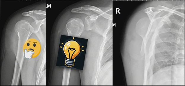
    Posterior shoulder dislocations are notoriously difficult to spot, often described as clinically and radiographically occult. In fact, misses are so common, they could be called, colloquially, the standard of care. Not that they have to be, however.

    Manickam “Nicks” Kumaravel, team doctor for the NFL’s Houston Texans, highlighted why posterior dislocations remain so frequently missed in chest and shoulder trauma during his ARRS Annual Meeting lecture.

    Glow On! On a standard AP view, look for the classic light bulb appearance of the humeral head. If a post-fall image simply doesn’t look right, voilà, that’s your cue to dig deeper.

    N.B. You cannot rely on a single projection…

    • Scapular Y view is the critical orthogonal projection.
    • It clearly demonstrates the posterior displacement that AP can hide.

    RadFYI: To avoid a miss, maintain a high index of suspicion, always verifying with orthogonal views.

  • Fournier’s Gangrene: A Urologic Emergency

    Fournier’s Gangrene: A Urologic Emergency

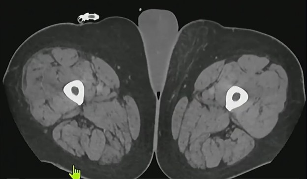
    Fournier’s gangrene is a rapidly progressing polymicrobial necrotizing fasciitis of the perineal, perianal, and genital regions. Because of its high mortality rate, immediate diagnosis and intervention are non-negotiable.

    As illustrated by Paul Nikolaidis, MD, during the ARRS Online Course Imaging of Pathologies of Male External Genitalia, this surgical emergency typically affects middle-aged men—particularly those with risk factors like diabetes, indwelling catheters, recent surgery, or immunosuppression from cancer treatments.

    • Down & Dirty? Look for dirty shadowing or ring-down artifacts caused by gas within the soft tissues, along with significant skin thickening.
    • Notable Normality: The testes, themselves, usually appear normal, as the infection follows fascial planes, rather than involving the scrotal contents directly.
    • Gold Map: CT comes in clutch in the ED to confirm the diagnosis and, more importantly, to show the extent of involvement for the surgical team.

    Whereas imaging is vital, roughly 10% of patients will not show visible gas on initial scans; therefore, clinical suspicion must remain high (even if those classic dirty shadows are absent).

    RadFYI: Early diagnosis is the only way to ensure survival. Once identified, management must include aggressive antibiotic therapy and immediate surgical debridement of all affected areas.

  • The Snow Blower and The Tuft Fracture

    The Snow Blower and The Tuft Fracture

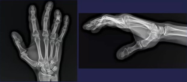
    A 43-year-old patient in Rochester, MN, presented to Mayo Clinic’s Ceylan Colak, MD, with complex hand trauma following a snowblower accident. While multiple phalangeal fractures were immediately apparent, as Dr. Colak recounted during the ARRS Online Course Imaging of Small Joints: A Review for Clinical Practice, a comminuted thumb fracture was initially overlooked due to the severity of the patient’s way more overt injuries.

    Distal Distraction: Distal phalangeal and tuft fractures are not just common; they account for nearly 50% of all hand fractures. In the setting of high-energy trauma—vis-à-vis a southeastern Minnesota snowblower injury—rads can easily fall victim to “satisfaction of search,” missing these small yet clinically significant injuries:

    • Initial Findings: Multiple displaced and non-displaced phalangeal fractures.
    • All Thumb: A comminuted, minimally displaced fracture of the thumb distal phalanx.
    • Call Kirschner: The patient ultimately required K-wire placement for stabilization of the multiple hand fractures.

    Bottom Line: When assessing a bad hand fracture, always perform a systematic check of each and every digit. Don’t let the most dramatic injuries distract you from the tuft; it’s where half of hand fractures happen.

  • AI-Powered Automation of Image Acquisition and Post-Processing

    AI-Powered Automation of Image Acquisition and Post-Processing

    AI-powered automation is moving upstream, shifting the focus from just lesion detection to the very beginning of the imaging process: acquisition and post-processing. Most imaging costs are tied to these upstream tasks. By automating them, Linda Moy, MD, explained at ARRS 2026, providers can improve image quality while simultaneously lowering costs and increasing operational efficiency…

    • On the DL: Deep learning (DL) reconstruction tools allow for noisier or lower-resolution initial imaging that AI then transforms into high-quality images. DL algos can reduce MRI scan times by >50%—while maintaining, or even improving, diagnostic quality.
    • Keep the Noise Down: AI denoising algorithms have demonstrated a 60% reduction in radiation dose, alongside a 39% reduction in image noise.
    • Table for 3D? Using three-dimensional ceiling cameras and infrared imaging to automate table height and positioning can save 32 seconds per exam, as well as reduce radiation dose by some 20%.
    • No Contrast: Synthetic AI images can now simulate contrast-enhanced MRI, without actual contrast agents. This is a breakthrough for pregnant patients, those with severely impaired renal function, or populations concerned about gadolinium deposition.

    These advancements significantly improve the patient experience, particularly for peds patients who may no longer require sedation, and claustrophobic patients who benefit from shorter time in the scanner.

    Mind the Mirage: Rads must remain vigilant regarding AI “hallucinations.” While rare, these tools can occasionally invent lesions that do not exist or remove true lesions during the reconstruction process.

    Bottom Line: AI reconstruction works seamlessly in the background to provide higher signal-to-noise ratios and reduced artifacts, allowing rads to focus on downstream diagnostic tasks with better data in less time.

  • Perirenal Fat Trap: Spotting Extra-Adrenal Myelipoma

    Perirenal Fat Trap: Spotting Extra-Adrenal Myelipoma

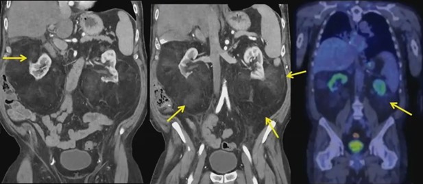
    When rads encounter symmetric macroscopic fat in the perirenal space, the initial instinct is often to fear well-differentiated liposarcoma or lymphoma. Distinguishing these from benign extra-adrenal perirenal myelipoma is critical to avoiding unnecessary clinical anxiety and aggressive surgical intervention.

    Impressive Abs! In this excerpt from the ARRS 2026 Challenging Abdominal Cases Instructional Course, Cooky Menias, MD, presents a patient with abdominal bloating who revealed a symmetric retroperitoneal process pressing on the kidneys. Despite the lack of subcutaneous fat, the patient exhibited significant intra-abdominal fat that really impressed on the renal parenchyma.

    The Diagnosis:

    • Extra-adrenal myelipoma is a rare, benign mesenchymal tumor composed of fat and hematopoietic cells.
    • They typically appear in the 5th to 6th decades of life.
    • The most common extra-adrenal locations are the perirenal and presacral spaces.

    The Pearls:

    • Fat Chance: Unlike perirenal fibrosis (IgG4-related disease) or Erdheim-Chester, which encase the kidneys and lack fat, myelipomas contain macroscopic fat and embed (i.e., “impress”) upon the renal tissue.
    • PET-CT Utility? While not diagnostic, PET-CT can be reassuring by showing a lack of significant uptake, helping to rule out high-grade lymphoma or aggressive malignancy.
    • Stress Test: These tumors may arise from ectopic adrenal tissue or reticular endothelial cells that become exuberant during chronic stress, endocrine dysfunction, or inflammation.

    Bottom Line: If you see symmetric macroscopic fat embedding the kidneys in an older patient with low metabolic activity on PET, think extra-adrenal myelipoma. While biopsy (even open biopsy) may be required for definitive comfort, recognizing this embedding fat pattern can narrow the differential significantly.

  • Remote Rules—Texas Telerad Wins in California Tax Court

    Remote Rules—Texas Telerad Wins in California Tax Court

    A landmark California Court of Appeal decision is shielding out-of-state remote professionals from Golden State taxation. In Garcia-Rojas v. California Franchise Tax Board, the court ruled that a Texas teleradiologist is not subject to California income tax, despite serving California patients.

    So, California attempted to use a complex corporate tax theory to claim income from a contractor who never physically entered the state.

    • The Provider: Dr. Xavier Garcia-Rojas, a Texas-based radiologist, performed contract reads for San Diego-based StatRad from 2018 to 2020.
    • The Dispute: The Franchise Tax Board (FTB) claimed Dr. Garcia-Rojas owed taxes because he used StatRad’s software, was licensed in California, and served Cali patients.
    • The Numbers: The doctor originally paid roughly $48,000 in taxes, penalties, and interest under protest before suing for a refund.

    Legal Pivot: The court rejected FTB’s attempt to apply the unitary business doctrine (usually reserved for multi-state corporations) to a single-person sole proprietorship.

    • The Rule: A nonresident providing remote professional services does not automatically become part of a unitary business just because their client is in California.
    • The Result: Because the actual labor (i.e., image interpretation) happened at a desk in Texas, the income is not California-sourced.

    As Bloomberg noted, Garcia-Rojas v. CAFTB sets a binding precedent for thousands of remotely contracted lawyers and consultants, as well as the surfeit of tech sector labor, working for California firms. Indeed, it confirms that location of labor—not the location of the client—is the deciding factor for individual income sourcing.

  • The Football Sign in Neonatal Imaging

    The Football Sign in Neonatal Imaging

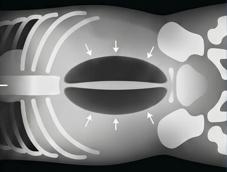
    The “football sign” is a pathognomonic indicator of massive pneumoperitoneum on supine abdominal radiographs, characterized by a large, oval radiolucent area shaped like an American football.

    SIP: In extremely low birth weight infants, this finding often signals spontaneous intestinal perforation (SIP), a condition distinct from necrotizing enterocolitis (NEC). Recognizing the difference is critical because SIP is often an isolated vascular insult, and as Susan D. John, MD, illustrated during Mastering Radiology: A Comprehensive Board Review for Residents and Refresher for Radiologists in Practice, some cases may heal without surgical intervention.

    Three Tips from Dr. John:

    1. Radiolucent Oval—Massive free air collects anterior to the abdominal viscera, distending the peritoneal cavity and creating a sharp interface with the parietal peritoneum.
    2. Laces Out—The air outlines longitudinal structures of the anterior abdominal wall, most commonly the falciform ligament in the right upper abdomen. It may also outline the median umbilical ligament (urachal vestige) or medial umbilical ligaments (umbilical arteries).
    3. Bowel Appearance—A key clue for SIP is that the bowel often appears normal in caliber without evidence of pneumatosis intestinalis, which would typically be present in NEC.

    Bottom Line: While frequently associated with spontaneous perforation in premature infants (ca. 24 weeks gestation), the football sign can also be seen in cases of iatrogenic trauma, such as rectal perforation resulting from the placement of a rectal tube. If the sign is suspected on a supine film, a decubitus view can be used to confirm the presence of free air.

  • Infiltrative Mass Faceoff: Oligodendroglioma vs. IDH-Mutant Astrocytoma

    Infiltrative Mass Faceoff: Oligodendroglioma vs. IDH-Mutant Astrocytoma

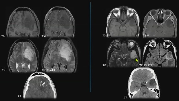
    Distinguishing between oligodendroglioma and IDH-mutant astrocytoma is a high-yield challenge for neurorads, as both typically present as infiltrative, T2-hyperintense masses with variable enhancement.

    1p/19q: While both tumors fall under the umbrella of IDH-mutant adult gliomas, their molecular signatures—and, consequently, imaging phenotypes—offer specific smoking guns for diagnosis.

    • Oligodendroglioma: Defined by IDH mutation and 1p/19q codeletion.
    • Astrocytoma: IDH-mutant but 1p/19q intact.

    As part of the dynamic, rapid-fire Neuroradiology Case Review during the 2026 ARRS Annual Meeting, Susana Calle, MD, pointed out some key discriminators…

    T2-FLAIR Mismatch: This is the strongest predictor for IDH-mutant astrocytoma, boasting a specificity of 90–100%.

    • The lesion shows homogeneous high signal on T2-weighted images but complete or near-complete signal suppression on T2-FLAIR.
    • The presence of this sign virtually rules out oligodendroglioma (although its absence does not exclude an astrocytoma diagnosis).

    Worm-like: If you see gyroform cortical calcifications, think oligodendroglioma.

    • Calcification occurs in up to 90% of oligodendrogliomas.
    • These tumors have a strong frontal lobe predominance and tend to be more cortically based than astrocytomas.
    • Astrocytomas are far less likely to calcify.

    Perfusion Paradox: Typically, high cerebral blood volume (CBV) indicates high-grade malignancy, but oligodendrogliomas are the exception.

    • Elevated CBV is often seen in oligodendrogliomas, regardless of grade, due to their characteristic “chicken-wire” fine intratumoral capillaries, rather than true neoangiogenesis.

    Bottom Line: Calcified cortical tumor in the frontal lobe? Sway toward oligodendroglioma. T2-FLAIR mismatch sign identified? Think IDH-mutant astrocytoma. Accurate noninvasive identification of these genotypes is critical because 1p/19q codeletion in oligodendrogliomas is associated with a better response to chemoradiation and significantly improved overall survival.