
Scimitar syndrome represents a distinctive form of right lung partial anomalous pulmonary venous return. It is classically associated with right lung hypoplasia, abnormalities of the right pulmonary artery, and a characteristic anomalous vein draining into the systemic venous system. As Abbey J. Winant, MD, MFA, illustrates in “Pediatric Thoracic Vascular Disorders: Congenital to Acquired Pathology,” CTA plays a central role in defining venous anatomy and identifying associated anomalies.
What Defines Scimitar Syndrome?
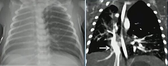
Scimitar syndrome (RLL PAPVR) with R Lung hypoplasia
The hallmark is a right lower lobe pulmonary vein draining anomalously—most often into the inferior vena cava, but occasionally into the inferior right atrium. This vein produces the classic “scimitar” appearance on chest radiography and cross-sectional imaging. Children often have concurrent right lung hypoplasia, which alters airway and vascular proportions.
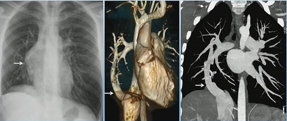
Scimitar Syndrome (“Scimitar Vein”)
Recognizing Associated Findings
In addition to partial anomalous pulmonary venous return, scimitar syndrome often presents with:
- Hypoplastic right lung
- Hypoplastic right pulmonary artery
- Systemic arterial supply to portions of the right lung
- Bronchial abnormalities, including bronchiectasis
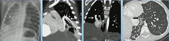
Scimitar Syndrome with Horseshoe Lung: Hypoplastic right lung, PAPVR to RA, horseshoe lung, R lung bronchiectasis
One of the most notable associations is horseshoe lung, seen in approximately 80% of cases. Horseshoe lung consists of a parenchymal isthmus connecting both lungs across the midline, usually posterior to the heart. When present, it further reinforces the diagnosis and alerts the radiologist to search for additional congenital anomalies.
Additional Congenital Abnormalities to Consider
Although not seen in every case, associated developmental abnormalities may include:
- Extralobar sequestration
- Vertebral anomalies
- Diaphragmatic defects
- Cardiac malformations
Their presence can significantly influence management, operative planning, and follow-up.
Why Does CTA Matter?
CTA provides the most comprehensive view of the venous drainage pattern, systemic arterial contributions, and bronchial architecture. It allows precise localization of anomalous veins and helps differentiate scimitar syndrome from other types of partial anomalous pulmonary venous return.
Bottom Line
Scimitar syndrome is more than an anomalous pulmonary vein. Its constellation of findings—right lung hypoplasia, anomalous venous return, and frequent association with horseshoe lung—requires careful, structured evaluation. CTA remains the best tool to clarify anatomy and guide clinical management.
Leave a Reply