Category: Latest Posts

  • Neutralizing Workplace Toxicity

    Neutralizing Workplace Toxicity

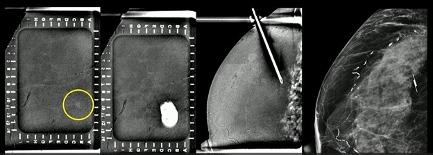
    During the Wellness Symposium at ARRS 2026, Sherry S. Wang, MBBS, from the University of Cincinnati shared her own, hard-won framework for identifying and navigating toxic professional environments.

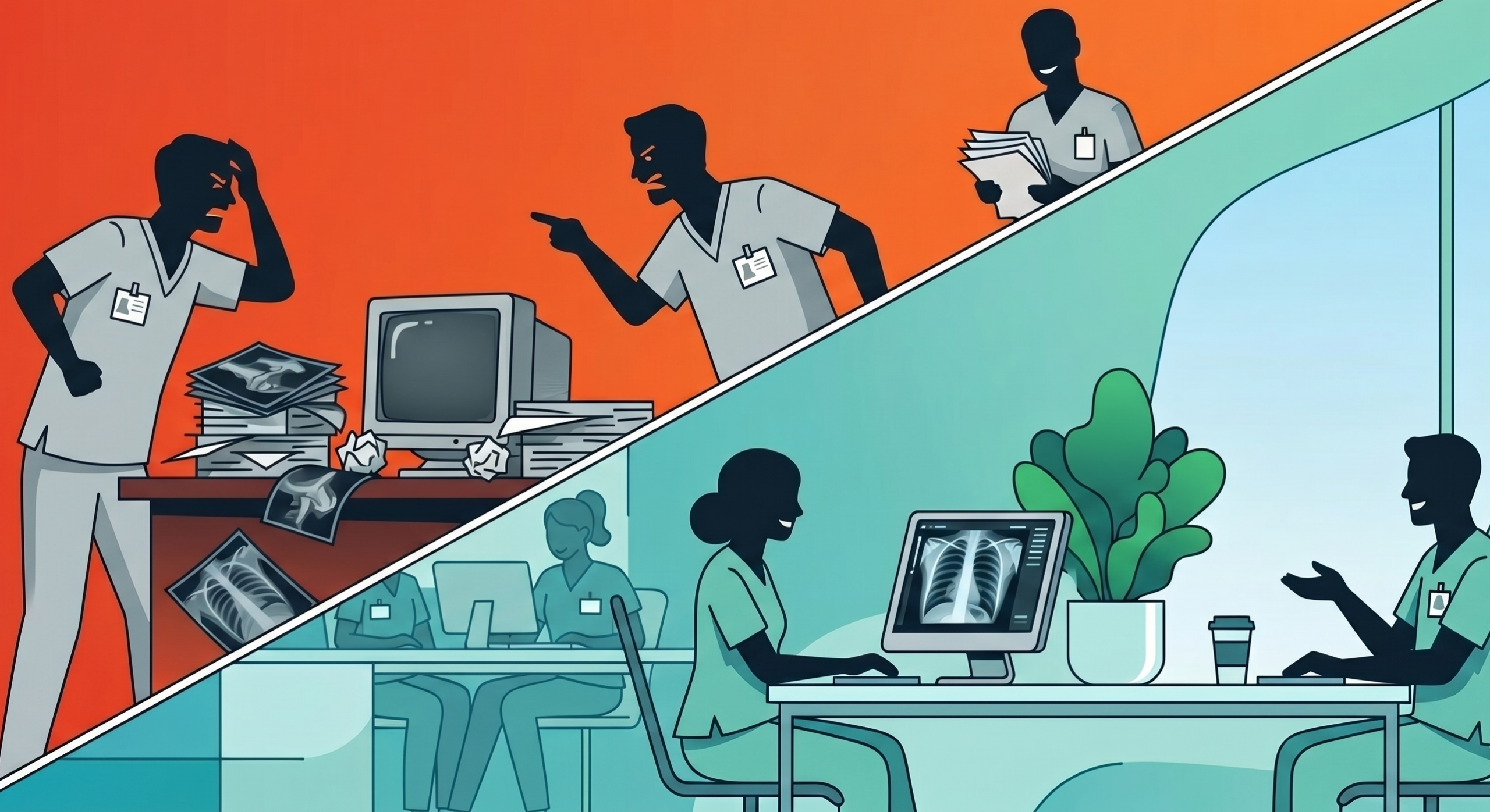
    Working Definition: A toxic workplace is defined by poor leadership, lack of transparency, unreasonable work volumes, and a culture of fear or retaliation. Because these factors can directly erode a rad’s confidence and performance right there at the workstation, addressing them is a professional necessity.

    Dr. Wang’s 5-Step Manual:

    1. Pattern Recognition—Differentiate between a one-time misunderstanding and repeated patterns of toxic behavior.
    2. People vs. Process—Determine if you are facing a people problem or a process problem, then assess if the organization is actually open to change.
    3. To Engage or Nah? Evaluate if the issue is worth the effort to fix and whether you can effectively protect yourself from retaliation.
    4. Writing & Speaking—If you feel safe, present documented evidence of the behavior to leadership. [N.B. If you’re too afraid to do so, you likely already have your answer.]
    5. Plan B, Always—Having an exit strategy is a courageous act of self-preservation. If you decide to leave, do so cordially, but prepare for potential retaliation during your notice period.

    Maybe It’s Me? Toxicity isn’t just an HR issue; it eats away at our wellbeing, impacting a rad’s sensitivity and specificity alike. And since we all contribute to institutional culture—be it a primary instigator or simply as silent bystanders who allow toxicity to persist—Dr. Wang challenged us to keep looking inward.

    Bottom Line: Merely surviving your reading room shift is no longer enough. Going forward, the goal should be detoxing our workplaces to make them that much more productive for each and every rad.

  • Uremic Encephalopathy with Lentiform Fork Sign

    Uremic Encephalopathy with Lentiform Fork Sign

    In patients presenting with renal failure, certain MRI patterns in the basal ganglia provide a leading history for a diagnosis of uremic encephalopathy. Specifically, the lentiform fork sign helps ID metabolic distress in the deep brain nuclei, particularly in the context of metabolic acidosis.

    Big Picture: While uremic encephalopathy most commonly affects the posterior parietooccipital cortical and subcortical regions, as Atul Agarwal, MD, of Indiana University duly noted during the ARRS Web Lecture Abnormalities of the Basal Ganglia, it can involve the basal ganglia, extensively, too.

    • Subcortical Structures: MRI typically shows expansile, abnormal signals on T1 and T2-weighted imaging involving the putamen and globus pallidus.
    • Brain Fork: The signature fork appears due to FLAIR hyperintensity in the external and internal capsules—those white matter pathways surrounding the lentiform nucleus.
    • Restrictions Apply: Imaging often reveals anticipated abnormal restricted diffusion in these regions.

    What to Watch For: This pattern is not exclusive to uremia; therefore, rads must consider several high-stakes mimics:

    • Metformin-Associated Encephalopathy: This can have a nearly identical appearance, making it a vital consideration for diabetic patients with renal impairment.
    • Atypical PRES: Whereas posterior reversible encephalopathy syndrome usually follows a standard pattern, acute hypertensive encephalopathy can present in this atypical form.
    • Metabolic/Ischemic Insults: Both hypoglycemia and ischemia share similar mechanisms that can produce comparable basal ganglia findings.

    Bottom Line: When you see the lentiform fork sign in a renal patient, metabolic acidosis is the likely culprit, but always cross-check for metformin use or hypertensive crisis.

  • Cold Comfort: Cryoablation of Vertebral Metastases

    Cold Comfort: Cryoablation of Vertebral Metastases

    Cryoablation is becoming the hottest tool for managing complex osseous metastases, utilizing alternating cycles of rapid freezing and thawing to achieve reliable cell death. And as Anderanik Tomasian, MD, of UC Irvine explained during the ARRS Online Course Minimally Invasive Musculoskeletal Interventional Oncology Masterclass, cryoablation is uniquely suited for tumors where heat-based methods like radiofrequency ablation (RFA) often fail.

    Why It Matters: Unlike RFA, which is ineffective for purely osteoblastic lesions due to high electrical impedance, cryoablation excels in treating these dense bone tumors. It also offers less intraprocedural pain and allows for the simultaneous use of multiple probes to cover large, complex geometries.

    Freezing is Believing: The Ice Ball

    • CT Visual: The primary advantage is your ability to see the hypoattenuating ice ball in real time.
    • Precision on the Margins: The visible edge of the ice ball typically represents 0°C; to guarantee cell death (which requires –40°C), the ice ball must extend 3–5 mm beyond the tumor boundary.
    • Heavy Caveat: While highly visible in soft tissue, the ice ball can be difficult to distinguish within the dense bone of a blastic lesion or normal bone.

    Targeted Case: In Dr. Tomasian’s case of a patient with non-small cell lung cancer, cryoablation was successfully used to target a painful L4 osteoblastic lesion. Because RFA cannot effectively penetrate such dense lesions, cryoablation was the preferred modality for pain palliation and local tumor control.

    Thermal Protection: Since bone cortex does not stop the expansion of an ice ball, adjacent vital structures are at risk.

    • Active: Techniques like pneumodissection or even hydrodissection are used to displace and insulate nerves or the spinal cord.
    • Passive: Strategies include placing thermocouples to monitor temperatures near sensitive structures, with active protection recommended if the temperature drops to 10°C.

    Bottom Line: Cryoablation is a versatile, less painful, and highly visible intervention that provides a critical alternative for patients with refractory bone pain—particularly those with large soft-tissue components or blastic disease.

  • Contrast-Enhanced Mammography–Guided Intervention—What Next?

    Contrast-Enhanced Mammography–Guided Intervention—What Next?

    83-year-old woman with invasive lobular carcinoma in left breast and enhancing right breast mass. No mammographic or ultrasonic correlate to right breast. Contrast-enhanced mammography–guided intervention localization per surgeon’s request.

    At some point of inflection, every new imaging modality will require its own image-guided biopsy capabilities. Why? Because as Haydee Ojeda-Fournier, MD, of UC San Diego Health reminded us in her ARRS Quick Byte video, suspicious findings may be occult on all other systems.

    Yes, and … Contrast-enhanced mammography (CEM) is transitioning from diagnostic instrument to a functional intervention platform—addressing Dr. Ojeda-Fournier’s “what next?” query for findings seen only on recombined images.

    Percentage Points:

    • 62% of CEM-detected lesions can be identified during a second look at digital breast tomosynthesis (DBT).
    • 76% of CEM findings have an ultrasound correlate; these correlates are more likely to be malignant.
    • ~60% of MRI-guided biopsies could potentially be performed via CEM, providing a faster, lower-cost alternative for impacted MR units.

    Case in Point: CEM-guided intervention remains a game-changer for patients who cannot tolerate MRI. For example, in the 83YO patient above with invasive lobular carcinoma who was not an MRI candidate, CEM-guided localization successfully targeted a contralateral mass that was mammographically and sonographically occult.

    In the projection below, you’ll notice some artifact from the needle. And after Dr. Ojeda-Fournier’s team deployed their wireless localizing device (based on geography), pathology came back benign. She’ll need a follow up, regardless.

    Final Points of Order:

    • Search in Teams: Using a combined scout (CEM+DBT) provides the most flexibility for choosing the optimal guidance modality.
    • Needle Up! While both horizontal and vertical approaches are possible, typically, the vertical needle approach is preferred because it samples a larger tissue volume in the z-axis and is less reliant on precise depth estimation.
    • Singular Sensation: A successful biopsy is defined as obtaining tissue within one procedure under a single compression, whether guided by recombined CEM, DBT, or stereotactic techniques.
  • Building the Perfect Workday

    Building the Perfect Workday

    Many factors contribute to burned out rads, of course, but one of the biggest has to be a fundamental loss of control over the daily work schedule, which can make long-term practice unsustainable. What was once considered a lifestyle specialty can quickly become an unsustainable grind.

    The Solution? During his presentation at ARRS 2026, Nicholas Galante, MD, advocated for abandoning traditional time management in favor of focus management.

    Proof of Concept: “Deep Work” Parkinson’s Law states that work will always expand to fill the time we give to it. So, to combat the kind of creep, Dr. Galante uses deep work to reclaim control of his time:

    • Breaking his daily clinical work into two, distinct 90-to-120-minute sessions, distractions are mitigated—allowing him to get into a flow state.
    • Between these two deep work sessions, he’ll unplug to handle administrative tasks, get some exercise, and run family errands.

    Bottom Line: Fighting rad burnout is less about managing time and much more about balancing times of high focus and releasing the expectation of work for work’s sake.

  • Tears of the Pancreas: Why Cyst Stability Isn’t Enough

    Tears of the Pancreas: Why Cyst Stability Isn’t Enough

    More than isolated findings, pancreatic cystic lesions can be a cry for help from the entire organ. In fact, the field defect suggests that the presence of a cyst indicates an increased risk of developing pancreatic ductal adenocarcinoma elsewhere in the gland.

    Look Around You! Rads frequently focus on the stability of a cystic lesion, but malignancy can arise in the surrounding pancreatic parenchyma—even when the cyst, itself, remains unchanged for months, even years.

    • Crying Metaphor: In his ARRS Quick Byte video, Atif Zaheer, MD, of Johns Hopkins dubs these lesions “tears of the pancreas,” warning of synchronous or metachronous cancer development.
    • Surveillance Trap: Whereas some guidelines suggest stopping surveillance for stable cysts after 5 years, Dr. Zaheer’s research indicates a continued risk of malignancy beyond the 5–10 year mark.
    • What’s the Protocol Here? Abbreviated MRI protocols designed for cyst follow-up must include sequences that specifically evaluate the parenchyma to avoid missing cancers arising in the uncinate process of the pancreatic head.

    Under New Management: Managing pancreatic cysts is shifting from a disease of technology toward a more personalized, multi-faceted approach, including:

    • Beyond Imaging: Effective risk stratification now involves clinical data, biochemical markers, and cyst fluid DNA sequencing for mutations in genes like PIK3CA and TP53.
    • AI Edge: Emerging deep-learning algorithms and radiomics are helping to create automated risk-prediction models to differentiate high-risk lesions from benign ones.
    • Patient Empowerment: With the 2020 Cures Act, patients have immediate access to their reports, increasing the need for structured, recommendation-based reporting that tracks imaging features over time.

    Bottom Line: Don’t just look at the cyst. Evaluating the health of the entire pancreatic gland is critical to detecting early invasive cancer that the field defect may be denying.

  • True vs. False Lumen in Acute Aortic Dissection

    True vs. False Lumen in Acute Aortic Dissection

    In matters of the heart, truth matters all the more. And identifying the real lumen remains critical for planning endovascular repairs, ensuring branch vessels are correctly supplied to prevent end-organ ischemia. Sure, continuity with the normal aorta remains the gold standard for sussing things out. But as Ferco Berger, MD, pinpointed during the ARRS Online Course “Imaging in the Emergency Department,” tracing it can be difficult in cases involving the aortic root or when only limited abdominal scans are available.

    False Witnesses:

    • Beak Sign: Acute angle formed where dissection flap meets outer wall, it’s the most useful and reliable indicator (present in nearly 100% of acute and chronic cases).
    • Larger Size: A false lumen typically distends and has a larger cross-sectional area than the real deal.
    • Cobweb Sign: Thin, linear defects representing remnants of media layer. Albeit rare, they are highly specific to false lumen.

    Truth Tellers:

    • Calcification: Generally, hardening along outer wall or eccentric flap is indicative of true lumen. [N.B. In chronic cases, false lumen can occasionally calcify.]
    • Wraparound: In transverse aortic arch, if one lumen appears to wrap around another, that inner lumen is invariably the truer one.
    • Density: In arterial phase, true lumen frequently show higher contrast density.

    Supply Chain: Differentiating the supply side here helps predict which organs are at risk:

    • True Lumen: Usually supplies celiac trunk, superior mesenteric artery, right renal artery.
    • False Lumen: Often supplies left renal artery and more prone to thrombus formation.

    Bottom Line: When continuity is unclear, look for the beak sign and larger caliber to identify the false lumen; double-check the arch wraparound and calcification patterns to confirm the true lumen.

  • The Buford Complex—Anatomy or Abnormality?

    The Buford Complex—Anatomy or Abnormality?

    Occurring in less than 2% of your shoulder patients, apropos, there are just two things you need to ID the Buford complex. As Robert J. French, Jr., MD, enumerated for the ARRS Online Course “Expanding the Field: Imaging of Sports-Related Injuries Beyond the Knee,” this normal anatomic variation is defined by an absent anterior superior labrum and a thickened middle glenohumeral ligament (MGHL).

    The Details:

    • Location: Labrum is missing in the 1- to 3-o’clock position (right shoulder) or 9- to 11-o’clock (left shoulder).
    • Reconstitution: Typically, labrum reappears at or below glenoid midpoint.
    • MGHL: Hypertrophied, it often appears as a discrete structure—similar in size to long head of biceps tendon.
    • Stability: Unlike pathologic lesions, biceps anchor and contiguous superior and anterior inferior labrum remain normal.

    Closed for Repairs: Mistaking this variation for a labral avulsion is a common pitfall. In fact, in one surgical error documented in AJR, a cordlike MGHL was unfortunately attached to the glenoid as a “fix,” resulting in severely restricted shoulder motion for the patient.

    The Planes:

    • Axial: Look for absence of low-signal-intensity labral tissue along anterior superior glenoid rim.
    • Sagittal: MGHL can be seen as a linear structure separate from the subscapularis tendon, inserting into biceps anchor.

    Bottom Line: If the superior labrum and biceps anchor are intact, yet the anterior superior labrum is missing alongside a thickened MGHL, think Buford complex—not a labral tear.

  • Decoding Dementia: The Lewy Body Problem

    Decoding Dementia: The Lewy Body Problem

    Dementia with Lewy bodies (DLB) mimics Alzheimer’s disease, indeed. And as RadNet neuroradiologist Suzie Bash, MD, discussed during the ARRS Online Course “Opportunities and Obligations in Imaging Patients with Alzheimer’s Disease,” specific multimodal patterns—occipital lobe involvement, particularly—can help hone your DDX.

    Big Pics: 73 y/o male presenting with visual hallucinations, resting tremors, and frequent falls (but only minimal memory loss) illustrates the clinical profile of classic DLB. Those bodies are defined by alpha-synuclein, sure, but imaging often reveals a much more mixed bag:

    • MRI: Showed atrophy in the hippocampi, as well as occipital and parietal lobes.
    • Amyloid PET: Positive, confirming beta-amyloid plaques can be present in DLB cases.
    • Tau PET: Revealed deposition in posterior cingulate gyrus and occipital/parietal lobes.
    • FDG PET: Confirmed hypometabolism in same posterior regions, a hallmark of the disease.

    Bigger Numbers:

    • 1 million: Americans living with DLB, compared to 6.7 million with Alzheimer’s.
    • 80%: Frequency of visual hallucinations as a presenting symptom in DLB.
    • 5–8 years: Typical survival rate following diagnosis.

    Bottom Line: Rads should pay closer attention to the occipital lobe. Whereas Alzheimer’s and DLB share temporal and parietal overlaps, the addition of occipital atrophy and hypometabolism is a strong indicator of Lewy body pathology.

  • What Lies Between—Navigating the Osseous and Membranous Labyrinth

    What Lies Between—Navigating the Osseous and Membranous Labyrinth

    No matter what happens here, suffice it to say that it almost always begins in the scala chambers. Whether identifying pathology or planning for surgery, as Amy Juliano, MD, of Massachusetts Eye and Ear Infirmary and Harvard Medical School delineated in her ARRS Quick Byte, rads need to lend their own ear toward the scala tympani.

    Three Examples: Understanding the compartmental anatomy of the osseous and membranous labyrinth allows rads to differentiate between tumors, inflammatory changes, and surgical targets.

    • Cochlear schwannoma: More often than not, these space-occupying, enhancing masses originate within the scala tympani. Contralateral comparison is key for detection.
    • Labyrinthitis ossificans: This pathological calcification of the lumen often follows meningitis (frequently bilateral), trauma/fracture, or prior surgery. It predominantly affects the scala tympani. Working theory? White blood cell recruitment from specific vasculature predisposes this chamber to ossification.
    • Cochlear implant: The surgical goal is to thread the electrode array into the scala tympani. Placing the implant in the scala vestibuli is a no-no.

    Bottom Line: Look first and frequently at the scala tympani, as that’s where clinical and pathological significance alike most likely lies.

  • What Is Academic Radiology’s Pain Threshold?

    What Is Academic Radiology’s Pain Threshold?

    During March’s AJR Live webinar on the increasing (albeit varying) attrition of our nation’s rad workforce, AJR Section Editor for Policy, Quality, and Practice Management Jonathan L. Mezrich, MD, relayed the following point of order from the audience to presenter Eric W. Christensen, PhD:

    “Increased corporatization of academic practices more focused on clinical productivity may also be contributing to academic attrition. The practices are no longer the place some may have signed up for.”

    Crowded House: Many rads choose academic paths specifically for the opportunity to teach and conduct research. That’s baked into the gig. And as Dr. Christensen acknowledged, when clinical demands start supplanting such pursuits, the job can become significantly less appealing.

    The Distinction: Dr. Christensen drew a clear line between the two distinct types of movement he’s seeing in the specialty:

    • Attrition—completely leaving the rad workforce
    • Turnover—switching from one practice to another

    The Numbers: Additional AJR research from Christensen et al. would indicate that the pain threshold for academic rads might be lower than for those in non-academic practices.

    • At lower clinical workload levels, academic rads are more likely to turnover than their non-academic peers.
    • Higher work volumes often interfere with the primary reasons they entered academia in the first place: instruction and investigation.

    Bottom Line: While the nature of the work is different, academic rads aren’t necessarily doing less work; rather, increasing clinical burdens are undermining their raison d’être and driving practice instability.

  • Spot the Sandwich: Refining Your Mesenteric Differential

    Spot the Sandwich: Refining Your Mesenteric Differential

    Lymphoma is the most common malignant tumor of the mesentery, occurring in 30% to 50% of patients with non-Hodgkin’s lymphoma. Because it often mimics sclerosing mesenteritis, identifying specific imaging clues is critical for an accurate diagnosis.

    Bread & Butter Tip: The “sandwich sign“occurs when mesenteric masses involve both leaves of the mesentery while relatively preserving the central fat and vasculature. This creates a layered appearance, kinda resembling a sammy.

    Key Differentiators for Lymphoma:

    • Lack of Calcification: Untreated lymphoma typically doesn’t calcify. If calcification is present within mesenteric masses, lymphoma can generally be eliminated from the differential.
    • Multifocal Disease: Lymphoma often presents with involvement beyond the mesentery, frequently extending into the retroperitoneum.
    • Node Size: A short-axis dimension of 10 mm or more is a major red flag that requires further workup.

    💯 Percent: Recent research indicates that the presence of just one of these worrisome features, significant node size or extra-mesenteric lymphadenopathy, yields 100% sensitivity and 100% NPV for malignancy.

    Bottom Line: Whereas lymphoma can share features with mesenteric panniculitis (e.g., relative mass effect, tumoral pseudocapsule delineation, the fat ring sign), the latter is dominated by fat necrosis and inflammation—rather than the bulky, “sandwiching” masses seen in malignant disease.